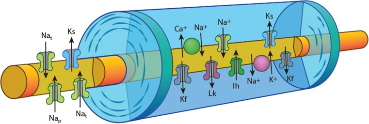
Fig. 1.
Schematic of axon structure and ion channel distribution, demonstrating the node of Ranvier with high density of voltage-gated Na+ channels [both transient (Nat) and persistent (Nap)] and slow K+ channels (Ks). The internode under the myelin sheath is depicted with fast K+ channels (Kf) located adjacent to the node, the hyperpolarization activated cation conductance (Ih), slow K+ channels, and voltage-independent leak conductances (Lk) along with the energy dependent Na+/K+ pump and the Na+/Ca2+ exchanger

