Abstract
Background:
A doughnut-shaped aneurysm, which is defined as a round-shaped aneurysm composed of an intraluminar thrombus and marginal parent artery, is an extremely uncommon subtype of partially thrombosed giant aneurysms. Surgical treatment of this characteristic aneurysm is technically challenging.
Case Description:
We report a rare case of a 79-year-old man with a symptomatic doughnut-shaped giant aneurysm at the A2 portion, which was successfully treated by outflow occlusion with an A3–A3 side-to-side anastomosis. Postoperative angiograms demonstrated no filling of the doughnut-shaped aneurysm and perfusion in the distal right anterior cerebral artery territory via the anastomosis. Follow-up magnetic resonance imaging 1 year after the surgery demonstrated significant diminution of the aneurysm.
Conclusions:
Outflow occlusion with distal revascularization could be an effective surgical option for such a unique aneurysm. To the best of our knowledge, this is the first report of outflow occlusion as a therapy for doughnut-shaped aneurysms.
Keywords: Anterior cerebral artery, bypass surgery, giant aneurysm, outflow occlusion, partially thrombosed aneurysm
INTRODUCTION
Giant aneurysms, those larger than 25 mm in diameter, are relatively uncommon and are often accompanied by thrombosis. Among such lesions, giant serpentine aneurysms have been described as a rare subcategory of partially thrombosed giant aneurysms.[6,8] They are uncommon fusiform arterial dilations in which the lumen extends longitudinally along the axis and curves of the parent artery, creating a serpentine pathway with separate inflow and outflow vessels. Doughnut-shaped aneurysms are giant round-shaped aneurysms composed of an intraluminar thrombus and marginal parent arteries.[6] Although complete obliteration of the aneurysm lumen with or without resection is an ideal treatment for such complex aneurysms, for some cases, it is difficult to achieve trapping and distal revascularization during surgery.[1] Here, we present a rare case of doughnut-shaped aneurysm that was successfully treated with outflow occlusion and A3–A3 side-to-side anastomosis.
CASE REPORT
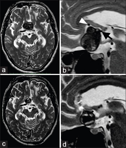
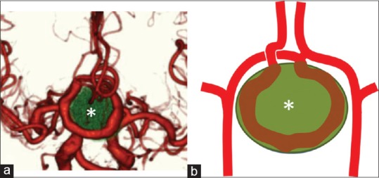
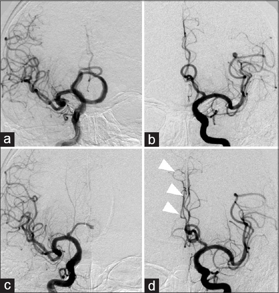
A 79-year-old man with no significant medical history presented with blurred vision for a few months. Native and contrast-enhanced magnetic resonance imaging (MRI) showed a heterogeneous round mass compressing the bilateral optic nerves and rectal gyri, which was 39 mm in maximum diameter, in the anterior interhemispheric fissure [Figure 1a and b]. Computed tomography angiography clearly revealed a round aneurysm composed of a wheeling A2 segment of the right anterior cerebral artery (ACA) and central thrombosis, suggesting a partially thrombosed aneurysm [Figure 2]. An outflow artery from the aneurysm arose from the top of the aneurysm under the genu of the corpus callosum, and an inflow artery was deeply located behind the aneurysm. Diagnostic cerebral angiography demonstrated it as an intraluminal filling defect shaped like a doughnut, which supplied no frontal branches [Figure 3a and b]. Considering the surgical risks and difficulties, we decided to perform outflow occlusion of the aneurysm with revascularization of the right distal ACA territory. A right-sided interhemispheric approach with a unilateral frontal craniotomy was performed. An A3–A3 side-to-side anastomosis was first performed. The outflow of the aneurysm was confirmed through the transcallosal route and was successfully occluded using a small aneurysm clip. The postoperative course was uneventful without any further neurological deterioration. Postoperative MRI showed no diffusion changes. Postoperative angiograms the day after the surgery demonstrated no filling of the doughnut-shaped aneurysm and perfusion in the distal right ACA territory via the anastomosis. Follow-up MRI 1 year after the surgery showed a significant decrease in the size of the aneurysm [Figure 1c and d].
Figure 1.

Preoperative axial (a) and mid-sagittal (b) T2-weighted images obtained on admission demonstrating a giant round mass at the frontal interhemispheric fissure, which was 39 mm in diameter. This lesion was largely thrombosed but incorporated flow void signals, which suggested a partially thrombosed aneurysm. Inflow (black arrow) and outflow (white arrowhead) arteries were located behind and at the upper surface of the aneurysm, respectively. Postoperative axial (c) and mid-sagittal (d) T2-weighted images obtained 1 year postoperatively showing significant reduction of the aneurysm
Figure 2.

A three-dimensional computed tomography angiogram (a) revealing a doughnut-shaped giant aneurysm at the A2 portion of the right anterior cerebral artery. The asterisk indicates intramural thrombosis. The entire schema of this case is a doughnut-shaped aneurysm (b)
Figure 3.

Preoperative right (a) and left (b) internal carotid angiograms demonstrating a giant aneurysm as a doughnut-shaped structure. Postoperative right (c) and left (d) internal carotid angiograms on the day after surgery revealing no filling of the aneurysm and right peripheral anterior cerebral arteries perfused by contralateral anterior cerebral arteries (arrowheads)
DISCUSSION
A giant serpentine aneurysm is defined as a partially thrombosed giant aneurysm with tortuous vascular channels forming a serpentine pathway with separate inflow and outflow tracts.[8] Pathogenesis is assumed by repeated dissection with intramural thrombosis,[1] and partial thrombosed aneurysms show various morphologies occasionally affected flow dynamics. Among these lesions, a round-shaped aneurysm with intramural thrombosis and a circumferentially flowing parent vessel is extremely rare. Rosta et al.[6] initially reported it as a doughnut-shaped aneurysm that is observed in only 1% of such intracranial giant aneurysms.
Optimal management of giant serpentine and doughnut-shaped aneurysms has not yet been established. In contrast to the usual saccular aneurysm, giant serpentine and doughnut-shaped aneurysms have separate inflow and outflow vessels; therefore, clipping the aneurysmal neck is unsuitable. For such cases, trapping of the involved segment with or without distal bypass is recommended. Proximal occlusion is also considered to be suitable for cases with surgical difficulty of trapping because proximal occlusion for aneurysms is believed to reduce the hemodynamic burden of the aneurysm, promote complete thrombosis in the aneurysm sac, and reduce the size of the aneurysm.[3] Recently, endovascular treatment has shown good results for large and giant aneurysms. However, the usefulness of coil embolization for partially thrombosed giant aneurysms remains controversial because of coil compaction and/or migration into the thrombus.
Outflow occlusion for the management of partially thrombosed aneurysms has been described in some reports.[4] In those, outflow occlusion with bypass for cases that are technically dangerous for proximal occlusion followed by rapid aneurysmal thrombosis. Horowitz et al.[4] described a mathematical model showing the intraluminal pressure changes that might be expected following outflow occlusion. They reported that the resulting variations in pressure should be less than those induced by normal daily activities and concluded that outflow occlusion would not be expected to increase the risk of an aneurysm rupture.
Recently, several groups have reported the efficacy and feasibility of bypass technique for the complex distal ACA aneurysm.[2,7] Bypass surgery to treat distal ACA aneurysms can be categorized as intracranial–intracranial (IC–IC) and extracranial–intracranial (EC–IC) types. IC–IC bypasses include in situ bypass (A3-A3 side-to-side anastomosis), reanastomosis, reimplantation, and bypass with graft placement. IC–IC bypass has several advantages. IC–IC bypass could provide enough hemodynamics to the target region without additional blood flow. Furthermore, IC–IC bypass do not require secondary incision and graft harvest. However, IC–IC bypass also has disadvantages. This maneuver is technically challenging in the narrow and deep working space in the interhemispheric fissure. In addition, if a bypass fails and occludes, both distal ACAs territories could develop serious ischemia. Conversely, EC–IC bypass has the disadvantage of requiring a secondary incision. However, EC–IC bypass has the advantage of an easier and safer procedure and a recovery option for the unexpected trouble in performing IC–IC anastomosis.[5] In our patient, because both inflow and outflow arteries were deeply situated between genu of corpus callosum and thrombosed giant aneurysm, bypass surgery around the aneurysm carries the risk of hemorrhagic and ischemic complication. Eventually, considering the availability in the depthless level at the same surgical field, A3–A3 side-to-side anastomosis was selected as the most appropriate revascularization.
In the present case, because the patient presented with visual symptoms due to aneurysmal mass effect, the best treatment might be aneurysm trapping with distal revascularization and aneurysmectomy. However, we anticipated that damage to the frontal lobe could not be avoided if occlusion of the inflow vessel was attempted in such a deep and narrow surgical field. Therefore, we chose outflow occlusion with an A3–A3 side-to-side anastomosis.
CONCLUSIONS
We experienced an extremely rare case of a doughnut-shaped giant aneurysm causing a mass effect. Surgical treatment of such an aneurysm is technically challenging. To the best of our knowledge, this is the first reported case of this entity that was successfully treated by outflow occlusion with distal vascular reconstruction.
Financial support and sponsorship
Nil.
Conflicts of interest
There are no conflicts of interest.
Footnotes
Contributor Information
Hidemichi Ito, Email: hdmcito@marianna-u.ac.jp.
Ryotaro Miyano, Email: r2miyano@marianna-u.ac.jp.
Taigen Sase, Email: sasetaigen@marianna-u.ac.jp.
Daisuke Wakui, Email: d2wakui@marianna-u.ac.jp.
Takashi Matsumori, Email: matsumori@marianna-u.ac.jp.
Hiroshi Takasuna, Email: hiroxneuro@marianna-u.ac.jp.
Kotaro Oshio, Email: koshio@marianna-u.ac.jp.
Yuichiro Tanaka, Email: tanaka@marianna-u.ac.jp.
REFERENCES
- 1.Day AL, Gaposchkin CG, Yu CJ, Rivet DJ, Dacey RG., Jr Spontaneous fusiform middle cerebral artery aneurysms: Characteristics and a proposed mechanism of formation. J Neurosurg. 2003;99:228–40. doi: 10.3171/jns.2003.99.2.0228. [DOI] [PubMed] [Google Scholar]
- 2.Dunn GP, Gerrard JL, Jho DH, Ogilvy CS. Surgical treatment of a large fusiform distal anterior cerebral artery aneusyrm with in situ end-to-side A3–A3 bypass graft and aneurysml trapping: Case report and review of the literature. Neurosurgery. 2011;68:E587–91. doi: 10.1227/NEU.0b013e3182036012. [DOI] [PubMed] [Google Scholar]
- 3.Hoh BL, Putman CM, Budzik RF, Carter BS, Oglivy CS. Combined surgical and endovascular techniques of flow alteration to treat fusiform and complex wide-necked intracranial aneurysms that are unsuitable for clipping or coil embolization. J Neurosurg. 2001;95:24–35. doi: 10.3171/jns.2001.95.1.0024. [DOI] [PubMed] [Google Scholar]
- 4.Horowitz MB, Yonas H, Jungreis C, Hung TK. Management of a giant middle cerebral artery fusiform serpentine aneurysm with distal clip application and retrograde thrombosis: Case report and review of the literature. Surg Neurol. 1994;41:221–5. doi: 10.1016/0090-3019(94)90126-0. [DOI] [PubMed] [Google Scholar]
- 5.Park ES, Ahn JS, Park JC, Kwon DH, Kwun BD, Kim CJ. STA-ACA bypass using the contralateral STA as an interposition graft for the treatment of complex ACA aneurysms: Report of two cases and a review of the literature. Acta Neurochir. 2012;154:1447–53. doi: 10.1007/s00701-012-1410-5. [DOI] [PubMed] [Google Scholar]
- 6.Rosta L, Battaglia R, Pasqualin A, Beltramello A. Italian cooperative study on giant intracranial aneurysms: 2. Radiological data. Acta Neurochir Suppl. 1998;42:53–9. doi: 10.1007/978-3-7091-8975-7_11. [DOI] [PubMed] [Google Scholar]
- 7.Sanai N, Zador Z, Lawton MT. Bypass surgery for complex brain aneurysms: An assessment of intracranial-intracranial bypass. Neurosurgery. 2009;65:670–83. doi: 10.1227/01.NEU.0000348557.11968.F1. [DOI] [PubMed] [Google Scholar]
- 8.Segal HD, McLaurin RL, Giant serpentine aneurysm. Report of two cases. J Neurosurg. 1977;46:115–20. doi: 10.3171/jns.1977.46.1.0115. [DOI] [PubMed] [Google Scholar]


