Abstract
Background
Pulmonary arterial hypertension (PAH) is a major cause of morbidity and mortality among patients with systemic sclerosis (SSc). Early detection and prompt treatment of PAH associated with SSc (SSc-PAH) result in better prognosis. We conducted echocardiographic study to presume the prevalence of PAH in Korean adult SSc patients and to diagnose SSc-PAH in their early stages with right heart catheterization (RHC).
Methods
We performed free of charge echocardiographic study including 37 adult SSc patients at the Chungnam National University Hospital. The possibility of PAH is determined by the estimation of pulmonary arterial pressure by peak tricuspid regurgitation velocity of > 3.0 m/s. Patients with possible PAH were recommended to undergo RHC to confirm the diagnosis.
Results
In 37 patients, 8 patients were suspected with PAH. Among them, 6 patients agreed to be examined with RHC, and 4 were confirmed with PAH. The prevalence of possible PAH was 21.6% (8 of 37 patients), and that of confirmed PAH was 10.8% (4 of 37 patients). Four patients who were confirmed with SSc-PAH through RHC have been treated with specific pulmonary vasodilators and maintained stable.
Conclusion
Eight patients (21.6%) were possible PAH and 4 (10.8%) were diagnosed as SSc-PAH by RHC after the echocardiographic screening study of 37 adult SSc patients.
Keywords: Systemic sclerosis, Pulmonary arterial hypertension, Echocardiography, Right heart catheterization, Screening
Introduction
Systemic sclerosis (SSc) is an uncommon connective tissue disease (CTD) of unknown cause and complex pathogenesis. The hallmarks of SSc include fibroblast dysfunction leading to increased deposition of extracellular matrix, vascular abnormality and production of autoantibodies.1) Pulmonary arterial hypertension (PAH) can be associated with SSc relatively frequently, and SSc associated PAH (SSc-PAH) is a leading cause of morbidity and mortality in patients with SSc.2) In one recent study regarding the SSc-related deaths, 26% of deaths resulted from PAH.3) Furthermore, compared to idiopathic PAH, SSc-PAH has a poorer response to the targeted therapy and a worse long-term survival rate.4),5),6)
Recent studies have shown that treatment in early stage resulted in better prognosis than advanced stage in patients with SSc-PAH.7),8) Because the echocardiographic study can estimate the pulmonary arterial pressure and is useful for screening in early stages, all patients with SSc are recommended to take an echocardiographic survey to find SSc-PAH.9) Because there have been several obstacles to performing annual echocardiographic screening in adult SSc patients in Korea, the precise prevalence of SSc-PAH is unknown. In this study, we planned to presume the prevalence of SSc-PAH in Korean adult patients with SSc through the echocardiography. If patients had the possibility of SSc-PAH without other causes of elevated pulmonary arterial pressure, they were recommended to receive a right heart catheterization (RHC) to confirm the diagnosis. Through this screening process, we hoped to give patients the opportunity to treat SSc-PAH in early stages.
Methods
Patients
All consecutive patients who had been diagnosed and managed with SSc were enrolled in this screening program from April 2013 to December 2015. The diagnosis of SSc was made following the 1980 American College of Rheumatology (ACR) preliminary criteria for the classification of SSc and the 2013 ACR/European league against rheumatism classification criteria for SSc.10),11) Patients who already had been diagnosed with PAH were excluded.
Method
Patients had more than one echocardiographic examination by two experienced cardiologists and a sonographer in this echocardiographic screening.
All participants underwent comprehensive 2-dimensional and Doppler echocardiography using an E9 echocardiographic machine with an M4S probe (GE Vingmed, Horten, Norway) and all echocardiographic images were stored digitally.
Echocardiographic dimensions were measured using M-mode echocardiography was performed on parasternal views. Left ventricular (LV) end-diastolic volume (LVEDV) and LV end-systolic volume values were measured using the biplane Simpson’s method on apical 4-chamber and 2-chamber views. LV ejection fraction were calculated as the division of stroke volume by LVEDV multiplied by 100. All conventional echocardiographic parameters were measured and averaged from 3 cardiac cycles.
The estimation of pulmonary artery systolic pressure was estimated from the maximal continuous-wave Doppler velocity of the tricuspid regurgitation (TR) jet plus estimated right atrial pressure with size of inferior vena cava and degree of change in caval diameter during respiration.12) TR Vmax value were averaged more than 3 beats and value more than 3.0 m/s (pulmonary arterial systolic pressure more than 41 mm Hg) was regarded as the possible PAH.
After the screening, the patients with possible PAH were recommended to undergo RHC to confirm the diagnosis. However, if patients had other causes of elevating pulmonary arterial pressure, the RHC was postponed after the correction of the causes. The PAH is defined as a mean pulmonary arterial pressure of ≥ 25 mm Hg at rest with a pulmonary capillary wedge pressure of < 15 mm Hg, and a pulmonary vascular resistance (PVR) > 3.0 Wood units in the absence of other causes of precapillary pulmonary hypertension (PH) such as PH due to lung diseases, chronic thromboembolic PH or other rare diseases.13) The study protocol had been reviewed and approved by the Institutional Review Board of Chungnam National University Hospital (IRB number is 2016-08-018). All participants gave their consent to collect and use their personal information.
Statistical analysis
Differences between non-PAH and confirmed PAH by RHC groups were analyzed. Statistical analysis was done with a commercial program (IBM SPSS Statistics for Windows, version 20.0, IBM Corp., Armonk, NY, USA). Data was expressed as mean standard deviation for continuous variables and as frequencies (percentages) for categorical variables. Statistical differences between groups were assessed using Fisher’s exact tests for categorical variables and Mann-Whitney U test for continuous variables. p < 0.05 was considered statistically significant.
Results
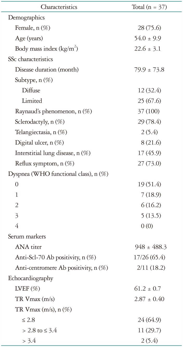
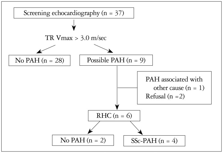
We enrolled 37 patents in screening program with echocardiography. All patients underwent more than one echocardiographic examination, and 7 patients received echocardiographic studies 2 times for their follow-up evaluation. Patients’ baseline characteristics are summarized in Table 1. Of the total 37 adult patients with SSc, 9 were suspected to have PAH. Out of the 9 patients, 1 was found to have increased pulmonary arterial pressure due to another cause, 2 refused to go through with the RHC, and 6 agreed to be examined through RHC. Four of the 6 patients who underwent the RHC were confirmed with PAH. The process and result of this study was summarized in the Fig. 1. The comparison of clinical and laboratory parameters according to the presence of PAH was summarized in the Supplementary Table 1. The characteristics of patients who underwent the RHC and their results of RHC are presented in Table 2 and 3. The percentage of possible PAH was 21.6 (8 of 37 patients). And that of confirmed PAH was 10.8 (4 of 37 patients). SSc-PAH patients seemed to be older than control patients. Three among 4 patients with confirmed SSc-PAH had limited type of SSc. Patients with confirmed SSc-PAH complained more severe grade of dyspnea. Two of 6 patients who underwent RHC were not diagnosed as PAH. One patient who was diagnosed as mildly elevated PH (mean PA pressure was 26 mm Hg) without elevated PVR (PVR was 231 dynes·sec·cm2) had been diagnosed as PAH about 11 months after the first RHC. She had been managed with oral sildenafil treatment. However, the patient expired from renal crisis about 14 months after the RHC.
Table 1. Baseline characteristics.
SSc: systemic sclerosis, WHO: World Health Organization, ANA: antinuclear antibody, LVEF: left ventricular ejection fraction, TR Vmax: maximal velocity of tricuspid regurgitation
Fig. 1. The flow chart of this study. TR Vmax: maximal velocity of tricuspid regurgitation, PAH: pulmonary arterial hypertension, RHC: right heart catheterization, SSc-PAH: pulmonary arterial hypertension associated with systemic sclerosis.
Table 2. The characteristics of patients with pulmonary arterial hypertension associated with SSc.
BMI: body mass index: WHO FC: World Health Organization-functional class, SSc: systemic sclerosis, ANA: antinuclear antibody, PFT: pulmonary function test, DLCO: diffusing capacity of the lungs for carbon monoxide, FVC: forced vital capacity, Pro-BNP: pro-b type natriuretic peptide, NC: not checked
Table 3. The results of echocardiography and RHC of patients with pulmonary arterial hypertension associated with systemic sclerosis.
RHC: right heart catheterization, LVEF: left ventricular ejection fraction, TR Vmax: maximal tricuspid regurgitation velocity, IVC: inferior vena cava, E/A: ratio of mitral E velocity by A velocity, E/e′: ratio of mitral E velocity by mitral annular e′ velocity, SPAP: systolic pulmonary artery pressure, mPAP: mean pulmonary artery pressure, PCWP: pulmonary capillary wedge pressure, PVR: pulmonary vascular resistance, CO: cardiac output
One patient who refused RHC had no pulmonary symptom and her initial TR Vmax was 3.1 m/sec. In the follow-up echocardiography after 32 months, her TR Vmax was decreased to 3.1 to 2.8 m/sec. The other patient complained of dyspnea with World Health Organization grade 1 with an initial TR Vmax of 3.6 m/sec.
Another patient, who was suspected to have PAH, had anemia and hypertension which can cause reactive PH. In this patient, the elevated TR Vmax was normalized with iron supply and antihypertensive medications within about 6 months.
The patients who were confirmed with PAH have been treated with specific pulmonary vasodilator medications including bosentan and conventional therapy. With these medications 2 patients have been improving and other 2 patients have maintained stable disease activity.
Discussion
In this pilot echocardiographic study among 37 Korean adult patients with SSc, we found 8 possible patients (21.6%) and 4 confirmed patients (10.8%) with SSc-PAH.
PH is one of the major causes of death in patients with CTD.14) In recently published data of Korean PAH registry including 625 patients, CTD was the most common etiology (49.8%).15) In CTD-associated PAH, SSc was the second common etiology of PAH secondary to systemic lupus erythematosus in Korea. CTD-associated PAH showed poor prognosis.15),16) Also, early treatment with specific vasodilators can decrease mortality in these patients. So, the screening study of PAH in patients with CTD is necessary. In this pilot study, we found the prevalence of PAH was 10.8%. We started PAH specific therapy in these patients, and 2 of them showed improvement and the other 2 remained stationary.
Because echocardiography is the best imaging tool for the screening of PAH, recent guidelines recommend yearly echocardiographic screening in high-risk patients of PAH.9) Such high-risk patients include those with bone morphogenic protein receptor type II mutation, first degree relatives of familial PAH patients, and SSc patients. However, the echocardiography has been used only in suspected patients with SSc because of its relatively high cost in Korea. Moreover, there is little data about the prevalence of SSc-PAH in Korea. Fortunately, Korean insurance organization has recently begun to cover echocardiographic screening.
SSc occurs more commonly in the African, Asian, and some Native American populations in the USA than in individuals of European descent.17) SSc associated mortality has been largely caused by renal disease, but with the advent of angiotensin-converting enzyme inhibitors to treat SSc renal crisis, SSc-PAH has emerged as a leading cause of morbidity and mortality in patients with SSc accounting for up to 30% of premature deaths.18) The treatment for PAH has evolved progressively in the past decade, increasing in complexity and in evidence for efficacy,9),19) especially because it has been known that treatment in the early stages result in better prognosis than in the advanced stages in patients with SSc-PAH.
In studies about SSc-PAH, the prevalence of PAH has been reported of up to 15% in patients with SSc.20),21) Although our study has small numbers of patients, the percentage of SSc-PAH was 10.8%, and the result was compatible with previous studies. However, large screening programs should be needed to evaluate precise prevalence rate in Korea.
Many patients with SSc-PAH are not diagnosed early because there is no symptom in early stages. Among patients with possible PAH that we screened with an echocardiography, 2 patients had no pulmonary or cardiologic symptoms in ordinary physical activity at the time of the screening. Because the patients did not have symptoms, they refused to receive RHC, unfortunately.
This study has several limitations. First, the study population is too small, and was carried out in only 1 center in Korea. However, we tried to include all patients with SSc in our institution. Further studies with a larger study population will be needed in order for this data to be generalized. Second, we can’t perform the RHC in 2 patients with suspected PAH.
In conclusion, we performed an echocardiographic study to 37 Korean adult patients with SSc. The percentage of possible PAH was 21.6 (8/37) and that of confirmed SSc-PAH with RHC was 10.8 (4/37). Because early treatment can have treatment benefits in SSc-PAH patients in their early stages, echocardiographic screening is crucial even if there is no symptom associated with PAH.
Acknowledgements
We deeply appreciated Seonhee Jeon in Handok pharmaceutical company for her enthusiastic assist in the arrangement of patients.
Supplementary materials
The online-only Data Supplement is available with this article at https://doi.org/10.4250/jcu.2016.24.4.312.
Comparison of clinical and laboratory characteristics according to the presence of PAH
References
- 1.Wollheim FA. Classification of systemic sclerosis. Visions and reality. Rheumatology(Oxford) 2005;44:1212–1216. doi: 10.1093/rheumatology/keh671. [DOI] [PubMed] [Google Scholar]
- 2.Dimitroulas T, Mavrogeni S, Kitas GD. Imaging modalities for the diagnosis of pulmonary hypertension in systemic sclerosis. Nat Rev Rheumatol. 2012;8:203–213. doi: 10.1038/nrrheum.2012.2. [DOI] [PubMed] [Google Scholar]
- 3.Tyndall AJ, Bannert B, Vonk M, Airò P, Cozzi F, Carreira PE, Bancel DF, Allanore Y, Müller-Ladner U, Distler O, Iannone F, Pellerito R, Pileckyte M, Miniati I, Ananieva L, Gurman AB, Damjanov N, Mueller A, Valentini G, Riemekasten G, Tikly M, Hummers L, Henriques MJ, Caramaschi P, Scheja A, Rozman B, Ton E, Kumánovics G, Coleiro B, Feierl E, Szucs G, Von Mühlen CA, Riccieri V, Novak S, Chizzolini C, Kotulska A, Denton C, Coelho PC, Kötter I, Simsek I, de la Pena Lefebvre PG, Hachulla E, Seibold JR, Rednic S, Stork J, Morovic-Vergles J, Walker UA. Causes and risk factors for death in systemic sclerosis: a study from the EULAR Scleroderma Trials and Research (EUSTAR) database. Ann Rheum Dis. 2010;69:1809–1815. doi: 10.1136/ard.2009.114264. [DOI] [PubMed] [Google Scholar]
- 4.Kawut SM, Taichman DB, Archer-Chicko CL, Palevsky HI, Kimmel SE. Hemodynamics and survival in patients with pulmonary arterial hypertension related to systemic sclerosis. Chest. 2003;123:344–350. doi: 10.1378/chest.123.2.344. [DOI] [PubMed] [Google Scholar]
- 5.Rubin LJ, Badesch DB, Barst RJ, Galie N, Black CM, Keogh A, Pulido T, Frost A, Roux S, Leconte I, Landzberg M, Simonneau G. Bosentan therapy for pulmonary arterial hypertension. N Engl J Med. 2002;346:896–903. doi: 10.1056/NEJMoa012212. [DOI] [PubMed] [Google Scholar]
- 6.Fisher MR, Mathai SC, Champion HC, Girgis RE, Housten-Harris T, Hummers L, Krishnan JA, Wigley F, Hassoun PM. Clinical differences between idiopathic and scleroderma-related pulmonary hypertension. Arthritis Rheum. 2006;54:3043–3050. doi: 10.1002/art.22069. [DOI] [PubMed] [Google Scholar]
- 7.Humbert M, Yaici A, de Groote P, Montani D, Sitbon O, Launay D, Gressin V, Guillevin L, Clerson P, Simonneau G, Hachulla E. Screening for pulmonary arterial hypertension in patients with systemic sclerosis: clinical characteristics at diagnosis and long-term survival. Arthritis Rheum. 2011;63:3522–3530. doi: 10.1002/art.30541. [DOI] [PubMed] [Google Scholar]
- 8.Galiè N, Rubin Lj, Hoeper M, Jansa P, Al-Hiti H, Meyer G, Chiossi E, Kusic-Pajic A, Simonneau G. Treatment of patients with mildly symptomatic pulmonary arterial hypertension with bosentan (EARLY study): a double-blind, randomised controlled trial. Lancet. 2008;371:2093–2100. doi: 10.1016/S0140-6736(08)60919-8. [DOI] [PubMed] [Google Scholar]
- 9.Galiè N, Humbert M, Vachiery JL, Gibbs S, Lang I, Torbicki A, Simonneau G, Peacock A, Vonk Noordegraaf A, Beghetti M, Ghofrani A, Gomez Sanchez MA, Hansmann G, Klepetko W, Lancellotti P, Matucci M, McDonagh T, Pierard LA, Trindade PT, Zompatori M, Hoeper M, Aboyans V, Vaz Carneiro A, Achenbach S, Agewall S, Allanore Y, Asteggiano R, Paolo Badano L, Albert Barberà J, Bouvaist H, Bueno H, Byrne RA, Carerj S, Castro G, Erol Ç, Falk V, Funck-Brentano C, Gorenflo M, Granton J, Iung B, Kiely DG, Kirchhof P, Kjellstrom B, Landmesser U, Lekakis J, Lionis C, Lip GY, Orfanos SE, Park MH, Piepoli MF, Ponikowski P, Revel MP, Rigau D, Rosenkranz S, Völler H, Luis Zamorano J. 2015 ESC/ERS Guidelines for the diagnosis and treatment of pulmonary hypertension: The Joint Task Force for the Diagnosis and Treatment of Pulmonary Hypertension of the European Society of Cardiology (ESC) and the European Respiratory Society (ERS): Endorsed by: Association for European Paediatric and Congenital Cardiology (AEPC), International Society for Heart and Lung Transplantation (ISHLT) Eur Heart J. 2016;37:67–119. [Google Scholar]
- 10.Subcommittee for scleroderma criteria of the American Rheumatism Association Diagnostic and Therapeutic Criteria Committee. Preliminary criteria for the classification of systemic sclerosis (scleroderma) Arthritis Rheum. 1980;23:581–590. doi: 10.1002/art.1780230510. [DOI] [PubMed] [Google Scholar]
- 11.van den Hoogen F, Khanna D, Fransen J, Johnson SR, Baron M, Tyndall A, Matucci-Cerinic M, Naden RP, Medsger TA, Jr, Carreira PE, Riemekasten G, Clements PJ, Denton CP, Distler O, Allanore Y, Furst DE, Gabrielli A, Mayes MD, van Laar JM, Seibold JR, Czirjak L, Steen VD, Inanc M, Kowal-Bielecka O, Müller-Ladner U, Valentini G, Veale DJ, Vonk MC, Walker UA, Chung L, Collier DH, Ellen Csuka M, Fessler BJ, Guiducci S, Herrick A, Hsu VM, Jimenez S, Kahaleh B, Merkel PA, Sierakowski S, Silver RM, Simms RW, Varga J, Pope JE. 2013 classification criteria for systemic sclerosis: an American college of rheumatology/European league against rheumatism collaborative initiative. Ann Rheum Dis. 2013;72:1747–1755. doi: 10.1136/annrheumdis-2013-204424. [DOI] [PubMed] [Google Scholar]
- 12.Grapsa J, Dawson D, Nihoyannopoulos P. Assessment of right ventricular structure and function in pulmonary hypertension. J Cardiovasc Ultrasound. 2011;19:115–125. doi: 10.4250/jcu.2011.19.3.115. [DOI] [PMC free article] [PubMed] [Google Scholar]
- 13.Hoeper MM, Bogaard HJ, Condliffe R, Frantz R, Khanna D, Kurzyna M, Langleben D, Manes A, Satoh T, Torres F, Wilkins MR, Badesch DB. Definitions and diagnosis of pulmonary hypertension. J Am Coll Cardiol. 2013;62(25 Suppl):D42–D50. doi: 10.1016/j.jacc.2013.10.032. [DOI] [PubMed] [Google Scholar]
- 14.Simonneau G, Robbins IM, Beghetti M, Channick RN, Delcroix M, Denton CP, Elliott CG, Gaine SP, Gladwin MT, Jing ZC, Krowka MJ, Langleben D, Nakanishi N, Souza R. Updated clinical classification of pulmonary hypertension. J Am Coll Cardiol. 2009;54(1 Suppl):S43–S54. doi: 10.1016/j.jacc.2009.04.012. [DOI] [PubMed] [Google Scholar]
- 15.Chung WJ, Park YB, Jeon CH, Jung JW, Ko KP, Choi SJ, Seo HS, Lee JS, Jung HO KORPAH Investigators. Baseline characteristics of the Korean registry of pulmonary arterial hypertension. J Korean Med Sci. 2015;30:1429–1438. doi: 10.3346/jkms.2015.30.10.1429. [DOI] [PMC free article] [PubMed] [Google Scholar]
- 16.Chung L, Liu J, Parsons L, Hassoun PM, McGoon M, Badesch DB, Miller DP, Nicolls MR, Zamanian RT. Characterization of connective tissue disease-associated pulmonary arterial hypertension from REVEAL: identifying systemic sclerosis as a unique phenotype. Chest. 2010;138:1383–1394. doi: 10.1378/chest.10-0260. [DOI] [PMC free article] [PubMed] [Google Scholar]
- 17.Mathai SC, Hassoun PM. Pulmonary arterial hypertension associated with systemic sclerosis. Expert Rev Respir Med. 2011;5:267–279. doi: 10.1586/ers.11.18. [DOI] [PMC free article] [PubMed] [Google Scholar]
- 18.Steen VD, Medsger TA. Changes in causes of death in systemic sclerosis, 1972-2002. Ann Rheum Dis. 2007;66:940–944. doi: 10.1136/ard.2006.066068. [DOI] [PMC free article] [PubMed] [Google Scholar]
- 19.Williams MH, Das C, Handler CE, Akram MR, Davar J, Denton CP, Smith CJ, Black CM, Coghlan JG. Systemic sclerosis associated pulmonary hypertension: improved survival in the current era. Heart. 2006;92:926–932. doi: 10.1136/hrt.2005.069484. [DOI] [PMC free article] [PubMed] [Google Scholar]
- 20.Mukerjee D, St George D, Coleiro B, Knight C, Denton CP, Davar J, Black CM, Coghlan JG. Prevalence and outcome in systemic sclerosis associated pulmonary arterial hypertension: application of a registry approach. Ann Rheum Dis. 2003;62:1088–1093. doi: 10.1136/ard.62.11.1088. [DOI] [PMC free article] [PubMed] [Google Scholar]
- 21.Mukerjee D, St George D, Knight C, Davar J, Wells AU, Du Bois RM, Black CM, Coghlan JG. Echocardiography and pulmonary function as screening tests for pulmonary arterial hypertension in systemic sclerosis. Rheumatology (Oxford) 2004;43:461–466. doi: 10.1093/rheumatology/keh067. [DOI] [PubMed] [Google Scholar]
Associated Data
This section collects any data citations, data availability statements, or supplementary materials included in this article.
Supplementary Materials
Comparison of clinical and laboratory characteristics according to the presence of PAH


