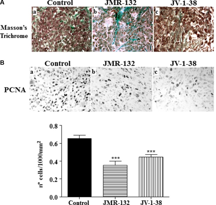
Figure 4. Effect of the GHRH antagonists JMR-132 (10 μg/day) and JV-1-38 (20 μg/day) on PC3 tumor proliferation.
(A) Histological sections from tumors stained with Masson's Trichrome. Control tumors presented scarce connective tissue between tumor cells (a); however, tumors from xenografts (b, c) presented more ground substance and areas of collagen fibers bundles. Original magnification X300. (B) Immunohistochemistry using a specific antibody against PCNA was performed. The highest number of proliferating cells was observed in control samples (black nuclei). Original magnification X300. The evaluation of the number of PCNA-immunoreactive nuclei is shown on graphical representation. Data in each bar are the means ± SEM. ***p < 0.001 vs. control.

