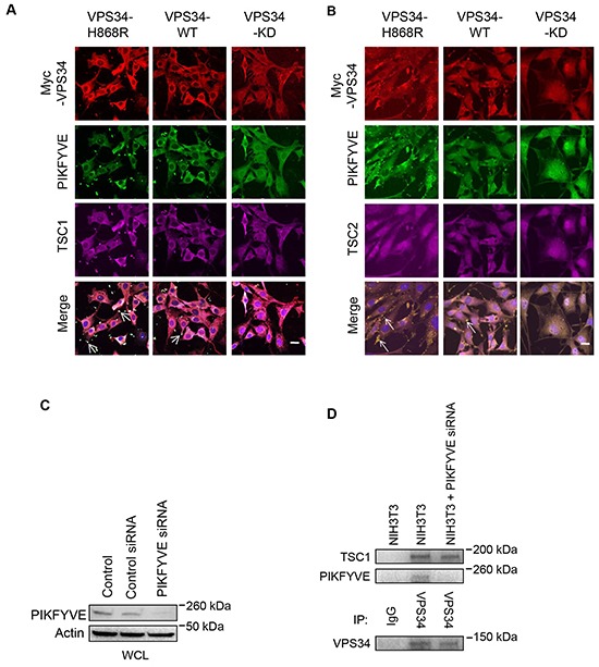
Figure 4. VPS34 co-localizes with TSC1 and PIKFYVE, but not with TSC2 at the plasma membrane.

A. NIH3T3 cells were transfected with Myc-tagged VPS34 proteins. Cells were immunostained for Myc-VPS34 (red), PIKFYVE (green) and TSC1 (magenta). Bar: 20 μm. The white color indicates colocalization of VPS34, PIKFYVE, and TSC1 (Arrows in merged images). B. Experimental procedures were essentially the same as that described in (A) except cells were immunostained for TSC2 (magenta). Yellow color indicates colocalization of VPS34 and PIKFYVE (arrows in merged images). Bar: 20 μm. All images in Figure 4 were captured on Zeiss LSM-510 Meta microscope. C. NIH3T3 cells were transfected with control and PIKFYVE siRNA, and Western blot analysis was performed to show PIKFYVE knockdown. D. NIH3T3 cells were transfected with PIKFYVE siRNA for 48h or left un-transfected. Indicated WCL were subjected to immunoprecipitation using anti-VPS34 antibody or control IgG. The levels of TSC1, PIKFYVE and VPS34 were detected by Western blotting in immunoprecipitated samples.
