Abstract
Context:
A number of controversies exist regarding appropriate treatment strategy for Vitamin D deficiency.
Aims:
The aim of this study was to investigate the efficacy of equivalent doses of oral cholecalciferol (60,000 IU weekly for 5 weeks) versus intramuscular (IM) cholecalciferol (300,000 IU) in correcting Vitamin D deficiency in apparently healthy volunteers working in a hospital.
Settings and Design:
Prospective randomized open-label single institution study.
Subjects and Methods:
This study enrolled 40 apparently healthy adults with Vitamin D deficiency into 2 arms. The oral cholecalciferol group (n = 20) received oral cholecalciferol 60,000 IU weekly for 5 weeks while the IM cholecalciferol group (n = 20) received a single injection of cholecalciferol 300,000 IU. The main outcome measure was serum 25-hydroxyvitamin D (25OHD) levels at baseline, 6 and 12 weeks after the intervention.
Statistical Analysis Used:
Differences in serum 25OHD and other biochemical parameters at baseline and follow-up were analyzed using general linear model.
Results:
Mean 25OHD level at baseline was 5.99 ± 1.07 ng/mL and 7.40 ± 1.13 ng/mL (P = 0.332) in the oral cholecalciferol and IM cholecalciferol group, respectively. In the oral cholecalciferol group, serum 25OHD level was 20.20 ± 1.65 ng/mL at 6 weeks and 16.66 ± 1.36 ng/mL at 12 weeks. The corresponding serum 25OHD levels in the IM cholecalciferol group were 20.74 ± 1.81 ng/mL and 25.46 ± 1.37 ng/mL at 6 and 12 weeks, respectively. At 12 weeks, the mean 25OHD levels in IM cholecalciferol group was higher as compared to the oral cholecalciferol group (25.46 ± 1.37 vs. 16.66 ± 1.36 ng/mL; P < 0.001).
Conclusions:
Both oral and IM routes are effective for the treatment of Vitamin D deficiency. 25-hydroxyvitamin D levels in the IM cholecalciferol group showed a sustained increase from baseline.
Keywords: Intramuscular cholecalciferol, oral cholecalciferol, Vitamin D deficiency, Vitamin D replacement
INTRODUCTION
Hypovitaminosis D is widely prevalent in most regions of the world.[1] Several studies have demonstrated that despite being replete in sunshine, Vitamin D deficiency is rampant across India.[2,3,4,5,6,7,8] Data from apparently healthy individuals suggest high prevalence of Vitamin D deficiency ranging from 70% to 100%.[9]
The studies have shown that serum 25-hydroxyvitamin D (25OHD) levels of around 30 ng/mL are optimal for bone health and extraskeletal effects.[10] Both cholecalciferol (Vitamin D3) and ergocalciferol (Vitamin D2) have been used for the treatment of Vitamin D deficiency. However, there is lack of consensus regarding the dosing schedule to achieve this level and also the route of administration. The Endocrine Society recommends all adults who are Vitamin D deficient be treated with 50,000 IU of Vitamin D2 or Vitamin D3 once a week for 8 weeks or its equivalent of 6000 IU of Vitamin D2 or Vitamin D3 daily to achieve a serum 25OHD above 30 ng/mL, followed by maintenance therapy of 1500–2000 IU/d.[11] This recommendation does not take into account the severity of deficiency or the body weight of the individual. A study from India has shown that despite normalization of serum 25OHD after 60,000 IU oral weekly dose schedule, at the end of 12 months, all the subjects became Vitamin D deficient, once again.[12] A number of controversies exist regarding appropriate treatment strategy for Vitamin D deficiency: vitamin D3 versus Vitamin D2, oral versus intramuscular (IM) administration, fixed or titrated dosing strategy, lower daily dose or higher intermittent dose.[13] In addition, the long-term bioavailability data of parenteral Vitamin D are scarce.
This study evaluated the efficacy and tolerability of oral cholecalciferol (60,000 IU) versus IM cholecalciferol (300,000 IU) in correcting Vitamin D deficiency in Vitamin D deficient apparently healthy individuals working in a tertiary care hospital.
SUBJECTS AND METHODS
Subject selection and study protocol
This was a prospective, randomized, open-label single institution study in which 40 adults with Vitamin D deficiency were studied. Subjects were otherwise healthy resident doctors, nursing staff, and employees of the hospital without any overt symptoms of Vitamin D deficiency and other metabolic bone diseases who volunteered to be enrolled in the study protocol. Vitamin deficiency was defined as serum 25OHD levels <30 ng/mL.[14] Recruitment was done in the months of September to November.
Subjects, with any disease, known to affect mineral metabolism were excluded from the study. Subjects with any chronic medical illnesses and chronic drug intake were excluded from the study. Before recruitment, a written informed written consent was taken from the study subjects and the study was approved by the Ethics Committee. Forty subjects with Vitamin D insufficiency/deficiency were recruited.
Twenty subjects selected by simple randomization done in the ratio of 1:1 (regardless of their Vitamin D status) were given oral cholecalciferol 60,000 IU weekly for 5 weeks, and other 20 were given injection of cholecalciferol 300,000 IU intramuscularly once. Single batch of both the oral and injection cholecalciferol was procured from identical pharmaceutical companies and used for the supplementation.
Investigations
Fasting blood samples were collected at baseline and at 6 and 12 weeks of intervention. Serum calcium (albumin-adjusted), phosphorus, alkaline phosphatase (ALP), 25OHD, and intact parathyroid hormone (iPTH) levels were measured at each visit. Serum calcium, phosphorus, and ALP levels were determined the same day. Sera for 25OHD and PTH were stored at −40° centigrade until measurement. Serum iPTH was determined by chemiluminescence method. Serum 25OHD was determined by radioimmunoassay method.
Statistical analysis
Numeric variables are presented using mean and standard error of mean. Data analysis was done using SPSS (version 14; SPSS, Inc., Chicago, IL, USA). Differences in serum 25OHD and other biochemical parameters at baseline and follow-up were analyzed using general linear model: mixed-effect regression model for between group comparison and repeated-measures ANOVA for within-group comparisons. P ≤ 0.05 was considered to be statistically significant.
RESULTS
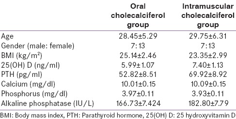
Forty subjects were enrolled into the study in groups of 20 each for the oral and IM cholecalciferol group. Table 1 compares the baseline characteristics of the intervention groups (oral vs. IM cholecalciferol). There was no difference in the baseline characteristics between the two groups. None of the subjects had baseline hypercalcemia.
Table 1.
Baseline characteristic of the study population
Difference in 25-hydroxyvitamin D values over time between two groups
The mean serum 25OHD level at baseline was 5.99 ± 1.07 ng/mL and 7.40 ± 1.13 ng/mL in the oral and IM cholecalciferol group, respectively and was not statistically significant (P = 0.332). Serum 25OHD level increased to 20.20 ± 1.65 ng/mL at 6 weeks followed by decline to 16.66 ± 1.36 ng/mL at 12 weeks in the oral cholecalciferol group. Increase in serum 25OHD levels at 6 and 12 weeks from baseline was statistically significant (P < 0.001). However, there was no significant difference in the mean serum 25OHD at weeks 6 and 12 in the oral cholecalciferol group. In the IM cholecalciferol group serum, 25OHD levels increased to 20.74 ± 1.81 ng/mL and 25.46 ± 1.37 ng/mL at 6 and 12 weeks, respectively. Within-group comparison using ANOVA showed a statistically significant difference in serum 25OHD levels at 6 and 12 weeks when compared to the baseline [Table 2]. At 12 weeks, the mean serum 25OHD levels in IM cholecalciferol group were higher as compared to the oral D3 group (25.46 ± 1.37 vs. 16.66 ± 1.36 ng/mL; P < 0.001). Figure 1 shows the trend in 25OHD levels during the study period.
Table 2.
25 hydroxyvitamin D and other parameters before and after intervention
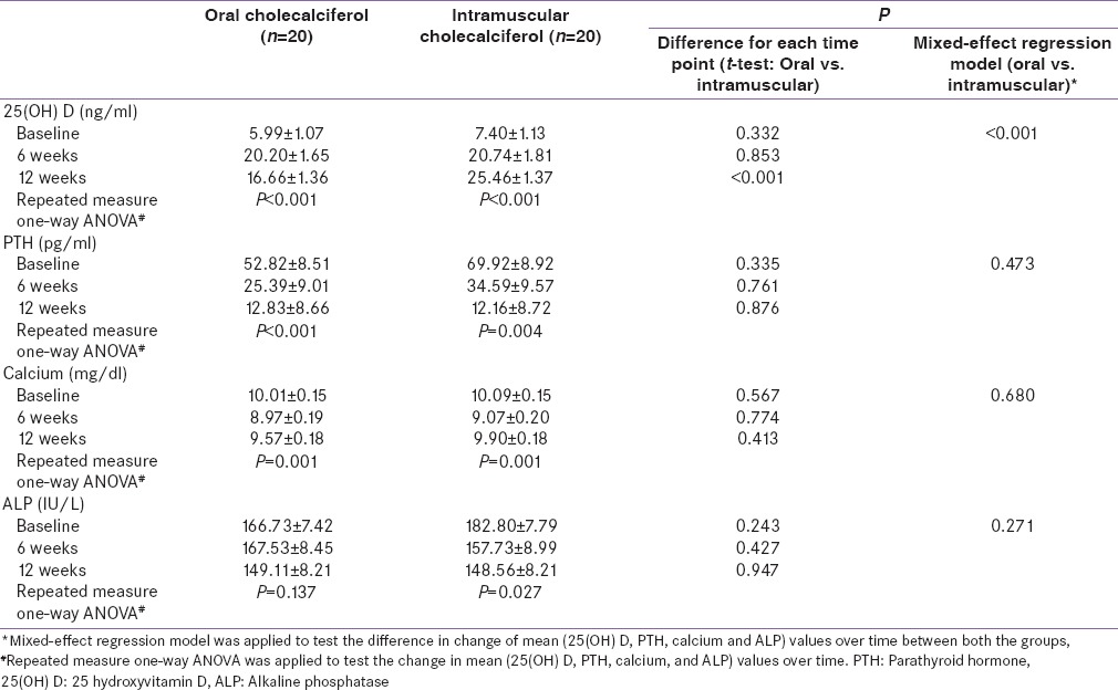
Figure 1.
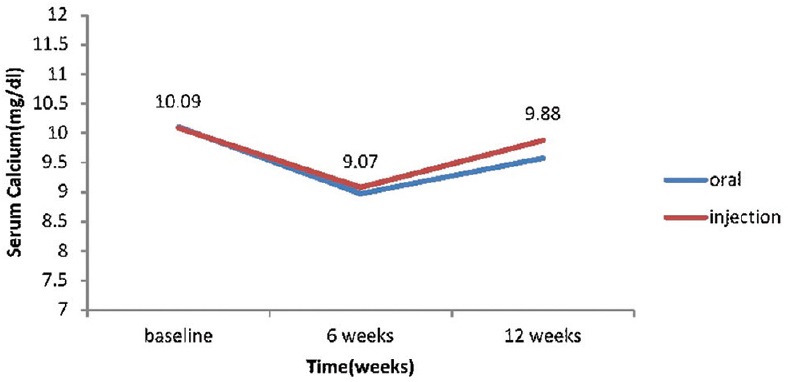
Serum calcium values at baseline and during follow-up in oral cholecalciferol and intramuscular cholecalciferol group
Difference in calcium values over time between two groups
Within-group comparison in both oral cholecalciferol and IM, cholecalciferol group showed significant changes in the serum calcium levels from baseline [Table 2]. The mean serum calcium was 10.01 ± 0.15 mg/dL and 10.09 ± 0.15 mg/dL, respectively in the oral cholecalciferol and IM cholecalciferol group (P = 0.567) and showed significant decline at 6 weeks followed by rise at 12 weeks. The mixed-effect regression model did not show any statistically significant difference in the mean calcium levels between the oral cholecalciferol and IM cholecalciferol at 12 weeks (P = 0.680). Figure 2 shows the trend in serum calcium values during the study period.
Figure 2.
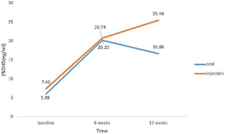
25-hydroxyvitamin D values at baseline and during follow-up in oral cholecalciferol and intramuscular cholecalciferol group
Difference in alkaline phosphatase values over time between two groups
Within-group comparison in the oral cholecalciferol group did not reveal any statistically significant change in the mean ALP values at baseline, 6, and 12 weeks (166.73 ± 7.42, 167.53 ± 8.45, and 149.11 ± 8.21 IU/L, respectively; P = 0.137). However, the ALP levels showed a statistically significant progressive decline in IM cholecalciferol group (182.80 ± 7.79, 157.73 ± 8.99, 148.56 ± 8.21 IU/L, respectively; P = 0.027). There was no difference in the change in ALP levels at 12 weeks in between the oral cholecalciferol and IM cholecalciferol group (P = 0.271).
Difference in parathyroid hormone values over time between two groups
Mean PTH levels at baseline in the oral cholecalciferol and IM cholecalciferol group were 52.82 ± 8.51 and 69.92 ± 8.92 pg/mL, respectively (P = 0.335). Both oral and IM route led to significant reductions in PTH from baseline at 6 and 12 weeks [Table 2]. Between-group comparison did not show any difference in PTH at 12 weeks between the oral and IM cholecalciferol group (P = 0.473). Figure 3 shows the iPTH levels at baseline and during follow-up in oral cholecalciferol and IM cholecalciferol group.
Figure 3.
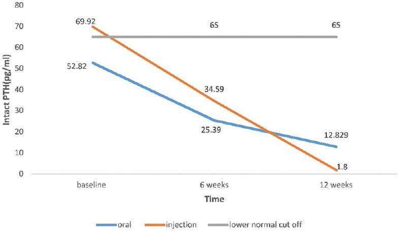
Intact parathyroid hormone levels at baseline and during follow-up in oral cholecalciferol and intramuscular cholecalciferol group
No adverse reaction such as injection site abscess, erythema, or cellulitis was reported during the study in either treatment groups. Serum calcium levels remained within normal limits in all patients at each time point in both studies.
DISCUSSION
Considerable confusion exists regarding the appropriate method for treatment of Vitamin D deficiency. The availability of multiple Vitamin D3 preparations (oral, parenteral) and lack of globally accepted repletion regimens further compound the problem. This study evaluated the response of different routes of Vitamin D3 administration (oral vs. IM) on serum 25OHD levels in apparently healthy adults with Vitamin D deficiency. In this study, both intervention groups showed improvement in serum 25OHD levels at the completion of the study. However, the IM cholecalciferol group showed a significant rise in 25OHD levels as compared to the oral cholecalciferol group.
The mean serum 25OHD level at baseline in the oral cholecalciferol group was 7.40 ± 1.13 ng/mL and increased to 20.20 ± 1.65 ng/mL at 6 weeks and then decreased to 16.66 ± 1.36 ng/mL at 12 weeks. Whyte et al. showed that the levels of 25(OH) D rise rapidly and peak about 1 week after dosing, and their peak is not sustained, if supplementation is not continued or if not started on maintenance dosage.[15] The mean 25OHD level at baseline in IM D3 group was 5.99 ± 1.07 ng/mL and increased to 20.74 ± 1.81 ng/mL at 6 weeks (nearly 3 times the baseline value) and 25.46 ± 1.37 ng/mL (nearly 4 times the baseline value) at 12 weeks. In this arm, about six subjects out of 20 achieved levels of > 30 ng/mL and 8 out of 20 achieved the level of 20 ng/mL. Mean serum 25OHD levels remained below 30 ng/mL in both oral and IM cholecalciferol group.
The mean serum levels of 25OHD achieved at 6 weeks were comparable in oral cholecalciferol and IM cholecalciferol group; however at 12 weeks, IM cholecalciferol group showed a higher 25OHD level (25.46 ± 1.37 vs. 16.66 ± 1.36). Mawer et al. have surmised that Vitamin D, that is, given orally associates with lipoproteins and enters the liver where some of it is metabolized by hepatic 25-hydroxylase and some gets inactivated. This can explain the greater but more transient serum 25OHD increases after a single oral dose of cholecalciferol.[16] Our results are similar to other studies comparing two different routes for Vitamin D supplementation. Cipriani et al. showed that oral dose of 600,000 IU of D2 or D3 is initially more effective in increasing serum 25OHD than the equivalent IM dose and is rapidly metabolized.[17] The depot IM preparation gets deposited in the injection site producing a slow and sustained release.[15]
Zabihiyeganeh et al. demonstrated that considerable efficacy and safety of two different oral and injectable regimens utilizing a total dose of 300,000-IU Vitamin D3 in treating hypovitaminosis D.[18] They concluded that in the short-term oral preparations are effective in correction of Vitamin D deficiency. In our study too, the serum 25OHD levels at 6 weeks were comparable in the oral and IM D3 group. Leventis and Kiely compared oral and IM D3 replacement regimes (single dose 300,000 IU) in Vitamin D deficient subjects.[19] They concluded that 300,000-IU bolus of Vitamin D2 or D3 was safe, well tolerated, and resulted in sustained serum 25OHD response and efficacious PTH suppression. Diamond et al. showed that an annual IM injection of 600,000 IU cholecalciferol was safe and resulted in the normalization of 25OHD levels in all the participants and remained above 50 nmol/L throughout the study.[20] Tellioglu et al. demonstrated that in Vitamin D deficient/insufficient elderly, a single megadose of cholecalciferol increased Vitamin D levels significantly, and the majority of the patients reached optimal levels.[21] At the end of the study period, serum 25OHD levels were ≥30 ng/mL in all patients in IM group and in 83.3% of the patients in the oral group.
The studies have demonstrated that compliance to oral Vitamin D replacement is usually low.[22,23] In adults with severe malabsorption or those in whom concordance with oral therapy is suspect, an IM dose of 300,000 IU calciferol monthly for 3 months followed by the same dose once or twice a year is suggested as an alternative treatment approach.[24] Single dose injectable Vitamin D is likely to improve patient compliance. In India, this will be cost-effective too as the cost of single injection of Vitamin D is approximately equal to one sachet of Vitamin D which is once a week therapy. This can pose a significant economic burden in a country like India, especially in lower socioeconomic population, where all family members might require treatment. The pharmacokinetics of IM D3 administration and lack of 25OHD fluctuations after IM administration makes it a suitable therapeutic option in individuals with obesity, malabsorption, and in individuals with problems related to compliance.[25] However, excessive doses and injudicious use of parenteral route might be associated with issues, such as hypercalcemia, hypercalciuria, and Vitamin D toxicity.[26] Two large community-based randomized controlled trials assessing the impact of annual doses of Vitamin D in elderly compared to placebo reported increased fracture rates in the Vitamin D supplement group.[27,28] The authors speculated that high serum levels of Vitamin D or metabolites following the large annual dose followed by decline in the levels, or both might be causal factor involved in increasing the fracture risk.[27] Another reason forwarded was improved mobility following improvement in myopathy but the persistence of mineralization defect leading to increased risk of fracture.[28]
Patients with Vitamin D deficiency often have elevated iPTH levels. In this study, the mean PTH level was not clearly elevated despite the presence of severe Vitamin D deficiency (69.92 ± 8.9 pg/mL vs. 52.83 ± 8.5 pg/mL). However, PTH values were in the high normal range and may be considered high looking at the age of the subjects.[29] Both arms showed statistically significant suppression of PTH from baseline. Several studies have shown that despite hypovitaminosis D, the PTH may not be elevated above the upper limit of normal in some individuals.[30,31] The possible reasons for lack of PTH elevation in many patients with low circulating 25OHD level have been the subject of intense speculation, but no clear explanations have emerged.
There are certain limitations to the present study. The subjects were not blinded to the treatment and duration of follow-up was small. A longer follow-up could have better characterized the time course of decline or rise in 25OHD levels with time. Although we did not evaluate subjects for hypercalciuria using urinary calcium measurement, most recent studies from India and Turkey have failed to show any significant hypercalciuria in Vitamin D treated subjects.[32,33]
CONCLUSIONS
Both oral and IM routes are effective for the treatment of Vitamin D deficiency. In the IM cholecalciferol group, serum 25OHD levels showed a sustained increase from baseline. A larger randomized control trial utilizing a larger dose and longer duration of follow-up is needed to further characterize the pros and cons of the oral and IM route.
Financial support and sponsorship
Nil.
Conflicts of interest
There are no conflicts of interest.
REFERENCES
- 1.Mithal A, Wahl DA, Bonjour JP, Burckhardt P, Dawson-Hughes B, Eisman JA, et al. Global Vitamin D status and determinants of hypovitaminosis D. Osteoporos Int. 2009;20:1807–20. doi: 10.1007/s00198-009-0954-6. [DOI] [PubMed] [Google Scholar]
- 2.Arya V, Bhambri R, Godbole MM, Mithal A. Vitamin D status and its relationship with bone mineral density in healthy Asian Indians. Osteoporos Int. 2004;15:56–61. doi: 10.1007/s00198-003-1491-3. [DOI] [PubMed] [Google Scholar]
- 3.Sachan A, Gupta R, Das V, Agarwal A, Awasthi PK, Bhatia V. High prevalence of Vitamin D deficiency among pregnant women and their newborns in Northern India. Am J Clin Nutr. 2005;81:1060–4. doi: 10.1093/ajcn/81.5.1060. [DOI] [PubMed] [Google Scholar]
- 4.Marwaha RK, Tandon N, Reddy DR, Aggarwal R, Singh R, Sawhney RC, et al. Vitamin D and bone mineral density status of healthy schoolchildren in Northern India. Am J Clin Nutr. 2005;82:477–82. doi: 10.1093/ajcn.82.2.477. [DOI] [PubMed] [Google Scholar]
- 5.Harinarayan CV. Prevalence of Vitamin D insufficiency in postmenopausal South Indian women. Osteoporos Int. 2005;16:397–402. doi: 10.1007/s00198-004-1703-5. [DOI] [PubMed] [Google Scholar]
- 6.Sahu M, Bhatia V, Aggarwal A, Rawat V, Saxena P, Pandey A, et al. Vitamin D deficiency in rural girls and pregnant women despite abundant Sunshine in Northern India. Clin Endocrinol (Oxf) 2009;70:680–4. doi: 10.1111/j.1365-2265.2008.03360.x. [DOI] [PubMed] [Google Scholar]
- 7.Zargar AH, Ahmad S, Masoodi SR, Wani AI, Bashir MI, Laway BA, et al. Vitamin D status in apparently healthy adults in Kashmir Valley of Indian subcontinent. Postgrad Med J. 2007;83:713–6. doi: 10.1136/pgmj.2007.059113. [DOI] [PMC free article] [PubMed] [Google Scholar]
- 8.Harinarayan CV, Ramalakshmi T, Prasad UV, Sudhakar D. Vitamin D status in Andhra Pradesh: A population based study. Indian J Med Res. 2008;127:211–8. [PubMed] [Google Scholar]
- 9.Ritu G, Gupta A. Vitamin D deficiency in India: Prevalence, causalities and interventions. Nutrients. 2014;6:729–75. doi: 10.3390/nu6020729. [DOI] [PMC free article] [PubMed] [Google Scholar]
- 10.Bischoff-Ferrari HA, Giovannucci E, Willett WC, Dietrich T, Dawson-Hughes B. Estimation of optimal serum concentrations of 25-hydroxyvitamin D for multiple health outcomes. Am J Clin Nutr. 2006;84:18–28. doi: 10.1093/ajcn/84.1.18. [DOI] [PubMed] [Google Scholar]
- 11.Holick MF, Binkley NC, Bischoff-Ferrari HA, Gordon CM, Hanley DA, Heaney RP, et al. Evaluation, treatment, and prevention of Vitamin D deficiency: An Endocrine Society clinical practice guideline. J Clin Endocrinol Metab. 2011;96:1911–30. doi: 10.1210/jc.2011-0385. [DOI] [PubMed] [Google Scholar]
- 12.Goswami R, Gupta N, Ray D, Singh N, Tomar N. Pattern of 25-hydroxyvitamin D response at short (2 month) and long (1 year) interval after 8 weeks of oral supplementation with cholecalciferol in Asian Indians with chronic hypovitaminosis D. Br J Nutr. 2008;100:526–9. doi: 10.1017/S0007114508921711. [DOI] [PubMed] [Google Scholar]
- 13.Francis R, Aspray T, Fraser W, Gittoes N, Javaid K. Vitamin D and Bone Health: A Practical Clinical Guideline for Patient Management. The National Osteoporosis Society. Camerton, Bath: 2013. [Last accessed on 2015 Dec 10]. Available from: https://www.nos.org.uk/document.doc?id=1352 . [DOI] [PubMed] [Google Scholar]
- 14.Heaney RP, Davies KM, Chen TC, Holick MF, Barger-Lux MJ. Human serum 25-hydroxycholecalciferol response to extended oral dosing with cholecalciferol. Am J Clin Nutr. 2003;77:204–10. doi: 10.1093/ajcn/77.1.204. [DOI] [PubMed] [Google Scholar]
- 15.Whyte MP, Haddad JG, Jr, Walters DD, Stamp TC. Vitamin D bioavailability: Serum 25-hydroxyvitamin D levels in man after oral, subcutaneous, intramuscular, and intravenous Vitamin D administration. J Clin Endocrinol Metab. 1979;48:906–11. doi: 10.1210/jcem-48-6-906. [DOI] [PubMed] [Google Scholar]
- 16.Mawer EB, Backhouse J, Holman CA, Lumb GA, Stanbury SW. The distribution and storage of Vitamin D and its metabolites in human tissues. Clin Sci. 1972;43:413–31. doi: 10.1042/cs0430413. [DOI] [PubMed] [Google Scholar]
- 17.Cipriani C, Romagnoli E, Pepe J, Russo S, Carlucci L, Piemonte S, et al. Long-term bioavailability after a single oral or intramuscular administration of 600,000 IU of ergocalciferol or cholecalciferol: Implications for treatment and prophylaxis. J Clin Endocrinol Metab. 2013;98:2709–15. doi: 10.1210/jc.2013-1586. [DOI] [PubMed] [Google Scholar]
- 18.Zabihiyeganeh M, Jahed A, Nojomi M. Treatment of hypovitaminosis D with pharmacologic doses of cholecalciferol, oral vs. intramuscular; an open labeled RCT. Clin Endocrinol (Oxf) 2013;78:210–6. doi: 10.1111/j.1365-2265.2012.04518.x. [DOI] [PubMed] [Google Scholar]
- 19.Leventis P, Kiely PD. The tolerability and biochemical effects of high-dose bolus Vitamin D2 and D3 supplementation in patients with Vitamin D insufficiency. Scand J Rheumatol. 2009;38:149–53. doi: 10.1080/03009740802419081. [DOI] [PubMed] [Google Scholar]
- 20.Diamond TH, Ho KW, Rohl PG, Meerkin M. Annual intramuscular injection of a megadose of cholecalciferol for treatment of Vitamin D deficiency: Efficacy and safety data. Med J Aust. 2005;183:10–2. doi: 10.5694/j.1326-5377.2005.tb06879.x. [DOI] [PubMed] [Google Scholar]
- 21.Tellioglu A, Basaran S, Guzel R, Seydaoglu G. Efficacy and safety of high dose intramuscular or oral cholecalciferol in Vitamin D deficient/insufficient elderly. Maturitas. 2012;72:332–8. doi: 10.1016/j.maturitas.2012.04.011. [DOI] [PubMed] [Google Scholar]
- 22.Sanfelix-Genovés J, Gil-Guillén VF, Orozco-Beltran D, Giner-Ruiz V, Pertusa-Martínez S, Reig-Moya B, et al. Determinant factors of osteoporosis patients’ reported therapeutic adherence to calcium and/or Vitamin D supplements: A cross-sectional, observational study of postmenopausal women. Drugs Aging. 2009;26:861–9. doi: 10.2165/11317070-000000000-00000. [DOI] [PubMed] [Google Scholar]
- 23.Díez A, Carbonell C, Calaf J, Caloto MT, Nocea G. Observational study of treatment compliance in women initiating antiresorptive therapy with or without calcium and Vitamin D supplements in Spain. Menopause. 2012;19:89–95. doi: 10.1097/gme.0b013e318223bd6b. [DOI] [PubMed] [Google Scholar]
- 24.Pearce SH, Cheetham TD. Diagnosis and management of Vitamin D deficiency. BMJ. 2010;340:b5664. doi: 10.1136/bmj.b5664. [DOI] [PubMed] [Google Scholar]
- 25.Vieth R. Why the minimum desirable serum 25-hydroxyvitamin D level should be 75 nmol/L (30 ng/ml) Best Pract Res Clin Endocrinol Metab. 2011;25:681–91. doi: 10.1016/j.beem.2011.06.009. [DOI] [PubMed] [Google Scholar]
- 26.Kaur P, Mishra SK, Mithal A. Vitamin D toxicity resulting from overzealous correction of Vitamin D deficiency. Clin Endocrinol (Oxf) 2015;83:327–31. doi: 10.1111/cen.12836. [DOI] [PubMed] [Google Scholar]
- 27.Sanders KM, Stuart AL, Williamson EJ, Simpson JA, Kotowicz MA, Young D, et al. Annual high-dose oral Vitamin D and falls and fractures in older women: A randomized controlled trial. JAMA. 2010;303:1815–22. doi: 10.1001/jama.2010.594. [DOI] [PubMed] [Google Scholar]
- 28.Smith H, Anderson F, Raphael H, Maslin P, Crozier S, Cooper C. Effect of annual intramuscular Vitamin D on fracture risk in elderly men and women – A population-based, randomized, double-blind, placebo-controlled trial. Rheumatology (Oxford) 2007;46:1852–7. doi: 10.1093/rheumatology/kem240. [DOI] [PubMed] [Google Scholar]
- 29.Haden ST, Brown EM, Hurwitz S, Scott J, El-Hajj Fuleihan G. The effects of age and gender on parathyroid hormone dynamics. Clin Endocrinol (Oxf) 2000;52:329–38. doi: 10.1046/j.1365-2265.2000.00912.x. [DOI] [PubMed] [Google Scholar]
- 30.Sahota O, Gaynor K, Harwood RH, Hosking DJ. Hypovitaminosis D and “functional hypoparathyroidism”-the NoNoF (Nottingham Neck of Femur) study. Age Ageing. 2001;30:467–72. doi: 10.1093/ageing/30.6.467. [DOI] [PubMed] [Google Scholar]
- 31.Pignotti GA, Genaro PS, Pinheiro MM, Szejnfeld VL, Martini LA. Is a lower dose of Vitamin D supplementation enough to increase 25(OH) D status in a sunny country? Eur J Nutr. 2010;49:277–83. doi: 10.1007/s00394-009-0084-0. [DOI] [PubMed] [Google Scholar]
- 32.Garg MK, Marwaha RK, Khadgawat R, Ramot R, Obroi AK, Mehan N, et al. Efficacy of Vitamin D loading doses on serum 25-hydroxyvitamin D levels in school going adolescents: An open label non-randomized prospective trial. J Pediatr Endocrinol Metab. 2013;26:515–23. doi: 10.1515/jpem-2012-0390. [DOI] [PubMed] [Google Scholar]
- 33.Emel T, Dogan DA, Erdem G, Faruk O. Therapy strategies in Vitamin D deficiency with or without rickets: Efficiency of low-dose stoss therapy. J Pediatr Endocrinol Metab. 2012;25:107–10. doi: 10.1515/jpem-2011-0368. [DOI] [PubMed] [Google Scholar]


