Abstract
Kidney size may be altered in renal diseases, and the detection of kidney size alteration has diagnostic and prognostic values. We hypothesized that radiographic kidney size, the kidney length to the second lumbar vertebra (L2) length ratio, in normal Miniature Schnauzer dogs may be overestimated due to their shorter vertebral length. This study was conducted to evaluate radiographic and ultrasonographic kidney size and L2 length in clinically normal Miniature Schnauzers and other dog breeds to evaluate the effect of vertebral length on radiographic kidney size and to reestablish radiographic kidney size in normal Miniature Schnauzers. Abdominal radiographs and ultrasonograms from 49 Miniature Schnauzers and 54 other breeds without clinical evidence of renal disease and lumbar vertebral abnormality were retrospectively evaluated. Radiographic kidney size, in the Miniature Schnauzer (3.31 ± 0.26) was significantly larger than that in other breeds (2.94 ± 0.27). Relative L2 length, the L2 length to width ratio, in the Miniature Schnauzer (1.11 ± 0.06) was significantly shorter than that in other breeds (1.21 ± 0.09). However, ultrasonographic kidney sizes, kidney length to aorta diameter ratios, were within or very close to normal range both in the Miniature Schnauzer (6.75 ± 0.67) and other breeds (7.16 ± 1.01). Thus, Miniature Schnauzer dogs have breed-specific short vertebrae and consequently a larger radiographic kidney size, which was greater than standard reference in normal adult dogs. Care should be taken when evaluating radiographic kidney size in Miniature Schnauzers to prevent falsely diagnosed renomegaly.
Keywords: dog, kidney size, L2, Miniature Schnauzer, radiography
Kidney size may be altered in renal diseases. The detection of alterations in kidney size has diagnostic and prognostic values, and multiple imaging modalities have been used to evaluate kidney size [8, 18, 21, 27, 29, 30]. Advanced imaging, such as computed tomography (CT), magnetic resonance imaging (MRI) and scintigraphy, may provide morphologic information, but the use of these modalities is only limitedly available, time- and cost-consuming, and requires general anesthesia [1, 7, 31]. In general practice, radiography and ultrasonography are the method of choice, in which linear measurements and the calculated volume of kidneys can be acquired. As kidney volume is better than linear measurements to assess changes in kidney size, several methods for the ultrasonographic evaluation of kidney volume have been reported [3,4,5, 12, 17, 23, 24]. However, they tend to underestimate kidney volume and are complex for practical use [1, 3,4,5, 12, 17, 18, 23, 24, 31]. Previous studies have shown that kidney length is also well correlated with actual kidney size [3, 5, 6, 13, 15, 24, 25, 31]. For practical term, the use of the ultrasonographic ratio of kidney length to aortic luminal diameter (K/Ao) with 5.5–9.1 of a reference range in normal dogs is a consistent and simple way to assess kidney size [22]. However, the difficulty in obtaining a standard plane accurately is the greatest limitation of the ultrasonographic evaluation of kidney size [1, 3, 5, 9, 17, 18, 24, 31].
The radiographic measurement of kidney size is quick and simple and less likely to be affected by an observer’s skill. In addition, one study reported that radiographic kidney length was better correlated with actual length than ultrasonographic kidney length [6]. Radiographic kidney size is determined by comparing kidney length to the length of the second lumbar vertebra (K/L2), and a ratio of 2.5–3.5 has been accepted as normal in adult dogs and is commonly used in general practice [6, 9, 11, 13, 19, 20, 27]. There have been several reports on radiographic kidney size in normal dogs; however, little has been reported on breed variations [6, 9, 13, 19, 20]. A recent study compared radiographic kidney sizes in dogs with different skull types (brachycephalic, mesaticephalic and dolichocephalic) and with different body weights (0–10 kg, >10–30 kg and >30 kg) [20]. The study showed significant differences between skull types, especially between brachycephalic and dolichocephalic dogs, and between small (≤10 kg) and large breed dogs (>30 kg). However, the normal reference ratio of 2.5–3.5 was still valid and among breed and body weight, which factor had a greater effect on radiographic kidney size was not determined in spite of the fact that breed and weight are interrelated.
It is hypothesized that normal Miniature Schnauzer dogs have a larger radiographic kidney size than other dog breeds because of a shorter vertebral length and the upper reference value of 3.5 is too low for them as maximum normal radiographic kidney size and that ultrasonographic K/Ao ratios in normal Miniature Schnauzer dogs may be similar to those in other dog breeds. The objectives of this study were to evaluate radiographic kidney size (K/L2) and L2 vertebral length and ultrasonographic kidney size (K/Ao) in clinically normal Miniature Schnauzer dogs and other dog breeds, to compare those values between groups, to evaluate the effect of vertebral length on radiographic kidney size and to reestablish radiographic kidney size in normal Miniature Schnauzer dogs.
MATERIALS AND METHODS
Abdominal radiographs and ultrasonograms from Miniature Schnauzer dogs and other dog breeds, which presented to the Department of Radiology at the Seoul National University Veterinary Medical Teaching Hospital from February 2012 to April 2014 and from February 2014 to April 2014, respectively, were retrospectively evaluated. Dogs with no evidence of clinical and subclinical renal disease by physical examination, clinical sign, serum chemistry (blood urea nitrogen, creatinine, calcium, phosphorus, glucose, total protein and albumin) and electrolytes, no ultrasonographic abnormalities of kidneys (normal cortical and medullary echogenicity, smooth contour, no focal lesion or pyelectasia) and with normal lumbar vertebral column were included. A normal lumbar vertebral column was defined as one with 7 segments, no transitional vertebra and no evidence of disease that could affect vertebral length. In addition, at least one kidney had to be measureable on either ventrodorsal or right lateral view. Dogs with systemic hypertension, immune-mediated disease, diabetic mellitus, urine specific gravity <1.015, proteinuria (confirmed by the urine protein:creatinine ratio or significant protein detection on a dip stick) or age under 1 year old were excluded from the study. Taking the effect of body weight on kidney size into account, dogs with a body weight above 15 kg were also intentionally excluded.
Radiographs were obtained using a direct digital radiography system with a focal film distance of 100 cm. kVp and mAs varied depending on the size of the dog. Ultrasound scans were performed using 4–9 or 4–10 MHz microconvex and 5–12 or 5–13 MHz linear probes (SA-9900, Medison, Seoul, Korea; and Aloka Prosound 7, Hitachi Aloka Medical Ltd., Tokyo, Japan). Ventrodorsal and right lateral radiographs and ultrasonograms were retrieved and reviewed in all dogs [14].
Radiographic measurements: The radiographs were evaluated in random order with a DICOM workstation (INFINITT PACS, INFINITT Healthcare Co., Ltd., Seoul, Korea) using electronic calipers. On ventrodorsal radiographs, kidney length and the length and width of L2 were measured. On right lateral radiographs, kidney length and the length and height of L2 were measured. Kidney length was measured as the maximum distance between the cranial and caudal poles of the kidney [13, 19, 20]. L2 length was measured at the level of midpoint parallel to the long axis of the vertebral body on ventrodorsal radiographs and at the level of the origin of the transverse processes parallel to the long axis of the vertebral body on right lateral radiographs [20]. The width and height of L2 were measured at the level where the caudal borders of the transverse processes meet the vertebral body on ventrodorsal and right lateral radiographs, respectively [13]. All measurements were made three times by one observer (J.S.), and mean values were obtained.
Radiographic kidney size: In each dog, both the left and right kidney sizes were calculated by dividing the kidney length by the L2 length on both ventrodorsal and right lateral radiographs [6, 9, 11, 13, 19, 20, 27].
Radiographic L2 length: In each dog, the radiographic L2 length was calculated as the ratios of L2 length to width (L2L/L2W) on ventrodorsal radiographs and L2 length to height (L2L/L2H) on right lateral radiographs.
Ultrasonographic kidney size: In each dog, the ultrasonographic kidney size was calculated by dividing the kidney length by the aortic luminal diameter using recorded values from preserved images in which both the left and right kidney lengths on the dorsal plane and aortic luminal diameter on the longitudinal plane had been measured by original examiners [22].
Group: Measurements and calculated ratios were compared between Miniature Schnauzer dogs and other dog breeds. The group of other dog breeds was further divided into either the brachycephalic or nonbrachycephalic group and either the chondrodystrophoid or nonchondrodystrophoid group.
Intra- and interobserver repeatability and reproducibility of radiographic measurements: Abdominal radiographs from 20 randomly selected dogs were evaluated by three observers (J.S., S.Y. and J.L.). Kidney length and the length, width and height of L2 were measured three times by each observer independently. Each of three measurements was compared to evaluate the intraobserver repeatability in each observer, and the mean values of three measurements in each observer were compared to evaluate interobserver reproducibility.
Statistical analysis: Descriptive statistics were calculated for all measurements of the left and right kidneys and L2. Data were presented as the mean ± standard deviation (SD). Normality of the data was assessed using the Shapiro-Wilk test. Left and right kidney sizes on either the ventrodorsal or right lateral radiograph and mean kidney sizes on the ventrodorsal and right lateral radiographs in each group were compared using an independent samples t-test. Left and right kidney sizes on ultrasonograms were compared using a paired samples t-test. Radiographic kidney size, radiographic L2 length and ultrasonographic kidney size in the Miniature Schnauzer group and the group of other dog breeds, as well as the effect of sex on radiographic kidney size, were compared using an independent samples t-test. Radiographic kidney size and L2 length in brachycephalic, nonbrachycephalic, chondrodystrophoid and nonchondrodystrophoid groups were compared with those in the Miniature Schnauzer group and each other using one-way ANOVA. Correlations of radiographic kidney size with age, body weight and radiographic L2 length were assessed using the Pearson’s correlation coefficient. Correlations of body weight with L2 length, width and height and radiographic L2 length were also assessed using the Pearson’s correlation coefficient. Intra- and interobserver repeatability and reproducibility were evaluated using the intraclass correlation coefficient (ICC). A P value <0.05 was considered statistically significant. All statistical tests were performed by one of the authors (J.S.) using SPSS (IBM SPSS Statistics for Windows, Version 21.0, IBM Corp., Armonk, NY, U.S.A.).
RESULTS
Forty-nine Miniature Schnauzer dogs and 54 other dog breeds without clinical evidence of renal disease and lumbar vertebral abnormality were included. Other dog breeds consisted of Maltese (n=12), Shih Tzu (n=8), Poodle (n=5), Dachshund (n=4), Bichon Frisé (n=3), Cocker Spaniel (n=3), Pekingese (n=3), Pug (n=3), Yorkshire Terrier (n=3), Chihuahua (n=2), Miniature Pinscher (n=2), Pomeranian (n=2), Beagle (n=1), Bulldog (n=1), Papillon (n=1) and Spitz (n=1).
Shih Tzu, Pekingese, Pug, Chihuahua and Bulldog were included in the brachycephalic group (n=17), and the others were included in the nonbrachycephalic group (n=37). The chondrodystrophoid group (n=31) included Shih Tzu, Poodle, Dachshund, Bichon Frisé, Cocker Spaniel, Pekingese, Pug, Beagle and Bulldog, and the others were included in the nonchondrodystrophoid group (n=23).
Mean ages and body weights were 10.08 ± 3.05 (range 1–17) years and 8.21 ± 1.78 (range 4.9–14.7) kg in the Miniature Schnauzer group, and 8.80 ± 3.32 (range 1–17) years and 5.90 ± 3.26 (range 1.9–15) kg in other dog breeds. There were 5 intact males, 23 castrated males, 7 intact females and 14 spayed females in the Miniature Schnauzer group and 4 intact males, 26 castrated males, 15 intact females and 9 spayed females in other dog breeds.
Radiographic measurements: In the Miniature Schnauzer group, 46 left kidneys (93.9%) and 41 right kidneys (83.7%) were measureable on ventrodorsal radiographs, and 45 left kidneys (91.8%) and 28 right kidneys (57.1%) were measureable on right lateral radiographs. In other dog breeds, 52 left kidneys (96.3%) and 36 right kidneys (66.7%) were measureable on ventrodorsal radiographs, and 46 left kidneys (85.2%) and 35 right kidneys (64.8%) were measureable on right lateral radiographs. L2 length, width and height were measurable on both ventrodorsal and right lateral radiographs from all of dogs.
Radiographic kidney size: Radiographic kidney sizes calculated from the measurements in the Miniature Schnauzer group and other dog breeds are summarized in Table 1. In each group, the ratios of left kidney length to L2 length were not significantly different from those of right kidney length to L2 length on both ventrodorsal and right lateral radiographs. In addition, the mean ratios of left and right kidneys on ventrodorsal views (K/L2VD) were not significantly different from those on right lateral views (K/L2RLAT). Consequently, further statistical evaluation was performed using the mean ratios of K/L2VD and K/L2RLAT (K/L2MEAN).
Table 1. Radiographic kidney size in the Miniature Schnauzer and other dog breeds.
| Median | Mean ± SD | Range | ||
|---|---|---|---|---|
| Miniature Schnauzer | ||||
| K/L2VD (n=48) | 3.34 | 3.36 ± 0.26a) | 2.83–3.95 | |
| K/L2RLAT (n=48) | 3.28 | 3.27 ± 0.27a) | 2.65–3.93 | |
| K/L2MEAN (n=49) | 3.29 | 3.31 ± 0.26a) | 2.74–3.94 | |
| Other dog breeds | ||||
| K/L2VD (n=53) | 2.96 | 2.97 ± 0.28 | 2.51–3.63 | |
| K/L2RLAT (n=49) | 2.93 | 2.91 ± 0.27 | 2.48–3.60 | |
| K/L2MEAN (n=54) | 2.94 | 2.94 ± 0.27 | 2.49–3.62 | |
SD: standard deviation, K: kidney length, L2: second lumbar vertebra length, VD: ventrodorsal radiograph, RLAT: right lateral radiograph, MEAN: mean ratio of K/L2VD and K/L2RLAT, a) Significant difference vs. other dog breeds (P<0.001).
The K/L2MEAN ratios in the Miniature Schnauzer group (3.31 ± 0.26) were significantly higher than those in other dog breeds (2.94 ± 0.27) (P<0.001). Interestingly, the K/L2MEAN ratios in 12 of 49 (24.5%) Miniature Schnauzer dogs were above 3.5, whereas those in only 2 of 54 (3.7%) dogs in other dog breeds were above 3.5 (one of Maltese dogs and a Chihuahua).
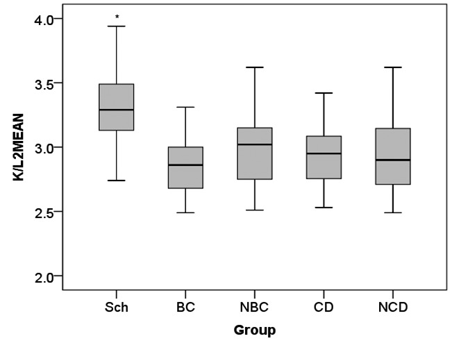
Among other dog breeds, the K/L2MEAN ratios were 2.86 ± 0.28 in the brachycephalic group, 2.98 ± 0.27 in the nonbrachycephalic group, 2.94 ± 0.24 in the chondrodystrophoid group and 2.95 ± 0.33 in the nonchondrodystrophoid group. The K/L2MEAN ratios in the Miniature Schnauzer group were significantly higher than the other 4 groups (P<0.001). There were no significant differences between those 4 groups (Fig. 1).
Fig. 1.

Boxplot of the mean kidney length to second lumbar vertebra length ratio (K/L2MEAN) in different breed groups. Sch, Miniature Schnauzer; BC, brachycephalic breed; NBC, nonbrachycephalic breed; CD, chondrodystrophoid breed; and NCD, nonchondrodystrophoid breed.
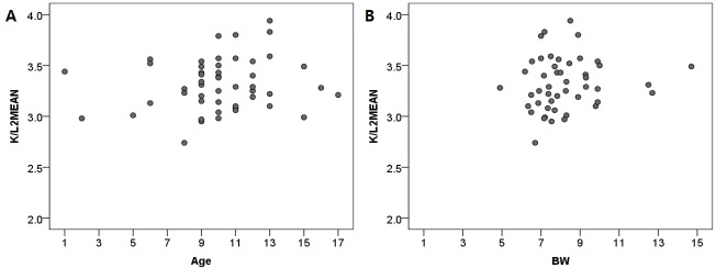
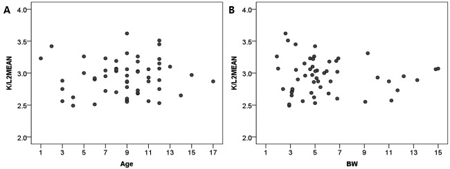
The K/L2MEAN ratios in the Miniature Schnauzer group significantly differed between male and female dogs regardless of whether the dogs were neutered or not (P=0.015), whereas no such difference was found in other dog breeds (P=0.094). However, mean differences were relatively small in both groups: 0.18 in the Miniature Schnauzer group and 0.13 in other dog breeds. There was no significant difference between intact (intact male and female) and neutered (neutered male and female) dogs both in the Miniature Schnauzer group (P=0.216) and other dog breeds (P=0.394). There were no significant relationships between the K/L2MEAN ratios and ages and between the K/L2MEAN ratios and body weights both in the Miniature Schnauzer group (R=0.150, P=0.304; and R=0.136, P=0.362, respectively) and other dog breeds (R=0.052, P=0.709; and R=−0.068, P=0.630, respectively) (Figs. 2 and 3).
Fig. 2.
Scatterplot of the mean kidney length to second lumbar vertebra length ratio (K/L2MEAN) in Miniature Schnauzer dogs in relation to age (A) and body weight (BW) (B).
Fig. 3.
Scatterplot of the mean kidney length to second lumbar vertebra length ratio (K/L2MEAN) in other dog breeds in relation to age (A) and body weight (BW) (B).
Radiographic L2 length: L2 length, width and height were significantly correlated with body weight in the Miniature Schnauzer group (R=0.643, P<0.001; R=0.556, P<0.001; and R=0.503, P<0.001, respectively) and other dog breeds (R=0.873, P<0.001; R=0.817, P<0.001; and R=0.815, P<0.001, respectively). Taking the effect of body weight on L2 length into account, L2 length was divided by either L2 width (L2L/L2W) or height (L2L/L2H), and both L2L/L2W and L2L/L2H values were constant regardless of body weight in the Miniature Schnauzer group (R=0.055, P=0.712; and R=−0.019, P=0.897, respectively) and other dog breeds (R=−0.080, P=0.574; and R=−0.224, P=0.111, respectively). L2L/L2W and L2L/L2H ratios were 1.11 ± 0.06 and 2.50 ± 0.18 in the Miniature Schnauzer group, and 1.21 ± 0.09 and 2.66 ± 0.32 in other dog breeds. Although both values were significantly correlated with radiographic kidney size (R=0.563, P<0.001; and R=0.250, P=0.011), correlations were relatively low. As L2L/L2W values were better correlated with radiographic kidney size, L2L/L2W values were selected for further statistical analysis.
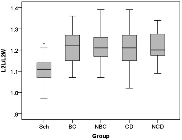
The ratios of L2L/L2W were significantly lower in the Miniature Schnauzer group than other dog breeds (P<0.001). Among other dog breeds, the L2L/L2W ratios were 1.20 ± 0.10 in the brachycephalic group, 1.21 ± 0.08 in the nonbrachycephalic group, 1.20 ± 0.10 in the chondrodystrophoid group and 1.22 ± 0.06 in the nonchondrodystrophoid group. There were no significant differences between those 4 groups. Only the Miniature Schnauzer group had significantly lower values compared to each group (P<0.001, respectively) (Fig. 4).
Fig. 4.

Boxplot of the second lumbar vertebra length to width ratio (L2L/L2W) in different breed groups. Sch, Miniature Schnauzer; BC, brachycephalic breed; NBC, nonbrachycephalic breed; CD, chondrodystrophoid breed; and NCD, nonchondrodystrophoid breed.
Ultrasonographic kidney size: The left and right kidney lengths were significantly different in both the Miniature Schnauzer group (P<0.001) and other dog breeds (P=0.004), and right kidneys were larger in both groups. However, mean differences were just 1.88 mm in the Miniature Schnauzer group and 1.13 mm in other dog breeds; therefore, the left and right kidney lengths were considered similar in practical terms. Thus, mean values were used to calculate K/Ao ratios. K/Ao ratios were 6.75 ± 0.67 in the Miniature Schnauzer group and 7.16 ± 1.01 in other dog breeds. Even though there was a significant difference between those groups (P=0.015), K/Ao ratios in all the dogs in both groups were within or very close to normal range: those in 2 Miniature Schnauzer dogs (5.48 and 5.49) and 2 other dog breeds (5.48 and 9.15) were out of, but very close to, normal range.
Intra- and interobserver repeatability and reproducibility of radiographic measurements: For all the measurements from 20 randomly selected dogs, intra- and interobserver repeatability and reproducibility showed excellent correlation (ICCs of all measurements >0.9).
DISCUSSION
In this study, the radiographic kidney size in Miniature Schnauzer dogs was significantly larger than that of other dog breeds. The standard normal radiographic kidney size in adult dogs may not be valid for Miniature Schnauzer dogs, as radiographic kidney sizes in 24.5% of Miniature Schnauzer dogs were greater than 3.5. On the other hand, the radiographic kidney size in other dog breeds was consistent with previous reports [6, 9, 13, 19, 20].
A recent study compared the radiographic kidney sizes in dogs with different skull types (brachycephalic, mesaticephalic and dolichocephalic) and different body weights (0–10 kg, >10–30 kg and >30 kg) [20]. The study showed significant differences between skull types and body weights, especially between brachycephalic and dolichocephalic dogs and between small breed (≤10 kg) and large breed dogs (>30 kg). However, the effect of breed on kidney size irrespective of body weight or the effect of body weight on kidney size irrespective of breed was not fully explained in spite of the fact that breed and weight are interrelated. In this study, only small- to medium-sized dogs (≤15 kg) were included to prevent differences resulting from body weight, and no differences between breeds except for Miniature Schnauzers as well as between body weights were identified. Thus, the radiographic kidney size was considered to be influenced by body weight rather than breed. One previous study reported a negative correlation between body weight and kidney volume per kilogram body weight [4].
There was a small but significant difference between dogs with different sexes in the Miniature Schnauzer group regardless of their neutering state, as males had larger kidneys. However, no such difference was found in other dog breeds. Previous studies also reported inconsistent results: one study reported that kidney size was slightly larger in male dogs, another feline study reported that intact cats had larger kidneys, and another canine studies reported that there were no differences between sexes [4, 19, 20, 22, 28]. The results of this study and previous studies may indicate that sex has a minor effect on kidney size. There was no correlation between age and kidney size, which was consistent with previous reports [9, 19, 20].
Whereas L2 vertebral length, width and height were related to body weight, L2L/L2W and L2L/L2H ratios were not correlated with body weight. However, like kidney size, L2 lengths were variable between individuals, so that the correlation between radiographic L2 length and radiographic kidney size was significant but relatively low. In spite of that, L2L/L2W ratios were significantly lower in Miniature Schnauzer dogs than in other dog breeds. In addition, there were no significant differences between other dog breeds, and even chondrodystrophoid breeds have a similar L2 length to other dog breeds, which indicates that Miniature Schnauzer dogs have breed-specific short vertebra.
Furthermore, ultrasonographic kidney sizes (K/Ao ratios), which were not affected by L2 length, in all the dogs in the Miniature Schnauzer and other dog breeds were within or very close to normal range, supporting the notion that the large radiographic kidney size in Miniature Schnauzer dogs was attributed to their short L2 length, not their long kidney length. Thus, care should be taken when using vertebral length to assess organ size in Miniature Schnauzer dogs, such as heart size and colon diameter on radiographs and kidney size on ultrasonograms, as well as radiographic kidney size [2, 16, 26].
This study has several limitations. The radiographic evaluation of organ size has inherent disadvantages of variable degrees of magnification and distortion. One study reported that a considerable degree of magnification may result depending on the distances between an object and film: a 10-cm distance led to 19% magnification [25]. However, a number of previous studies reported relatively consistent radiographic kidney size in various breeds of dogs, which may indicate that variable degrees of magnification of kidney and L2 lengths may not lead to significant deviations from the standard normal ranges of radiographic kidney size [6, 9, 11, 13, 19, 20]. In addition, one human study reported that renal inclination did not lead to significant error in the radiographic measurement of kidney length in most cases [10]. Second, actual kidney size was not measured by necropsy, and more accurate methods to measure kidney size using CT and MRI were not used. In this study, ultrasonographic kidney size using the K/Ao ratio was used as a reference. However, the K/Ao ratio was not a gold standard used to evaluate kidney size. Moreover, to assume actual kidney size, whether the K/Ao ratio on ultrasonograms is superior to the K/L2 ratio on radiographs has not been studied. However, previous studies reported that radiographic and ultrasonographic kidney lengths were closely correlated with actual kidney size, and in this study, using the K/Ao ratio was sufficient to prove that absolute kidney length was not the factor leading to significant differences in radiographic kidney size by comparing kidney length with different anatomic structures, L2 and the aorta. Third, this study included dogs without clinical evidence of renal disease, and thus, dogs with subclinical renal disease may be included in this study. Finally, changes in radiographic kidney size in Miniature Schnauzer dogs with renal disease and comparison with each breed were not studied, and further study research is needed.
In conclusion, Miniature Schnauzer dogs have breed-specific short vertebrae and consequently a larger radiographic kidney size. In this study, the normal radiographic kidney size of Miniature Schnauzer dogs was 3.31 ± 0.26, which was greater than the standard reference in normal adult dogs. Thus, care should be taken when evaluating radiographic kidney size in Miniature Schnauzer dogs to prevent falsely diagnosed renomegaly. Further studies in Miniature Schnauzer dogs with known renal disease are necessary.
REFERENCES
- 1.Bakker J., Olree M., Kaatee R., de Lange E. E., Moons K. G. M., Beutler J. J., Beek F. J. A.1999. Renal volume measurements: accuracy and repeatability of US compared with that of MR imaging. Radiology 211: 623–628. doi: 10.1148/radiology.211.3.r99jn19623 [DOI] [PubMed] [Google Scholar]
- 2.Barella G., Lodi M., Sabbadin L. A., Faverzani S.2012. A new method for ultrasonographic measurement of kidney size in healthy dogs. J. Ultrasound 15: 186–191. doi: 10.1016/j.jus.2012.06.004 [DOI] [PMC free article] [PubMed] [Google Scholar]
- 3.Barr F. J.1990. Evaluation of ultrasound as a method of assessing renal size in the dog. J. Small Anim. Pract. 31: 174–179. doi: 10.1111/j.1748-5827.1990.tb00762.x [DOI] [Google Scholar]
- 4.Barr F. J., Holt P. E., Gibbs C.1990. Ultrasonographic measurements of normal renal parameters. J. Small Anim. Pract. 31: 180–184. doi: 10.1111/j.1748-5827.1990.tb00764.x [DOI] [Google Scholar]
- 5.Barrera R., Duque J., Ruiz P., Zaragoza C.2009. Accuracy of ultrasonographic measurements of kidney of dogs for clinical use. Revista Cientifica 19: 576–583. [Google Scholar]
- 6.Churchill J. A., Feeney D. A., Fletcher T. F., Osborne C. A., Polzin D. J.1999. Effects of diet and aging on renal measurements in uninephrectomized geriatric bitches. Vet. Radiol. Ultrasound 40: 233–240. doi: 10.1111/j.1740-8261.1999.tb00354.x [DOI] [PubMed] [Google Scholar]
- 7.Daniel G. B., Mitchell S. K., Mawby D., Sackman J. E., Schmidt D.1999. Renal nuclear medicine: a review. Vet. Radiol. Ultrasound 40: 572–587. doi: 10.1111/j.1740-8261.1999.tb00883.x [DOI] [PubMed] [Google Scholar]
- 8.Dennis R., Kirberger R. M., Barr F., Wrigley R. H.2010. Urogenital tract. pp. 297–330. In: Handbook of Small Animal Radiology and Ultrasound: Techniques and Differential Diagnoses. (Dennis, R., Kirberger, R. M., Barr, F. and Wrigley, R. H. eds.), Elsevier Saunders, St. Louis. [Google Scholar]
- 9.England G. C. W.1996. Renal and hepatic ultrasonography in the neonatal dog. Vet. Radiol. Ultrasound 37: 374–382. doi: 10.1111/j.1740-8261.1996.tb01246.x [DOI] [Google Scholar]
- 10.Farrant P., Meire H. B.1978. Ultrasonic measurement of renal inclination; its importance in measurement of renal length. Br. J. Radiol. 51: 628–630. doi: 10.1259/0007-1285-51-608-628 [DOI] [PubMed] [Google Scholar]
- 11.Feeney D. A., Thrall D. E., Barber D. L., Culver D. H., Lewis R. E.1979. Normal canine excretory urogram: effects of dose, time, and individual dog variation. Am. J. Vet. Res. 40: 1596–1604. [PubMed] [Google Scholar]
- 12.Felkai C. S., Voros K., Vrabely T., Karsai F.1992. Ultrasonographic determination of renal volume in the dog. Vet. Radiol. Ultrasound 33: 292–296. doi: 10.1111/j.1740-8261.1992.tb00146.x [DOI] [Google Scholar]
- 13.Finco D. R., Stiles N. S., Kneller S. K., Lewis R. E., Barrett R. B.1971. Radiologic estimation of kidney size of the dog. J. Am. Vet. Med. Assoc. 159: 995–1002. [PubMed] [Google Scholar]
- 14.Grandage J.1975. Some effects of posture on the radiographic appearance of the kidneys of the dog. J. Am. Vet. Med. Assoc. 166: 165–166. [PubMed] [Google Scholar]
- 15.Griffiths G. J., Cartwright G., McLachlan S. F.1975. Estimation of renal size from radiographs: is the effort worthwhile? Clin. Radiol. 26: 249–256. doi: 10.1016/S0009-9260(75)80053-5 [DOI] [PubMed] [Google Scholar]
- 16.Jung Y., Jeong J., Kim M., Lee H., Kim N., Lee K.2012. Is it true that the Miniature Schnauzer have larger vertebral heart scale? −vertebral heart scale (VHS) in normal Miniature Schnauzer dogs. J. Vet. Clin. 29: 148. [Google Scholar]
- 17.Konde L. J., Wrigley R. H., Park R. D., Lebel J. L.1984. Ultrasonographic anatomy of the normal canine kidney. Vet. Radiol. 25: 173–178. doi: 10.1111/j.1740-8261.1984.tb02138.x [DOI] [Google Scholar]
- 18.Lamb C. R.1990. Abdominal ultrasonography in small animals: intestinal tract and mesentery, kidneys, adrenal glands, uterus and prostate. J. Small Anim. Pract. 31: 295–304. doi: 10.1111/j.1748-5827.1990.tb00811.x [DOI] [Google Scholar]
- 19.Lee R., Leowijuk C.1982. Normal parameters in abdominal radiology of the dog and cat. J. Small Anim. Pract. 23: 251–269. doi: 10.1111/j.1748-5827.1982.tb01664.x [DOI] [Google Scholar]
- 20.Lobacz M. A., Sullivan M., Mellor D., Hammond G., Labruyère J., Dennis R.2012. Effect of breed, age, weight and gender on radiographic renal size in the dog. Vet. Radiol. Ultrasound 53: 437–441. doi: 10.1111/j.1740-8261.2012.01937.x [DOI] [PubMed] [Google Scholar]
- 21.MacKay E. M.1932. Kidney weight, body size and renal function. Arch. Intern. Med. (Chic.) 50: 590–594. doi: 10.1001/archinte.1932.00150170082008 [DOI] [Google Scholar]
- 22.Mareschal A., d’Anjou M. A., Moreau M., Alexander K., Beauregard G.2007. Ultrasonographic measurement of kidney-to-aorta ratio as a method of estimating renal size in dogs. Vet. Radiol. Ultrasound 48: 434–438. doi: 10.1111/j.1740-8261.2007.00274.x [DOI] [PubMed] [Google Scholar]
- 23.Nyland T. G., Fisher P. E., Gregory C. R., Wisner E. R.1997. Ultrasonographic evaluation of renal size in dogs with acute allograft rejection. Vet. Radiol. Ultrasound 38: 55–61. doi: 10.1111/j.1740-8261.1997.tb01604.x [DOI] [PubMed] [Google Scholar]
- 24.Nyland T. G., Kantrowitz B. M., Fisher P., Olander H. J., Hornof W. J.1989. Ultrasonic determination of kidney volume in the dog. Vet. Radiol. Ultrasound 30: 174–180. doi: 10.1111/j.1740-8261.1989.tb00771.x [DOI] [Google Scholar]
- 25.Olesen S., Genster H. G.1970. Estimation of renal volume and function in dogs from the radiological appearance. Invest. Urol. 7: 363–370. [PubMed] [Google Scholar]
- 26.Schwarz T.2013. The large bowel. pp. 705–725. In: Textbook of Veterinary Diagnostic Radiology, 6th ed. (Thrall, D. E. ed.), Elsevier Saunders, St. Louis. [Google Scholar]
- 27.Seiler G. S.2013. The kidneys and ureters. pp. 812–824. In: Textbook of Veterinary Diagnostic Radiology, 6th ed. (Thrall, D. E. ed.), Elsevier Saunders, St. Louis. [Google Scholar]
- 28.Shiroma J. T., Gabriel J. K., Carter R. L., Scruggs S. L., Stubbs P. W.1999. Effect of reproductive status on feline renal size. Vet. Radiol. Ultrasound 40: 242–245. doi: 10.1111/j.1740-8261.1999.tb00355.x [DOI] [PubMed] [Google Scholar]
- 29.Soldo D., Brkljacic B., Bozikov V., Drinkovic I., Hauser M.1997. Diabetic nephropathy. Comparison of conventional and duplex Doppler ultrasonographic findings. Acta Radiol. 38: 296–302. [DOI] [PubMed] [Google Scholar]
- 30.Thornton G. W.1962. Radiographs in the diagnosis of disease conditions of the urinary system. Vet. Radiol. 3: 1–12. doi: 10.1111/j.1740-8261.1962.tb01819.x [DOI] [Google Scholar]
- 31.Tyson R., Logsdon S. A., Werre S. R., Daniel G. B.2013. Estimation of feline renal volume using computed tomography and ultrasound. Vet. Radiol. Ultrasound 54: 127–132. doi: 10.1111/vru.12007 [DOI] [PubMed] [Google Scholar]


