Abstract
Proliferative and necrotising otitis externa (PNOE) is a very rare disease affecting the ear canals and concave pinnae of kittens. This report describes a 5-month-old cat with PNOE. Histopathological examination confirmed the diagnosis. Treatment was initiated with local injection of methylprednisolone acetate into the lesions. The cat was subsequently treated with clobetasol propionate cream, a potent topical glucocorticoid ointment. The cat showed marked improvement. While topical treatment with tacrolimus, an immunosuppressive agent, is reported to be an effective therapy, to the best of our knowledge, this is the first report to treat PNOE with local corticosteroid therapy.
Keywords: clobetasol propionate, methylprednisolone acetate, Persian cat, proliferative and necrotizing feline otitis externa
Proliferative and necrotizing otitis externa (PNOE) is a very rare skin disease of the cat, so that it has been described only in few reports and in a chapter of a book [2]. Although a few reports about PNOE have been published [1, 4, 9], the reported therapeutic options seem to be limited. Tacrolimus (FK506), is an immunosuppressive agent with a similar mechanism of action to cyclosporine, the most commonly used for PNOE [1, 4, 9]. On the other hand, glucocorticoid therapy has been reported to be ineffective or only partially effective [4]. Here, we describe the first juvenile case of PNOE in Japan, which was treated with local corticosteroid therapy alone.
A 5-month-old castrated male Golden Chinchilla Persian cat presented with chronic otitis. This condition was first documented at the time of purchase (4 months old). Treatment for suspected ear mites and ear infection was provided at a veterinary hospital, but there was no improvement of the otitis. The owner then took the cat to a different veterinary clinic, where it was treated with routine ear cleaning, oral prednisolone and oral amoxicillin. Although this treatment was continued, the pruritus and the lesions did not improve. Instead, unusual crusted, pigmented and friable lesions developed in both ear canals.
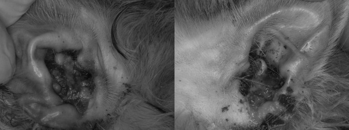
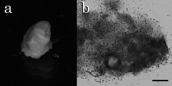
At the time of referral, the cat was in good health. Both concave pinnae were covered with large tan to dark brown coalescing crusted plaques (Fig. 1). On palpation, the lesions were found to occupy the vertical part of the ear canal, with friable material from the plaques and a thick exudate occluding the canals. Gentle finger pressure resulted in the extrusion of purulent or caseous exudate from the plaques in the ear canals (Fig. 2a). A smear of the exudate revealed amorphous debris with corneocytes, inflammatory cells and cocci (Fig. 2b).
Fig. 1.
Lesions of both ears at the initial visit.
Fig. 2.
Caseous exudate from an ear lesion (a) and a smear of the exudate (b). Wright-Giemsa stain. Bar=20 µm.
The complete blood count and serum biochemistry tests were within normal limits. Both feline leukemia virus (FeLV) and feline immunodeficiency virus (FIV) were negative. Bacterial culture of the exudate revealed methicillin-resistant coagulase-negative Staphylococcus, while culture for dermatophytes was negative.
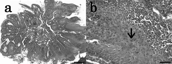
To confirm the diagnosis, two 4-mm skin punch biopsy specimens were obtained from the lesions over the ear canals. Histopathologic examination revealed severe parakeratotic hyperkeratosis with neutrophilic crusts. The epidermis and the outer root sheaths of the hair follicles showed marked papillomatous hyperplasia, as well as acanthosis and spongiosis (Fig. 3a). Numerous pyknotic and hypereosinophilic keratinocytes (apoptotic cells) were observed in the epidermis and within the outer root sheaths of the hair follicles (Fig. 3b). The superficial dermis was edematous, and the vessels were dilated, associated with infiltration by various inflammatory cells (neutrophils, lymphocytes, mastocytes and macrophages). Based on these clinical and histopathological features, a diagnosis of PNOE was made.
Fig. 3.
Histopathological features of the biopsy specimens. (a) Severe papillomatous hyperplasia of the epidermis and external root sheath of a hair follicle. Hematoxylin and eosin. Bar=500 µm. (b) Dyskeratotic keratinocyte in the hyperplastic hair follicle wall (arrow). Hematoxylin and eosin. Bar=50 µm.
Treatment was initiated with local injection of methylprednisolone acetate (Depo-Medrol; Pfizer Animal Health, Tokyo, Japan) into lesions of both ears at a total dosage of 2 mg/kg. Thereafter, 0.05% clobetasol propionate cream (Dermovate; GlaxoSmithKline, Tokyo, Japan), which is a potent topical steroid, was applied once daily to the lesions, and an ear cleaner (Epi-Otic; Virbac, Tokyo, Japan) was applied in both ears twice a week. Based on the results of the bacterial and sensitivity testing, doxycycline (Vibramycin; Pfizer Animal Health) was administered at 5 mg/kg orally twice daily for 4 weeks in order to treat the methicillin-resistant Staphylococcal infection. At the 2-week recheck, the PNOE lesions showed marked involution, and bacterial otitis was also improved. The lesions and proliferative tissue both resolved after 2 months of treatment. Subsequently, episodes of pruritus and recurrent bacterial otitis were occasionally observed. However, no recurrence of PNOE was noted during follow-up for 1 year.
PNOE is a rare disease that has only been mentioned in four reports [1, 2, 4, 9]. The pathogenesis of PNOE is currently being explored. Severe acanthosis of the epidermis and hair follicles is one of the primary histological features. Epidermal and follicular hyperplastic changes are also observed in the feline dermatitis associated with viral infections. However, polymerase chain reaction analysis [2] and immunohistochemical staining [4] have ruled out infection by herpesvirus, calicivirus or papillomavirus. Instead, Videmont et al. [9] demonstrated apoptosis of keratinocytes and infiltration of CD3+ T cells into the epidermis in PNOE, and they suggested that the infiltrating T cells might induce keratinocyte apoptosis. Thus, they proposed a T cell-mediated pathogenesis of this disease, which provides an explanation for the efficacy of tacrolimus treatment.
The present case was FIV and FeLV negative, and had no history of upper respiratory tract disease, rhinitis or conjunctivitis associated with herpesvirus or calicivirus infection. Moreover, administration of corticosteroids can cause viral reactivation and shedding [6], but there was no exacerbation of the present lesions with corticosteroid treatment. This supports the lesions being unrelated to viral infection.
The histological features of viral infection are also characteristic. In feline dermatitis associated with herpesvirus or calicivirus infection, there are pustulosis and ulcerative or necrotic dermatitis [6]. In addition, multinucleated keratinocytic giant cells are observed along with amphophilic intranuclear inclusion bodies in feline herpesvirus dermatitis [3]. Histological features of papillomavirus infection, such as virus-associated feline Bowenoid carcinoma in situ, resemble the epithelial changes seen in PNOE. In papillomavirus infection, keratinocytes with dark condensed nuclei surrounded by a clear cytoplasmic halo (koilocytosis) have been reported in the Bowenoid lesions [7]. None of these pathological changes described above were seen in the present case, making a viral etiology highly unlikely.
The reported treatment options for PNOE are limited. Gross et al. described spontaneous regression of PNOE after 12–24 months [2], while the other three reports indicated that tacrolimus is effective for this condition [1, 2, 4]. In particular, Mauldin et al. reported that corticosteroid therapy only achieved partial improvement of this disease. Similar to tacrolimus, corticosteroids suppress the activity of inflammatory cells, including T cells. Corticosteroids also decrease keratinocyte hyperplasia by inhibiting proliferation and cytokine production by these cells [8], and epidermal hyperplasia is one of the pathological features of PNOE. Therefore, we considered that the appropriate administration route and potency of steroid therapy might be important for achieving improvement of PNOE.
In fact, the present case showed marked improvement after intralesional injection of methylprednisolone acetate (MPA) and topical application of clobetasol propionate, implying the efficacy of local corticosteroid treatment.
Corticosteroids are generally available as oral, injectable and topical formulations. Treatment of PNOE using oral and topical steroids was previously reported to be ineffective. In the present case, we used MPA, an injectable steroid formulation. It belongs to the class of steroid esters, including triamcinolone acetonide, which are slow-release formulations generally used for local treatment of joint disease in humans and horses [5]. Although the active moiety of MPA is methylprednisolone, which is approximately equal in potency to the intermediate-acting corticosteroid prednisolone, intralesional injection of MPA should achieve higher concentrations within the lesions than oral or topical treatment and this might explain why it was effective for PNOE.
Clobetasol propionate is classified as a superpotent steroid according to the World Health Organization criteria. The steroid ointments used in the other reports on PNOE were betamethasone diproprionate (potent), betamethasone valerate (potent), hydrocortisone (mid-strength) and dexamethasone (mild) [2, 4, 9]. Because the other agents were found to be ineffective, we selected clobetasol propionate (superpotent) in this case.
In conclusion, this was the first reported case of PNOE in Japan, and the combination of intralesional MPA injection with topical application of clobetasol propionate was supposed to be effective therapy. To the best of our knowledge, this is the first report of PNOE responding to local corticosteroid therapy alone.
REFERENCES
- 1.Borio S., Massari F., Abramo F., Colombo S.2013. Proliferative and necrotising otitis externa in a cat without pinnal involvement: video-otoscopic features. J. Feline Med. Surg. 15: 353–356. doi: 10.1177/1098612X12468838 [DOI] [PMC free article] [PubMed] [Google Scholar]
- 2.Gross T. L., Ihrke P. J., Walder E. J., Affolter V. K.2005. Necrotizing diseases of the epidermis. pp. 79–91. In: Skin Diseases of the Dog and Cat, 2nd ed., Blackwell Science Ltd., Oxford. [Google Scholar]
- 3.Hargis A. M., Ginn P. E.1999. Feline herpesvirus 1-associated facial and nasal dermatitis and stomatitis in domestic cats. Vet. Clin. North Am. Small Anim. Pract. 29: 1281–1290. doi: 10.1016/S0195-5616(99)50126-5 [DOI] [PubMed] [Google Scholar]
- 4.Mauldin E. A., Ness T. A., Goldschmidt M. H.2007. Proliferative and necrotizing otitis externa in four cats. Vet. Dermatol. 18: 370–377. doi: 10.1111/j.1365-3164.2007.00614.x [DOI] [PubMed] [Google Scholar]
- 5.McIlwraith C. W.2010. The use of intra-articular corticosteroids in the horse: what is known on a scientific basis? Equine Vet. J. 42: 563–571. doi: 10.1111/j.2042-3306.2010.00095.x [DOI] [PubMed] [Google Scholar]
- 6.Miller W. H., Griffin C. G., Campbell K. L.2013. Viral disease. pp. 343–351. In: Muller and Kirk’s Small Animal Dermatology, 7th ed., Saunders, Philadelphia. [Google Scholar]
- 7.Munday J. S., Willis K. A., Kiupel M., Hill F. I., Dunowska M.2008. Amplification of three different papillomaviral DNA sequences from a cat with viral plaques. Vet. Dermatol. 19: 400–404. doi: 10.1111/j.1365-3164.2008.00710.x [DOI] [PubMed] [Google Scholar]
- 8.Sanders S., Busam K. J., Halpern A. C., Nehal K. S.2002. Intralesional corticosteroid treatment of multiple eruptive keratoacanthomas: case report and review of a controversial therapy. Dermatol. Surg. 28: 954–958. [DOI] [PubMed] [Google Scholar]
- 9.Vidémont E., Pin D.2010. Proliferative and necrotising otitis in a kitten: first demonstration of T-cell-mediated apoptosis. J. Small Anim. Pract. 51: 599–603. doi: 10.1111/j.1748-5827.2010.00999.x [DOI] [PubMed] [Google Scholar]


