Abstract
Background
The emergence and spread of Carbapenem-resistant Escherichia coli (CREC) is becoming a serious problem in Chinese hospitals, however, the data on this is scarce. Therefore, we investigate the risk factors for healthcare-associated CREC infection and study the incidence, antibiotic resistance and medical costs of CREC infections in our hospital.
Methods
We conducted a retrospective, matched case–control–control, parallel study in a tertiary teaching hospital. Patients admitted between January 2012 and December 2015 were included in this study. For patients with healthcare-associated CREC infection, two matched subject groups were created; one group with healthcare-associated CSEC infection and the other group without infection.
Results
Multivariate conditional logistic regression analysis demonstrated that prior hospital stay (<6 months) (OR:3.96; 95%CI:1.26–12.42), tracheostomy (OR:2.24; 95%CI: 1.14–4.38), central venous catheter insertion (OR: 8.15; 95%CI: 2.31–28.72), carbapenem exposure (OR: 12.02; 95%CI: 1.52–95.4), urinary system disease (OR: 16.69; 95%CI: 3.01–89.76), low hemoglobin (OR: 2.83; 95%CI: 1.46–5.50), and high blood glucose are associated (OR: 7.01; 95%CI: 1.89–26.02) with CREC infection. Total costs (p = 0.00), medical examination costs (p = 0.00), medical test costs (p = 0.00), total drug costs (p = 0.00) and ant-infective drug costs (p = 0.00) for the CREC group were significantly higher than those for the no infection group. Medical examination costs (p = 0.03), total drug costs (p = 0.03), and anti-infective drug costs (p = 0.01) for the CREC group were significantly higher than for the CSEC group. Mortality in CREC group was significantly higher than the CSEC group (p = 0.01) and no infection group (p = 0.01).
Conclusion
Many factors were discovered for acquisition of healthcare-associated CREC infection. CREC isolates were resistant to most antibiotics, and had some association with high financial burden and increased mortality.
Keywords: Healthcare-associated infection, Risk factors, CREC, CSEC
Background
Carbapenems have long served as reliable and potent agents against Gram-negative bacilli [1]. Carbapenems are most consistently active against members of the Enterobacteriaceae family [2], however, few treatment options exist for carbapenem-resistant Enterobacteriaceae (CRE) infection, which can result in high mortality [3]. In recent years, carbapenem-resistant Escherichia coli (CREC), as one class of CRE, has become a major threat in hospitals worldwide [4–7]. Carbapenem resistance in E.coli is an emerging problem that is mainly caused by plasmid-encoded carbapenemases [8–13]. As a result of the emergence of carbapenemases [14], antimicrobial resistance is increasing in most hospitals, and has become a global healthcare problem. CREC strains should be closely monitored because of their potential trend to spread in both hospital and community settings [15].
There are several previous studies on the risk factors for CRE infection [16, 17], but few published studies have specifically evaluated the risk factors for CREC acquisition, especially in China. Therefore, we performed a retrospective study to evaluate the risk factors for healthcare-associated infection (HAI) caused by CREC among in-patients in a teaching hospital in central south China, thus, we could do better in decreasing the incidence of CREC infection.
The case–control–control study design of this study, which utilizes two separate case–control analyses, has become a standard method for the specific identification of risk factors that are uniquely connected to infection by antimicrobial-resistant pathogens [18, 19]. We studied the risk factors for CREC infection through the case–control–control design. In addition, CREC is often resistant to multiple antibiotics; therefore, we investigated the antibiotic resistance and economic burden of CREC infections.
Methods
Study design and setting
We conducted a retrospective, parallel, case–control–control study to identify the incidence, risk factors, antibiotic resistance, and medical costs associated with the acquisition of healthcare-associated CREC infection among hospitalized patients treated at Xiangya Hospital, a 3500-bed general hospital in Changsha, Hunan Province, Central South China. The CREC infection group was compared with a no infection group to assess the risk factors for acquisition of CREC infection; meanwhile, the CREC group was compared with the CSEC infection group to evaluate reasons for antibiotic resistance.
Subjects with CREC or CSEC isolated from multiple sites, or on multiple dates, were counted only once, and the data from the first infection was included in the study. Healthcare-acquired CREC or CSEC infection was defined as isolation 48 hours after admission to the hospital. Healthcare-associated infection (HAI) was defined according to the CDC/NHSN surveillance criteria in patients with samples from any specimen source site positive for CR-EC or CS-EC; meanwhile, the patients with CR-EC or CS-EC colonization and community-associated infection (CAI) were ruled out.
Study population
Patients from whom CREC were isolated from clinical cultures from any source between January 1, 2012 and December 31, 2015 were included in this study. For each CREC patient, we randomly selected two controls from hospitalized patients who were admitted within the same period with CSEC isolated, and the two groups were matched for age and sex. Additionally, we selected two controls from the in-patients admitted within the study period with no bacterial infection, and the two groups were matched for age and sex.
Microbiological identification and susceptibility testing
An automated broth microdilution method (Vitek 2; bioMérieux, Marcy-l′Étoile, France) was used to perform identification and susceptibility testing. Carbapenem resistance was determined using the disk diffusion method. All isolates with resistance, or intermediate susceptibility to carbapenem were defined as resistant isolates. Clinical and Laboratory Standards Institute document M100-S22 (January 2012) was used for interpretation of the antimicrobial susceptibility testing and ESBL testing, and CREC was defined as E.coli resistant to at least one of the carbapenems (imipenem, meropenem, or ertapenem).
Using current EUCAST breakpoints, imipenem MICs of CR-KP isolates ranged from 2 to >32 μg/ml (breakpoint for resistance and intermediate susceptibility MIC ≥ 2 μg/ml); meropenem MICs from 4 to > 32 μg/ml (breakpoint for resistance and intermediate susceptibility MIC ≥ 4 μg/ml); all the isolates had ertapenem MICs in the resistant range (breakpoint for resistance and intermediate susceptibility MIC ≥ 1 μg/ml).
Data collection
Data were obtained from patients’ medical records, and relative data were recorded on structured abstraction forms. Variables analyzed as possible predictors included demographics (age, sex, marital status, and ward class); clinical departments where strains were isolated; and the history of admission before the infection (within 6 months prior to E.coli infection); length of hospital, intensive care unit (ICU) stay before E.coli infection; specimen source site (blood, bile, etc.); invasive procedures (urinary catheter insertion, mechanical ventilation, etc.) within 1 month prior to E.coli infection; surgical procedures within 1 month prior to E.coli infection; administration of drugs (glucocorticoids and immunosuppressive agents), radiotherapy and chemotherapy within 1 month prior to E.coli infection; specific co-morbidities included many system diseases (respiratory, central nervous, etc.); exposure (greater than, or equal to, one day) to antimicrobials (cephalosporins, carbapenems, etc.) within 3 months prior to CREC identification.
We also noted any related laboratory results when healthcare-aquired isolation of E.coli was recorded in the inspection system, and recorded the drug sensitivity test results obtained from the microbiology laboratory and the economic costs associated with these patients as noted in the financial system. The economic costs included total costs, medical examination costs, medical test costs, total drug costs and anti-infective drug costs.
Statistical analysis
Continuous variables were presented as mean ± SD, and we used t-tests for comparisons. As the results of the age and average costs of the data for the three groups showed non-normal distribution, they were compared with the median, and the data for two groups were compared using the Wilcoxon rank-sum test. We presented categorical variables as numbers and percentages, and compared percentages using the chi-square test or Fisher’s exact test.
We performed univariate analyses for each of the variables using conditional logistic regression to compare the cases and controls in terms of risk factor analysis. The association between independent variables is shown as the odds ratio with 95% confidence intervals, and variables for which the P value was less than 0.05 in the univariate analysis were included in a conditional logistic regression model for multivariate analysis. Multivariate logistic regression models were used to compare each case group and control group. A forward elimination process was used, and adjusted odds ratios and 95% confidence intervals were calculated.
A two-tailed P value of less than 0.05 was considered to show statistical significance, and statistical analyses were performed using SPSS 17.0 (SPSS, Inc, Chicago, IL, USA).
Results
Incidence of CREC infection
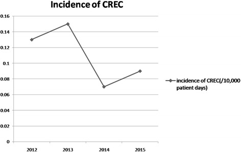
During the 4-year study period, CREC was isolated from 49 patients who met the criteria for healthcare-associated infection (HAI), including fourteen patients in 2012 (0.13/10,000 patient days), seventeen patients in 2013 (0.15/10,000 patient days), eight patients in 2014 (0.06/10,000 patient days), and ten patients in 2015 (0.10/10,000 patient days). The incidence of CREC infection over the 4-year study presented in Fig. 1.
Fig. 1.

Title: The incidence of carbapenem-resistant E.coli (CREC). Legend: The incidence of carbapenem-resistant E.coli (CREC) in 2012, 2013, 2014, and 2015 are presented in the figure; we can observe the change of the incidence in the four years from the figure
Specimen source site and specimen source ward
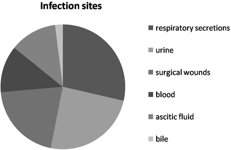
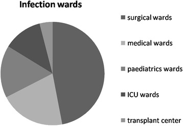
A total of 49 patients were included in the case group. CREC was most frequently recovered from respiratory secretions (28.6%), followed by urine (24.5%), surgical wounds (20.4%), blood (12.2%), ascitic fluid (12.2%), and bile (2.0%) (Fig. 2). When a positive culture result was obtained, patients infected with CREC were most frequently staying in surgical wards (46.9%), followed by medical wards (20.4%), pediatric wards (16.3%), ICU wards (12.2%), and the transplant center (4%) (Fig. 3).
Fig. 2.

Title: The infection sites of carbapenem-resistant E.coli (CREC). Legend: The proportion of carbapenem-resistant E.coli (CREC) strains recovered from the sites are presented in the figure, we can observe the regularity of the pathogens distributed
Fig. 3.

Title: The infection wards of carbapenem-resistant E.coli (CREC). Legend: The proportion of carbapenem-resistant E.coli (CREC) strains collected in which ward are presented in this figure, we can observe the regularity of the pathogens distributed
Resistance rate to antibiotics
The antibiotic susceptibility patterns of the isolates from the case and control patients are shown in Table 1. All CREC strains were resistant to ampicillin, ampicillin- sulbactam, cefazolin, ceftriaxone, and cefepime, followed by aztreonam, ceftazidime, ciprofloxacin, levofloxacin, piperacillin/tazobactam, trimethoprim and sulfamethoxazole, cefotetan, cefoperazone/sulbactam, tobramycin, and gentamicin; drug resistance rate to nitrofurantoin and amikacin was relatively low.
Table 1.
The antibiotic-resistanceof the two groups {carbapenem-resistant E.coli (CREC) and carbapenem-susceptible E.coli (CSEC)}
| Case (n = 49) | Control (n = 98) | p | |
|---|---|---|---|
| Ampicillin | 47/47 (100%) | 90/96 (94%) | 0.08 |
| Piperacillin/tazobactam | 38/48 (79%) | 10/96 (10%) | 0.00 |
| Ampicillin/sulbactam | 40/40 (100%) | 75/88 (85%) | 0.01 |
| Cefoperazone/sulbactam | 29/45 (64%) | 13/96 (14%) | 0.00 |
| Cefazolin | 42/42 (100%) | 75/92 (82%) | 0.00 |
| Ceftazidime | 37/39 (95%) | 48/87 (55%) | 0.00 |
| Ceftriaxone | 47/47 (100%) | 75/97 (77%) | 0.00 |
| Cefepime | 49/49 (100%) | 50/96 (52%) | 0.00 |
| Cefotetan | 22/31 (71%) | 2/85 (2%) | 0.00 |
| Aztreonam | 47/49 (96%) | 62/97 (54%) | 0.00 |
| Tobramycin | 26/45 (58%) | 51/94 (54%) | 0.69 |
| Amikacin | 3/48 (6%) | 8/98 (8%) | 0.68 |
| Gentamicin | 26/48 (54%) | 49/98 (50%) | 0.64 |
| Ciprofloxacin | 41/46 (89%) | 64/96 (67%) | 0.00 |
| Levofloxacin | 41/49 (84%) | 60/96 (63%) | 0.01 |
| Trimethoprim + sulfamethoxaz ole | 36/49 (73%) | 58/96 (60%) | 0.05 |
| Nitrofurantoin | 18/44 (41%) | 20/98 (20%) | 0.01 |
NOTE. Categorical variables are no/total no (%), case is carbapenem-resistant E.coli (CREC), control is carbapenem-susceptible E.coli (CSEC)
Univariate and multivariateanalyses regarding the risk factors of the CREC and CSEC groups
Results of the univariate and multivariate analyses from the comparison of the CREC and CSEC groups regarding the risk factors for healthcare-acquired CREC are shown in Table 2. The univariate conditional logistic regression analysis demonstrated that prior hospital stay (<6 months), urinary catheter insertion, tracheostomy, central venous catheter insertion, gastric tube insertion, urinary system disease, cephalosporins exposure, carbapenems exposure, antifungal agents exposure, glycopeptides and oxazolidinones exposure, low hemoglobin, low blood albumin, and high blood glucose were all risk factors for healthcare-acquired CREC infection. The multivariate conditional logistic regression analysis demonstrated that prior hospital stay (<6 months), incision of trachea, central venous catheter insertion, urinary system disease, low hemoglobin, and high blood glucose were all risk factors for healthcare-acquired CREC infection.
Table 2.
Univariate and multivariate analyses regarding the risk factors of the carbapenem-resistant E.coli (CREC) and carbapenem-susceptible E.coli (CSEC) groups
| Variable | Study group | Univariate | Multivariable | |||||
|---|---|---|---|---|---|---|---|---|
| Case (n = 49) | Control (n = 98) | OR | 95% CI | P | OR | 95%CI | P | |
| Demographic characteristics | ||||||||
| Sex, male (%) | 20 (41%) | 40 (41%) | 0.57 | |||||
| Age {year, median (range)} | 51 (0–82) | 53 (0–91) | 0.69 | |||||
| Related to hospitalization | ||||||||
| Prior hospital stay (<6 months) | 8 (77%) | 57 (58%) | 2.48 | 1.10-5.56 | 0.03 | 3.96 | 1.26-12.42 | 0.02 |
| ICU stay (<6 months) | 18 (36%) | 22 (22%) | 1.48 | 0.98-2.24 | 0.06 | |||
| Operation history | 26 (53%) | 28 (29%) | 2.53 | 1.28-5.01 | 0.01 | |||
| Urinary catheter insertion | 32 (65%) | 59 (60%) | 1.55 | 1.04-2.32 | 0.03 | |||
| Mechanical ventilation | 16 (32%) | 18 (18%) | 1.80 | 0.97-3.35 | 0.06 | |||
| Tracheostomy | 12 (24%) | 10 (10%) | 1.64 | 1.09-2.45 | 0.02 | 2.24 | 1.14-4.38 | 0.02 |
| Bronchofibroscope use | 7 (14%) | 0 (0%) | 72.96 | 0.53-9980.04 | 0.09 | |||
| Central venous catheter insertion | 15 (30%) | 7 (7%) | 4.48 | 1.72-11.67 | 0.00 | 8.15 | 2.31-28.72 | 0.00 |
| Gastric tube insertion | 28 (57%) | 37 (37%) | 1.53 | 1.09-2.16 | 0.01 | |||
| Wound drainage tube use | 18 (36%) | 27 (27%) | 1.46 | 0.93-2.29 | 0.09 | |||
| Underlying disorder | ||||||||
| Central nervous diseases | 17 (34%) | 35 (35%) | 0.96 | 0.47-1.96 | 0.52 | |||
| Respiratory diseases | 7 (14%) | 18 (18%) | 0.74 | 0.28-1.92 | 0.35 | |||
| Circulatory diseases | 11 (22%) | 24 (24%) | 0.89 | 0.39-2.01 | 0.48 | |||
| Endocrine diseases | 7 (14%) | 11 (11%) | 1.32 | 0.48-3.64 | 0.39 | |||
| Hematological diseases | 3 (6%) | 7 (7%) | 0.85 | 0.21-3.43 | 0.56 | |||
| Digestive system diseases | 9 (18%) | 23 (23%) | 0.73 | 0.31-1.74 | 0.31 | |||
| Urinary system diseases | 11 (36%) | 8 (27%) | 3.61 | 1.23-10.61 | 0.02 | 16.69 | 3.01-89.76 | 0.00 |
| Autoimmune diseases | 3 (6%) | 5 (5%) | 1.21 | 0.28-5.30 | 0.54 | |||
| Burn | 10 (20%) | 10 (10%) | 2.26 | 0.87-5.86 | 0.08 | |||
| Antimicrobials agents exposure | ||||||||
| Cephalosporins a | 36 (73%) | 52 (53%) | 2.45 | 1.16-5.18 | 0.01 | |||
| Carbapenemsb | 19 (38%) | 19 (19%) | 1.91 | 1.19-3.04 | 0.01 | |||
| Antifungal agentsc | 17 (35%) | 9 (9%) | 1.63 | 1.15-2.32 | 0.01 | |||
| Anti-anaerobic agentsd | 2 (4%) | 3 (3%) | 1.34 | 0.22-8.34 | 0.54 | |||
| Glycopeptidese and Oxazolidinones | 13 (26%) | 10 (10%) | 1.73 | 1.08-2.78 | 0.02 | |||
| Relative laboratory results | ||||||||
| Hemoglobin | 104 ± 26 | 114 ± 26 | 1.71 | 1.13-2.59 | 0.01 | 2.83 | 1.46-5.50 | 0.00 |
| Serum creatinine | 116 ± 151 | 115 ± 25 | 2.85 | 0.85-9.60 | 0.09 | |||
| Blood albumin | 32 ± 7 | 36 ± 7 | 1.65 | 1.05-2.57 | 0.03 | |||
| Blood glucose | 9 ± 7 | 6 ± 3 | 2.59 | 1.16-5.77 | 0.02 | 7.01 | 1.89-26.02 | 0.00 |
NOTE. Categorical variables are no/total no (%), and continuous variables are mean ± SD.CI:confidence interval, OR:odds ratio
a Cephalosporins include First, second, third and fourth generation cephalosporins
bCarbapenems include imipenem, meropenem, and ertapenem
cAntifungal agents include metronidazole and tinidazole
dAnti-anaerobic agents include fluconazole, itraconazole, voriconazole and caspofungin
eGlycopeptides include vancomycin, teicoplanin, and norvancomycin
Univariate and multivariate analyses regarding the risk factors in the CREC and no infection groups
The univariate and multivariate analyses results of the CREC and no infection groups are presented in Table 3. The univariate analysis results showed that prior hospital stay, ICU stay, operation history, urinary catheter insertion, mechanical ventilation, tracheostomy, central venous catheter insertion, bronchofiberscope use, gastric tube insertion, wound drainage tube use, urinary system disease, surgical trauma, cephalosporins exposure, carbapenem exposure, antifungal agents exposure, glycopeptides and oxazolidinones exposure, high white blood cell count, low hemoglobin, low blood albumin, and high blood glucose were all risk factors for CREC infection. Multivariate conditional logistic regression analysis demonstrated that, urinary catheter insertion, central venous catheter insertion and carbapenem exposure were all risk factors associated with the acquisition of CREC.
Table 3.
Univariate and multivariate analyses regarding the risk factors of the carbapenem-resistant E.coli (CREC) and no infection groups
| Variable | Study group | Univariate | Multivariable | |||||
|---|---|---|---|---|---|---|---|---|
| Case (n = 49) | Control (n = 98) | OR | 95% CI | P | OR | 95%CI | P | |
| Demographic characteristics | ||||||||
| Sex, male (%) | 20 (41%) | 37 (38%) | 0.43 | |||||
| Age {year, median (range)} | 51 (0–82) | 47 (0–82) | 0.34 | |||||
| Related to hospitalization | ||||||||
| Prior hospital stay (<6 months) | 38 (78%) | 59 (60%) | 2.36 | 1.05-5.30 | 0.04 | |||
| ICU stay (<6 months) | 18 (37%) | 10 (10%) | 3.05 | 1.60-5.82 | 0.00 | |||
| Operation history | 26 (53%) | 28 (29%) | 2.53 | 1.28-5.01 | 0.01 | |||
| Urinary catheter insertion | 32 (65%) | 19 (19%) | 5.34 | 2.58-11.06 | 0.00 | 7.14 | 2.37-21.49 | 0.00 |
| Mechanical ventilation | 16 (33%) | 18 (18%) | 12.45 | 2.92-53.05 | 0.00 | |||
| Tracheostomy | 12 (24%) | 1 (1%) | 7.45 | 1.33-41.65 | 0.02 | |||
| Central venous catheter insertion | 15 (31%) | 1 (1%) | 7.17 | 2.01-25.60 | 0.00 | 8.85 | 1.04-75.51 | 0.04 |
| Bronchofibroscope use | 7 (14%) | 2 (2%) | 6.74 | 1.52-29.83 | 0.01 | |||
| Gastric tube insertion | 28 (57%) | 4 (4%) | 19.25 | 2.77-133.69 | 0.00 | |||
| Wound drainage tube use | 18 (37%) | 27 (28%) | 3.04 | 1.60-5.78 | 0.00 | |||
| Underlying disorder | ||||||||
| Central nervous diseases | 17 (34%) | 23 (23%) | 1.73 | 0.82-3.67 | 0.11 | |||
| Respiratory diseases | 7 (14%) | 13 (13%) | 1.09 | 0.41-2.63 | 0.53 | |||
| Circulatory diseases | 11 (22%) | 21 (21%) | 1.06 | 0.46-2.43 | 0.52 | |||
| Endocrine diseases | 7 (14%) | 8 ( 8%) | 1.88 | 0.64-5.51 | 0.19 | |||
| Hematological diseases | 3 (6%) | 10 (10%) | 0.57 | 0.15-2.19 | 0.31 | |||
| Digestive system diseases | 9 (18%) | 16 (16%) | 1.15 | 0.47-2.84 | 0.46 | |||
| Urinary system diseases | 9 (18%) | 8 ( 28%) | 5.06 | 1.37-18.76 | 0.02 | 16.79 | 0.72-389.5 | 0.07 |
| Autoimmune diseases | 3 (6%) | 4 (4%) | 1.53 | 0.33-7.13 | 0.01 | |||
| Burn | 10 (20%) | 18.54 | 18.54 | 2.36-145.58 | 0.01 | |||
| Antimicrobials agents exposure | ||||||||
| Cephalosporins a | 36 (73%) | 28 (29%) | 6.92 | 3.20-14.97 | 0.00 | |||
| Carbapenemsb | 19 (38%) | 4 ( 4%) | 7.41 | 2.46-22.36 | 0.00 | 12.02 | 1.52-95.4 | 0.01 |
| Antifungal agentsc | 17 (35%) | 2 (2%) | 4.72 | 1.65-13.52 | 0.00 | |||
| Anti-anaerobic agentsd | 2 (4%) | 3 (3%) | 1.35 | 0.22-8.34 | 0.54 | |||
| Glycopeptidese and Oxazolidinones | 13 (27%) | 0 (0%) | 4.69 | 1.53-4.31 | 0.01 | |||
| Relative laboratory results | ||||||||
| White blood cellcount | 11 ± 7 | 7 ± 4 | 1.95 | 1.11-3.43 | 0.00 | |||
| Hemoglobin | 104 ± 26 | 122 ± 23 | 2.25 | 1.41-3.57 | 0.00 | |||
| Blood albumin | 32 ± 7 | 40 ± 5 | 4.03 | 2.15-7.58 | 0.00 | |||
| Blood glucose | 9 ± 7 | 5.5 ± 1.8 | 5.29 | 2.09-13.41 | 0.00 | |||
NOTE.Categorical variables are no/total no (%), and continuous variables are mean ± SD.CI:confidence interval, OR:odds ratio
aCephalosporins include First, second, third and fourth generation cephalosporins
bCarbapenems include imipenem, meropenem, and ertapenem
cAntifungal agents include metronidazole and tinidazole
dAnti-anaerobic agents include fluconazole, itraconazole, voriconazole and caspofungin
eGlycopeptides include vancomycin, teicoplanin, and norvancomycin
Medical costs and mortality of the three groups
Comparison of the CREC and CSEC groups, and the CREC and no infection groups, in terms of economic costs are shown in Table 4. Mortality in the CREC group was significantly higher than that in the other two groups. In addition, medical costs of CREC group (including total costs, medical examination costs, medical test costs and total drug costs and anti-infective drug costs) were statistically significantly higher than those for the no infection group. The medical examination costs, and total drug costs and anti-infective drug costs for the CREC group were also statistically significantly higher than those for the CSEC group.
Table 4.
Economic burden and mortality rate of the three groups
| Case (¥) | control 1 (¥) | control 2 (¥) | p1 | p2 | |
|---|---|---|---|---|---|
| Mortality | 6/49 (12%) | 1/96 (1%) | 1/96 (1%) | 0.01 | 0.01 |
| Total costs | 78,900 | 64,078 | 17,551 | 0.05 | 0.00 |
| examination costs | 2923 | 2571 | 1062 | 0.59 | 0.00 |
| Medical test costs | 6329 | 4649 | 1389 | 0.03 | 0.00 |
| Total drug costs | 42,586 | 29,051 | 6560.5 | 0.03 | 0.00 |
| Anti-infective drug costs | 8907 | 4820 | 122 | 0.01 | 0.00 |
NOTE. Categorical variables are no/total no (%), continuous variables are median, case is carbapenem-resistant E.coli (CREC) , control 1 indicates the carbapenem-susceptible E.coli (CSEC) group, and p1 indicates the p values for the comparison between carbapenem-resistant E.coli (CREC) and carbapenem-susceptible E.coli (CSEC). Control 2 indicates the no infection group, and p2 indicates the p values for the comparison between carbapenem-resistant E.coli (CREC) group and no infection group
Discussion
To our knowledge, few studies have evaluated the risk factors for the acquisition of CREC infection. Therefore, the aim of our matched case–control–control study was to assess the potential risk factors [20] for the acquisition of CREC in clinical specimens from hospitalized patients and to investigate the incidence, medical costs, and antibiotic resistance of the strains from these infections.
During our study period, the incidence of CREC infection was lower than 1/10,000 patient days; it was likely related to the presence of active antimicrobial stewardship teams in the hospital. Although the incidence of CREC is low in CRE, carbapenem resistance in Escherichia coli is also emerging worldwide; the reasons for the spread of CREC are likely limited infection control and antimicrobial control measures [21].
The CREC strains were resistant to at least three kind of antibiotics, the antibiotic resistance of the CREC group was more severe than that of the CSEC group. Compared with the strains from the CSEC patients, most of those from the CREC patients were resistant to cephalosporins, penicillin, aztreonam, ciprofloxacin, and levofloxacin, but the strains remained relatively susceptible to amikacin and nitrofurantoin. We could not have chosen a better way to treat CREC infections considering the above results and according to individual clinical conditions.
The results of our study show that the CREC group was associated with more expenses than the other two groups, particularly in terms of the medical examination costs, total drug costs, and anti-infective drug costs; thus, it appears that antibiotic resistance associated with a higher financial burden. The result is consistent with the study of Bartsch et al. [22]. In our study, although the mortality of the CREC group was significantly higher than that of the CSEC and no infection groups, mortality was not associated with carbapenem resistance [23].
In our study, the univariate analyses of the two case–control groups found many common risk factors, including prior hospital stay, invasive procedures such as urinary catheter insertion [24], incision of trachea, central venous catheter insertion, and gastric tube insertion, urinary system disease, and antibiotic exposure (cephalosporins, carbapenems, antifungal agents, glycopeptides and oxazolidinones). In addition, our study identified unique risk factors, for example, related laboratory results including low hemoglobin, low blood albumin, and high blood glucose. Multivariate analysis demonstrated a number of risk factors, including prior hospital stay (<6 months), tracheostomy, urinary catheter insertion, central venous catheter insertion, carbapenem exposure, urinary system disease, low hemoglobin, and high blood glucose.
The identification of prior hospital stay as risk factor is not unexpected [25]. The environment plays an important role in the spread of antimicrobial resistance, which is a limitless reservoir of antimicrobial resistance genes [26]. Patients who fulfill the variables of prior hospital stay and long total hospitalization time may have had more opportunities to be exposed to additional antibiotics and to other patients carrying antibiotic-resistant organisms [27]. Our result is in agreement with those of a previous study on antibiotic-resistant organisms, which also found these variables to be risk factors [28]. The results suggest that we need to strengthen the management of antibiotics for long-term inpatients and frequently hospitalized patients.
From these two comparisons, it is not surprising to find that invasive procedures, including urinary catheter insertion [24], incision of trachea, and central venous catheter insertion [29] are risk factors for the acquisition of CREC infection. This emphasizes the importance of safety practice in patient care, especially the management of devices. For example, the aseptic technique in catheter use is important as a strategy for the prevention of CREC infections.
There is a close association between healthcare-associated infection and antibiotic use [30–33], especially carbapenem exposure. Thus, in order to more accurately characterize the antibiotic exposure in our study, we assessed the treatment with antibiotics in the 3 months before infection for the case patients and control patients, in this timeframe for data collection is longer than that of other studies [4]. Our findings are in line with those of a recent study that showed the benefit of short-duration, high-dose antibiotic courses as a method to limit unnecessary antibiotic exposure, thus, reduce the risk of antibiotic resistance [34]. According to the suggestion, treatment with high doses and controlled durations is recommended to limit the risk of infections.
It is interesting that the related laboratory results including low hemoglobin and high blood glucose are risk factors for CREC infection, which is different from other studies. The low hemoglobin and high blood glucose are susceptibility risk factors for infection; therefore, special attention should be paid to patients that meet these criteria. We can closely monitor the infection index of these patients while reducing the exposure to risk factors for infection.
One limitation of our study is that we could not assess the patient-to-patient infection spread, we did not collect isolates for gene molecular epidemiologic analysis, thus, we could not assess if there were any outbreaks during the study period. The second is the small study sample size. Moreover, the financial burden is associated with total cost of patients after isolation of CREC or CSEC, of which the direct cost of CREC or CSEC infection was not considered.
Conclusion
Our results suggest that healthcare-acquired CREC infection may be related to prior hospital stay, tracheostomy, central venous catheter insertion, carbapenem exposure, and urinary system disease. Further, anemia and high blood glucose are important risk factors for the acquisition of CREC infection. Hospital infection control and the implementation of antimicrobial stewardship practices across the continuum of healthcare settings will hopefully help to curb the emergence and spread of CREC infections.
Acknowledgements
The authors thank all members of Infection Control Center of Xiangya Hospital for the technical supports and language proof reading.
Funding
This work was supported by the Research Fund of Hunan Provincial Health and Family Planning Commission (No. B2016107), Young Scientists Fund of Xiangya Hospital (2014Q05), and Xiangya Sinobio way Health Research Fund (No. xywm2015I11).
Availability of data and materials
The datasets supporting the conclusions of this article are included within the article.
Author’ contributions
XJM, CHL and AHW conceived the experiments, XJM, SDL, JPD, XH, PCZ, XRX, REG, YZ, and CCF conducted the experiments, and XJM analyzed the results. XJM wrote the draft manuscript. XJM, CHL and AHW finalized the manuscript. All authors reviewed and approved the final manuscript.
Authors’ information
All of the author come from infection Control Center of Xiangya Hospital Central South University in China.
Competing interests
The authors declare that they have no competing interests.
Consent for publication
Not applicable.
Ethics approval and consent to participate
This study was approved by the Ethics Committee of Xiangya Hospital Central South University (NO 201,510,052) and all participates consent to join in the study.
Abbreviations
- CAI
Community-associated infection.
- CRE
Carbapenem-resistant Enterobacteriaceae
- CREC
Carbapenem-resistant Escherichia coli
- CSEC
Carbapenem-susceptible escherichia coli
- HAI
Healthcare-associated infection
- ICU
Intensive care unit
Contributor Information
Xiujuan Meng, Email: mxj0324@163.com.
Sidi Liu, Email: 8071401745@qq.com.
Juping Duan, Email: 46151260@qq.com.
Xun Huang, Email: 940953180@qq.com.
Pengcheng Zhou, Email: 273741169@qq.com.
Xinrui Xiong, Email: 262277368@qq.com.
Ruie Gong, Email: 37545530@qq.com.
Ying Zhang, Email: 350208667@qq.com.
Yao Liu, Email: 1098021942@qq.com.
Chenchao Fu, Email: 183831284@qq.com.
Chunhui Li, Phone: +86 731 89753953, Email: lichunhui@csu.edu.cn.
Anhua Wu, Phone: +86 731 89753266, Email: xywuanhua@csu.edu.cn.
References
- 1.Feng Y, Yang P, Xie Y, Wang X, McNally A, Zong Z. Escherichia coli ofsequence type 3835 carrying bla NDM-1, bla CTX-M-15, bla CMY-42 and bla SHV-12. Sci Rep. 2015;5:12275. doi: 10.1038/srep12275. [DOI] [PMC free article] [PubMed] [Google Scholar]
- 2.Lartigue MF, Poirel L, Poyart C, Reglier-Poupet H, Nordmann P. Ertapenem resistance of Escherichia coli. Emerg Infect Dis. 2007;13(2):315–317. doi: 10.3201/eid1302.060747. [DOI] [PMC free article] [PubMed] [Google Scholar]
- 3.Lee BY, Bartsch SM, Wong KF, McKinnell JA, Slayton RB, Miller LG, Cao C, Kim DS, Kallen AJ, Jernigan JA, et al. The Potential Trajectory of Carbapenem-Resistant Enterobacteriaceae, an Emerging Threat to Health-Care Facilities, and the Impact of the Centers for Disease Control and Prevention Toolkit. Am J Epidemiol. 2016;183(5):471–479. doi: 10.1093/aje/kwv299. [DOI] [PMC free article] [PubMed] [Google Scholar]
- 4.Jeon M, Choi S, Kwak YG, Chung J, Lee S, Jeong J, Woo JH, Kim YS. Risk factors for the acquisition of carbapenem-resistant Escherichia coli among hospitalized patients. Diagn Microbiol Infect Dis. 2008;62(4):402–406. doi: 10.1016/j.diagmicrobio.2008.08.014. [DOI] [PubMed] [Google Scholar]
- 5.Gulmez D, Woodford N, Palepou MF, Mushtaq S, Metan G, Yakupogullari Y, Kocagoz S, Uzun O, Hascelik G, Livermore DM. Carbapenem-resistant Escherichia coli and Klebsiella pneumoniae isolates from Turkey with OXA-48-like carbapenemases and outer membrane protein loss. Int J Antimicrob Agents. 2008;31(6):523–526. doi: 10.1016/j.ijantimicag.2008.01.017. [DOI] [PubMed] [Google Scholar]
- 6.Urban C, Bradford PA, Tuckman M, Segal Maurer S, Wehbeh W, Grenner L, Colon Urban R, Mariano N, Rahal JJ. Carbapenem‐ResistantEscherichia coli HarboringKlebsiella pneumoniae Carbapenemase β‐Lactamases Associated with Long‐Term Care Facilities. Clin Infect Dis. 2008;46(11):e127–e130. doi: 10.1086/588048. [DOI] [PubMed] [Google Scholar]
- 7.Cui L, Zhao J, Lu J. Molecular characteristics of extended spectrum beta-lactamase and carbapenemase genes carried by carbapenem-resistant Enterobacter cloacae in a Chinese university hospital. Turkish Gunma J Med Sci. 2015;45(6):1321–1328. doi: 10.3906/sag-1407-62. [DOI] [PubMed] [Google Scholar]
- 8.Queenan AM, Bush K. Carbapenemases: the versatile beta-lactamases. Clin Microbiol Rev. 2007;20(3):440–458. doi: 10.1128/CMR.00001-07. [DOI] [PMC free article] [PubMed] [Google Scholar]
- 9.Cantón R, Akóva M, Carmeli Y, Giske CG, Glupczynski Y, Gniadkowski M, Livermore DM, Miriagou V, Naas T, Rossolini GM, et al. Rapid evolution and spread of carbapenemases among Enterobacteriaceae in Europe. Clin Microbiol Infect. 2012;18(5):413–431. doi: 10.1111/j.1469-0691.2012.03821.x. [DOI] [PubMed] [Google Scholar]
- 10.Shevchenko OV, Mudrak DY, Skleenova EY, Kozyreva VK, Ilina EN, Ikryannikova LN, Alexandrova IA, Sidorenko SV, Edelstein MV. First detection of VIM-4 metallo-beta-lactamase - producing Escherichia coli in Russia. Clin Microbiol Infect. 2012;18(7):E214–E217. doi: 10.1111/j.1469-0691.2012.03827.x. [DOI] [PubMed] [Google Scholar]
- 11.Nordmann P, Naas T, Poirel L. Global spread of Carbapenemase-producing Enterobacteriaceae. Emerg Infect Dis. 2011;17(10):1791–1798. doi: 10.3201/eid1710.110655. [DOI] [PMC free article] [PubMed] [Google Scholar]
- 12.Mushtaq S, Irfan S, Sarma JB, Doumith M, Pike R, Pitout J, Livermore DM, Woodford N. Phylogenetic diversity of Escherichia coli strains producing NDM-type carbapenemases. J Antimicrob Chemother. 2011;66(9):2002–2005. doi: 10.1093/jac/dkr226. [DOI] [PubMed] [Google Scholar]
- 13.Glasner C, Albiger B, Buist G, Tambic AA, Canton R, Carmeli Y, Friedrich AW, Giske CG, Glupczynski Y, Gniadkowski M, et al. Carbapenemase-producing Enterobacteriaceae in Europe: a survey among national experts from 39 countries, February 2013. Euro Surveill. 2013;18(28):9–15. doi: 10.2807/1560-7917.ES2013.18.28.20525. [DOI] [PubMed] [Google Scholar]
- 14.Tzouvelekis LS, Markogiannakis A, Psichogiou M, Tassios PT, Daikos GL. Carbapenemases in Klebsiella pneumoniae and other Enterobacteriaceae: an evolving crisis of global dimensions. Clin Microbiol Rev. 2012;25(4):682–707. doi: 10.1128/CMR.05035-11. [DOI] [PMC free article] [PubMed] [Google Scholar]
- 15.Schwaber MJ, Carmeli Y. Carbapenem-resistant Enterobacteriaceae: a potential threat. JAMA. 2008;300(24):2911–2913. doi: 10.1001/jama.2008.896. [DOI] [PubMed] [Google Scholar]
- 16.Teo J, Cai Y, Tang S, Lee W, Tan TY, Tan TT, Kwa AL. Risk factors, molecular epidemiology and outcomes of ertapenem-resistant, carbapenem-susceptible Enterobacteriaceae: a case-case–control study. Plos One. 2012;7(3):e34254. doi: 10.1371/journal.pone.0034254. [DOI] [PMC free article] [PubMed] [Google Scholar]
- 17.Nguyen ML, Toye B, Kanji S, Zvonar R. Risk Factors for and Outcomes of Bacteremia Caused by Extended-Spectrum ss-Lactamase-Producing Escherichia coli and Klebsiella Species at a Canadian Tertiary Care Hospital. Can J Hosp Pharm. 2015;68(2):136–143. doi: 10.4212/cjhp.v68i2.1439. [DOI] [PMC free article] [PubMed] [Google Scholar]
- 18.Paterson DL. Looking for risk factors for the acquisition of antibiotic resistance: a 21st-century approach. Clin Infect Dis. 2002;34(12):1564–1567. doi: 10.1086/340532. [DOI] [PubMed] [Google Scholar]
- 19.Kaye KS, Harris AD, Samore M, Carmeli Y. The case-case–control study design: addressing the limitations of risk factor studies for antimicrobial resistance. Infect Control Hosp Epidemiol. 2005;26(4):346–351. doi: 10.1086/502550. [DOI] [PubMed] [Google Scholar]
- 20.Logan LK, Meltzer LA, McAuley JB, Hayden MK, Beck T, Braykov NP, Laxminarayan R, Weinstein RA. Extended-Spectrum beta-Lactamase-Producing Enterobacteriaceae Infections in Children: A Two-Center Case-Case–control Study of Risk Factors and Outcomes in Chicago. Illinois. J Pediatric Infect Dis Soc. 2014;3(4):312–319. doi: 10.1093/jpids/piu011. [DOI] [PubMed] [Google Scholar]
- 21.Suwantarat N, Carroll KC. Epidemiology and molecular characterization of multidrug-resistant Gram-negative bacteria in Southeast Asia. Antimicrob Resist Infect Control. 2016;5(1):1–8. doi: 10.1186/s13756-016-0115-6. [DOI] [PMC free article] [PubMed] [Google Scholar]
- 22.Bartsch SM, McKinnell JA, Mueller LE, Miller LG, Gohil SK, Huang SS, Lee BY: Potential economic burden of carbapenem-resistant Enterobacteriaceae (CRE) in the United States. Clin Microbiol Infect. 2016;23(1):48.e9-48.e16. [DOI] [PMC free article] [PubMed]
- 23.Candevir UA, Kurtaran B, Inal AS, Komur S, Kibar F, Yapici CH, Bozkurt S, Gurel D, Kilic F, Yaman A, et al. Risk factors of carbapenem-resistant Klebsiella pneumoniae infection: a serious threat in ICUs. Med Sci Monit. 2015;21:219–224. doi: 10.12659/MSM.892516. [DOI] [PMC free article] [PubMed] [Google Scholar]
- 24.Ling ML, Tee YM, Tan SG, Amin IM, How KB, Tan KY, Lee LC. Risk factors for acquisition of carbapenem resistant Enterobacteriaceae in an acute tertiary care hospital in Singapore. Antimicrob Resist Infect Control. 2015;4(1):1–7. doi: 10.1186/s13756-014-0041-4. [DOI] [PMC free article] [PubMed] [Google Scholar]
- 25.Han JH, Nachamkin I, Zaoutis TE, Coffin SE, Linkin DR, Fishman NO, Weiner MG, Hu B, Tolomeo P, Lautenbach E. Risk factors for gastrointestinal tract colonization with extended-spectrum beta-lactamase (ESBL)-producing Escherichia coli and Klebsiella species in hospitalized patients. Infect Control Hosp Epidemiol. 2012;33(12):1242–1245. doi: 10.1086/668443. [DOI] [PMC free article] [PubMed] [Google Scholar]
- 26.Gonzalez-Zorn B, Escudero JA. Ecology of antimicrobial resistance: humans, animals, food and environment. INT MICROBIOL. 2012;15(3):101–109. doi: 10.2436/20.1501.01.163. [DOI] [PubMed] [Google Scholar]
- 27.Zhou P, Xiong X, Li C, Wu A. Association of Length of Stay With Contamination of Multidrug-Resistant Organisms in the Environment and Colonization in the Rectum of Intensive Care Unit Patients in China. Infect Control Hosp Epidemiol. 2016;37(1):120–121. doi: 10.1017/ice.2015.282. [DOI] [PubMed] [Google Scholar]
- 28.Harris AD, Smith D, Johnson JA, Bradham DD, Roghmann MC. Risk Factors for Imipenem-Resistant Pseudomonas aeruginosa among Hospitalized Patients. CLIN INFECT DIS. 2002;34(3):340–345. doi: 10.1086/338237. [DOI] [PubMed] [Google Scholar]
- 29.Kang CI, Wi YM, Lee MY, Ko KS, Chung DR, Peck KR, Lee NY, Song JH. Epidemiology and risk factors of community onset infections caused by extended-spectrum beta-lactamase-producing Escherichia coli strains. J CLIN MICROBIOL. 2012;50(2):312–317. doi: 10.1128/JCM.06002-11. [DOI] [PMC free article] [PubMed] [Google Scholar]
- 30.Cheah AL, Peel T, Howden BP, Spelman D, Grayson ML, Nation RL, Kong DC. Case-case–control study on factors associated with vanB vancomycin-resistant and vancomycin-susceptible enterococcal bacteraemia. BMC Infect Dis. 2014;14(1):1–8. doi: 10.1186/1471-2334-14-353. [DOI] [PMC free article] [PubMed] [Google Scholar]
- 31.Kritsotakis EI, Tsioutis C, Roumbelaki M, Christidou A, Gikas A. Antibiotic use and the risk of carbapenem-resistant extended-spectrum-{beta}-lactamase-producing Klebsiella pneumoniae infection in hospitalized patients: results of a double case–control study. J Antimicrob Chemother. 2011;66(6):1383–1391. doi: 10.1093/jac/dkr116. [DOI] [PubMed] [Google Scholar]
- 32.Patel N, Harrington S, Dihmess A, Woo B, Masoud R, Martis P, Fiorenza M, Graffunder E, Evans A, McNutt LA, et al. Clinical epidemiology of carbapenem-intermediate or -resistant Enterobacteriaceae. J Antimicrob Chemother. 2011;66(7):1600–1608. doi: 10.1093/jac/dkr156. [DOI] [PubMed] [Google Scholar]
- 33.Liu SW, Chang HJ, Chia JH, Kuo AJ, Wu TL, Lee MH. Outcomes and characteristics of ertapenem-nonsusceptible Klebsiella pneumoniae bacteremia at a university hospital in Northern Taiwan: a matched case–control study. J Microbiol Immunol Infect. 2012;45(2):113–119. doi: 10.1016/j.jmii.2011.09.026. [DOI] [PubMed] [Google Scholar]
- 34.Stein RA. When less is more: high-dose, short duration regimens and antibiotic resistance. INT J CLIN PRACT. 2008;62(9):1304–1305. doi: 10.1111/j.1742-1241.2008.01866.x. [DOI] [PubMed] [Google Scholar]
Associated Data
This section collects any data citations, data availability statements, or supplementary materials included in this article.
Data Availability Statement
The datasets supporting the conclusions of this article are included within the article.


