Abstract
Subcutaneous panniculitis-like T-cell lymphoma (SPTCL) is a rare form of skin lymphoma that is localized primarily to the subcutaneous adipose tissue without involvement of the lymph nodes. Clinically, the skin lesions mimic lipomas, while histologically they resemble panniculitis. We report a case of a young woman with SPTCL. She achieved complete remission after combination chemotherapy.
Subcutaneous panniculitis-like T-cell lymphoma (SPTCL), a rare form of skin lymphoma, preferentially infiltrates the subcutaneous adipose tissue. In 2008, it was categorized as a type of mature T-cell and natural killer cell lymphoma (1). We report a young woman who presented with multiple subcutaneous lesions and was diagnosed with SPTCL.
CASE DESCRIPTION
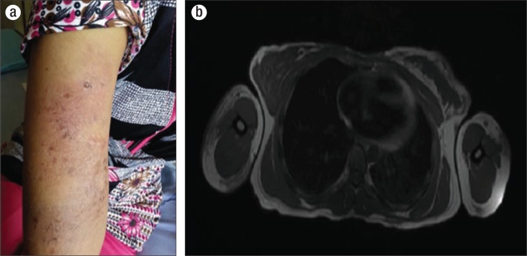
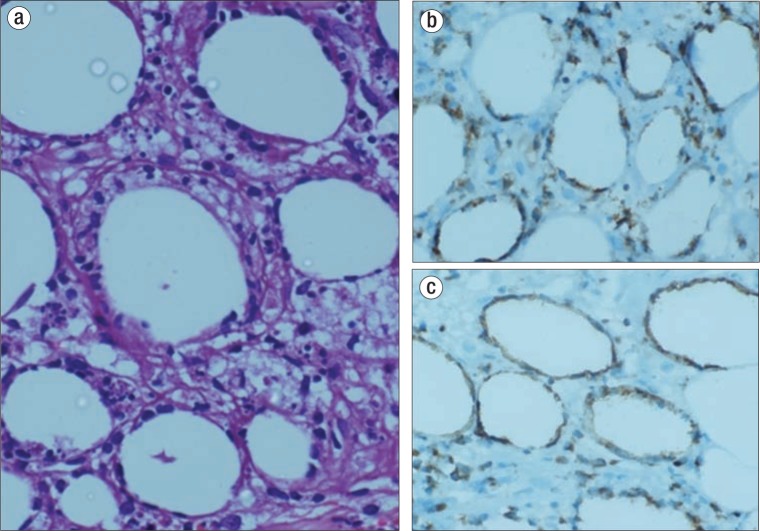
A 25-year-old woman presented with painless papulonodular skin lesions in the abdomen, chest, and limbs. The lesions had appeared 18 months earlier. She had a low-grade intermittent fever, had a poor appetite, and had lost 5 kg over the past 6 months. She had painless nodular subcutaneous skin lesions 1 to 2 cm in size involving all four limbs and the chest and abdomen (Figure 1a). The nodules were painless and erythematous. She had no other symptoms, organomegaly, or lymphadenopathy. A computed tomography scan showed subcutaneous soft tissue thickening with fat stranding in the upper chest, anterior abdominal wall, and upper arm bilaterally. T1- and T2-weighted magnetic resonance imaging of both arms showed intermediate-signal-intensity lesions in the skin and subcutaneous plane (Figure 1b). These lesions showed heterogeneous postcontrast enhancement, and underlying muscles showed normal signal intensities. Histopathological examination of a skin biopsy showed the subcutaneous tissue infiltrated by a neoplasm composed of atypical lymphoid cells with medium size scanty cytoplasm and a round nucleus with fine chromatin and small nucleoli. Angioinvasion was also present (Figure 2a). On immunohistochemistry, the tumor cells were positive for CD3, CD8, perforin, and granzyme and were negative for CD20, CD4, and CD56 (Figure 2b, 2c). The picture was diagnostic of SPTCL. Her hemoglobin was 8.8 g/dL; total leukocyte count, 5000/mm3; platelet count, 327000 /mm3; and erythrocyte sedimentation rate, 80 mm/1st hour. Her renal function, liver function, and serum electrolytes were normal. Her serum lactate dehydrogenase level was 1407 IU/L. A bone marrow study was normal. She was started on chemotherapy with cyclophosphamide, doxorubicin, vincristine, and prednisolone. Her skin lesions resolved after two courses, and she completed six courses of chemotherapy. At present, she is alive and in complete remission 18 months after therapy.
Figure 1.
(a) Erythematous skin lesions in the left upper limb. (b) Magnetic resonance imaging of the right and left arms, axial view, showing lesions in the skin and the subcutaneous plane of both arms.
Figure 2.
(a) Skin biopsy showing the subcutaneous tissue infiltrated with atypical lymphoid cells. (b) Tumor cells showing CD3 positivity. (c) Tumor cells showing perforin positivity.
DISCUSSION
SPTCL is a rare tumor of T-cell origin representing <1% of all non-Hodgkin's lymphomas (2). It was first described in 1991 in an eight-case series, but was not recognized as a distinct entity by the World Health Organization until 2001 (2, 3). It affects young adults, with a median age at diagnosis of 39 years.
The disease typically follows a distinctive, indolent course of recurrent, self-healing subcutaneous nodules. Often it presents as multiple painless subcutaneous nodules on the extremities and trunk. In its early phase, the nodules may resolve without treatment and new nodules may develop on the same or different skin locations. Diagnosis of SPTCL is a challenge, especially in the early stages when the symptoms mimic other more common conditions such as benign panniculitis, eczema, dermatitis, cellulitis, and other skin and soft tissue infections. About three-fourths of patients with SPTCL have multifocal cutaneous involvement (4). More serious conditions associated with SPTCL include serosal effusions, hemophagocytosis syndrome, and pancytopenia.
Imaging is useful for diagnosis and for monitoring response to therapy. Diagnosis of SPTCL is based on pathological examination of skin and subcutaneous tissue, clinical characteristics, immunohistochemistry staining patterns, and molecular analysis. There are two distinct types of SPTCL based on the T-cell receptor phenotype and immunohistochemistry characteristics. The first, T-cell receptor αβ, is characterized by an indolent, protracted course and is usually CD4−, CD8+, and CD56−. The second, T-cell receptor γδ, is associated with rapid clinical deterioration and coexisting hemophagocytosis. It is usually CD4−, CD8−, and CD56+ (5). Currently, the T-cell receptor αβ subtype is designated as SPTCL, whereas the T-cell receptor γδ is designated as cutaneous gamma/delta positive T-cell lymphoma (6).
Chemotherapy with cyclophosphamide, doxorubicin, vincristine, and prednisolone is the preferred regimen, with overall remission rates of 50% (7). In some cases, autologous bone marrow transplantation has been attempted. The overall 5-year survival rate for T-cell receptor αβ exceeds 80% (8). Our patient received six courses of combination chemotherapy and is alive with no evidence of disease 18 months after therapy.
References
- 1.Swerdlow SH, Campo E, Harris NL, Jaffe ES, Pileri SA, Stein H, Thiele J, Vardiman JW. WHO Classification of Tumours of Haematopoietic and Lymphoid Tissues. 4th ed. Lyon, France: IARC Press, 2008; pp. 294–295. [Google Scholar]
- 2.Jaffe ES, Harris NL, Stein H, Vardiman JW. Pathology and Genetics of Tumours of Haematopoietic and Lymphoid Tissues. World Health Organization Classification of Tumours. Lyon, France: IARC Press, 2001; pp. 351–352. [Google Scholar]
- 3.Gonzalez CL, Medeiros LJ, Braziel RM, Jaffe ES. T-cell lymphoma involving subcutaneous tissue: a clinicopathologic entity commonly associated with hemophagocytic syndrome. Am J Surg Pathol. 1991;15(1):17–27. doi: 10.1097/00000478-199101000-00002. [DOI] [PubMed] [Google Scholar]
- 4.Willemze R, Jaffe ES, Burg G, Cerroni L, Berti E, Swerdlow SH, Ralfkiaer E, Chimenti S, Diaz-Perez JL, Duncan LM, Grange F, Harris NL, Kempf W, Kerl H, Kurrer M, Knobler R, Pimpinelli N, Sander C, Santucci M, Sterry W, Vermeer MH, Wechsler J, Whittaker S, Meijer CJ. WHO-EORTC classification for cutaneous lymphomas. Blood. 2005;105(10):3768–3785. doi: 10.1182/blood-2004-09-3502. [DOI] [PubMed] [Google Scholar]
- 5.Takeshita M, Imayama S, Oshiro Y, Kurihara K, Okamoto S, Matsuki Y, Nakashima Y, Okamura T, Shiratsuchi M, Hayashi T, Kikuchi M. Clinicopathologic analysis of 22 cases of subcutaneous panniculitis-like CD56– or CD56+ lymphoma and review of 44 other reported cases. Am J Clin Pathol. 2004;121(3):408–416. doi: 10.1309/TYRG-T196-N2AP-LLR9. [DOI] [PubMed] [Google Scholar]
- 6.Parveen Z, Thompson K. Subcutaneous panniculitis-like T-cell lymphoma: redefinition of diagnostic criteria in the recent World Health Organization–European Organization for Research and Treatment of Cancer classification for cutaneous lymphomas. Arch Pathol Lab Med. 2009;133(2):303–308. doi: 10.5858/133.2.303. [DOI] [PubMed] [Google Scholar]
- 7.Medhi K, Kumar R, Rishi A, Kumar L, Bakhshi S. Subcutaneous panniculitislike T-cell lymphoma with hemophagocytosis: complete remission with BFM-90 protocol. J Pediatr Hematol Oncol. 2008;30(7):558–561. doi: 10.1097/MPH.0b013e31817588e8. [DOI] [PubMed] [Google Scholar]
- 8.Willemze R, Jansen PM, Cerroni L, Berti E, Santucci M, Assaf C, Canninga-van Dijk MR, Carlotti A, Geerts ML, Hahtola S, Hummel M, Jeskanen L, Kempf W, Massone C, Ortiz-Romero PL, Paulli M, Petrella T, Ranki A, Peralto JL, Robson A, Senff NJ, Vermeer MH, Wechsler J, Whittaker S, Meijer CJ EORTC Cutaneous Lymphoma Group. Subcutaneous panniculitis-like T-cell lymphoma: definition, classification, and prognostic factors: an EORTC Cutaneous Lymphoma Group Study of 83 cases. Blood. 2008;111(2):838–845. doi: 10.1182/blood-2007-04-087288. [DOI] [PubMed] [Google Scholar]


