Abstract
Chondroitin sulfate proteoglycans (CSPGs) inhibit the formation of axon collateral branches. The regulation of the axonal cytoskeleton and mitochondria are important components of the mechanism of branching. Actin-dependent axonal plasticity, reflected in the dynamics of axonal actin patches and filopodia, is greatest along segments of the axon populated by mitochondria. We report that CSPGs partially depolarize the membrane potential of axonal mitochondria, which impairs the dynamics of the axonal actin cytoskeleton and decreases the formation and duration of axonal filopodia, the first steps in the mechanism of branching. The effects of CSPGs on actin cytoskeletal dynamics are specific to axon segments populated by mitochondria. In contrast, CSPGs do not affect the microtubule content of axons, or the localization of microtubules into axonal filopodia, a required step in the mechanism of branch formation. We also report that CSPGs decrease the mitochondria-dependent axonal translation of cortactin, an actin associated protein involved in branching. Finally, the inhibitory effects of CSPGs on axon branching, actin cytoskeletal dynamics and the axonal translation of cortactin are reversed by culturing neurons with acetyl-L-carnitine, which promotes mitochondrial respiration. Collectively these data indicate that CSPGs impair mitochondrial function in axons, an effect which contributes to the inhibition of axon branching.
Keywords: axon sprouting, mitochondria, cortactin, actin patch, acetyl-l-carnitine
INTRODUCTION
Chondroitin sulfate proteoglycans (CSPGs) are a family of extracellular matrix components that regulate aspects of axon development and pathfinding (Silver and Silver, 2014). Moreover, failure of the CNS to repair and reestablish function after injury is in part attributed to the injury-induced expression of CSPGs around the lesion site (Bartus et al., 2012; Sharma et al., 2012; Silver and Silver, 2014; Ohtake and Li, 2015). CSPGs are also a major component of the perineuronal nets that regulate developmental neuronal plasticity (Miao et al., 2014; Dyck and Karimi-Abdolrezaee, 2015). An additional role of CSPGs is to suppress axon collateral branching in vivo (Barritt et al., 2006; Tom et al., 2009; Lee et al., 2010; Starkey et al., 2012; Lee et al., 2014; Lemarchant et al., 2014; Lang et al., 2015), and this rewiring of existing circuitry through axon branching is of major importance in the response of the nervous system to injury (Reviewed in Onifer et al., 2011; Wang and Sun, 2011; Dietz and Fouad, 2014; Kadomatsu and Sakamoto, 2014). While it is clear that CSPGs negatively regulate axon branching the mechanism used by CSPGs to suppress the formation of axon branches remains to be elucidated.
The production of an axon collateral branch requires localized reorganization of the cytoskeleton that can be broken down into sequential steps (Gallo, 2011; Gallo, 2013; Kalil and Dent, 2014). The first step is the formation of an axonal filopodium. Axonal filopodia arise from focal dynamic accumulations of actin filaments along the axon shaft termed actin patches. The targeting of one or more axonal microtubules into the filopodium is the next necessary event prior to the maturation of a filopodium into a branch. Filopodia containing microtubules have the potential to reorganize and develop polarity resulting in a nascent branch with a growth cone-like structure at its tip. Recent studies have identified a crucial role for axonal mitochondria in the formation of both axonal filopodia and their maturation into branches (Courchet et al., 2013; Spillane et al., 2013; Tao et al., 2014). Stalled mitochondria determine the sites along the axon where branches can form (Courchet et al., 2013; Spillane et al., 2013; Tao et al., 2014), and the respiration of mitochondria is required for branch formation (Spillane et al., 2013). Actin patches and filopodia are dependent on actin filament polymerization. Actin monomers must be ATP loaded in order to polymerize after which the ATP is hydrolyzed to ADP (Pollard and Cooper, 2009). Consequently, blockade of mitochondrial respiration blocks axonal actin dynamics (Ketschek and Gallo, 2010). In the context of nerve growth factor (NGF) induced branching of sensory axons mitochondria respiration is also required to establish localized sites of high intra-axonal protein synthesis that is in turn required for NGF-induced branching (Spillane et al., 2012; Spillane et al., 2013). The current study aims to elucidate the aspects of the axonal cytoskeletal dynamics underlying branching affected by CSPGs, and whether CSPGs may also be affecting axonal mitochondria thus contributing to the suppression of axon branching.
METHODS
Culturing and transfection of sensory neurons
Gallus Gallus fertilized eggs of unknown sex were obtained from Charles River Laboratories and incubated in a 1502 Sportsman incubator (GQF MFG Inc). Culturing was performed on laminin (25 μg/ml; Invitrogen), polylysine (100 μg/mL; Sigma) and chicken CSPGs (5 μg/mL; Millipore) coated substrata, in defined F12H (Invitrogen) serum-free medium with supplements as previously described in and detailed in (Lelkes P.I., 2006). Briefly, coverslips were coated with polylysine overnight, washed, coated with CSPGs or water for one hour, washed again and coated with laminin for an hour. Substrates were suspended in water when coating slides for all experiments involving CSPGs. NGF (R&D Systems) was used at 20 ng/ml and acetyl-L-Carnitine (Fisher Scientific) was used at 500 μM unless otherwise stated.
For axon length and branching determinations E7 DRG neurons were dissociated and cultured. All live imaging was performed on dissociated neurons. For analysis of axon branching in response to acute or chronic (24 hrs) NGF treatment we cultured E7 explants as previously detailed for this assay (Ketschek and Gallo, 2010; Spillane et al., 2012). Dissociated DRG cells were transfected using an Amaxa Nucleoporator (program G-13, 10 μg of plasmid) and chicken transfection reagents, as previously described by (Ketschek and Gallo, 2010). All experiments were performed 24 hrs after plating.
Immunocytochemistry
To image and quantify the axonal cytoskeleton cultures were simultaneously fixed and extracted using a combination of 0.25% glutaraldehyde and 0.1% triton X-100 in cytoskeleton preservation buffer (PHEM buffer) (see Gallo and Letourneau, 1998; Gallo and Letourneau, 1999) for 15 min. In all cases, following fixation/extraction the samples were washed with phosphate buffered saline (PBS) and then treated with 4 mg/mL sodium borohydride to quench glutaraldehyde autofluorescence. The samples were then washed in PBS and blocked in 10% goat serum in PBS containing 0.1% triton X-100 for 15 min, followed by washing three times with PBS. Tubulin and actin filaments were stained using an anti-α-tubulin antibody directly conjugated to fluorescein (Sigma; DM1A-FITC, 1:100) and rhodamine phalloidin (Invitrogen; per supplier’s directions) for 45 min. All antibodies were diluted in PBS containing 10% goat serum and 0.1% triton X-100.
Mitochondria labeling approaches
Axonal mitochondria were labeled with mitotracker red (1 nM) or green (25 nM) for 30 minutes followed by two washes with dye free medium (both from ThermoFisher). Similarly, labeling was performed with JC-1 (50 nM; ThermoFisher) for 30 min followed by two washes prior to image acquisition. The distal most three JC-1 or mitotracker red labeled mitochondria, which were not overlapping or clustering with other mitochondria, in the distal 20–70 μm of axons were quantified, excluding the distal 20 μm representative of the growth cones.
Imaging
Imaging was performed using a Zeiss 200M inverted microscope equipped with an enclosed heating stage. An Orca ER camera (Hamamatsu) was used for image acquisition and all digital aspects of imaging and microscope and camera control were performed using Zeiss Axiovision software. For determination of mitochondria morphology, mitotracker red and JC-1 signals images were acquired at 100x (1.3 numerical aperture) using 1×1 camera binning to obtain maximal spatial resolution. For all data sets involving the quantification of fluorescence intensities imaging parameters were used which did not result in any saturated pixels, as determined through consideration of the intensity histograms for images. Live imaging of mitotracker red labeled mitochondrial dynamics was performed using 2×2 binning using minimal light exposure resulting in clearly detectable mitochondria to minimize bleaching during imaging.
Image analysis
The mean intensity of mitotracker red was determined for individual mitochondria following subtraction of the signals in the adjacent cytoplasmic domain. For analysis of JC-1 both channels were acquired in image stacks and the ratio of the two determined within the outline of the mitochondrion.
FRAP analysis of translational reporters was performed as previously detailed (Spillane et al., 2012; Spillane et al., 2013). MyrGFP hotspots were determined from line scans of images generated using ImageJ. For each line scan, the numerical values of intensity were exported into Excel and the mean determined. A hotspot was defined as a peak in the signal which was at least twice the mean for the linescan (determined from the Y-value data sets obtained from ImageJ).
Statistical analysis
The data were analyzed using Instat 3 software (GraphPad Inc.). If any data set in an experimental design was not normal (Kolmogorov-Smirnov test), then non-parametric analysis was performed (Mann-Whitney test for two group comparisons, and nonparametric ANOVA with Dunn’s posthoc tests). Parametric data are presented as means and standard error of measurements. If all sets were normally distributed then Welch t-tests were used, with paired test used for pre-post experimental designs, or ANOVA with Bonferroni post-hoc tests. Categorical data sets were analyzed through the Fischer’s test using the raw categorical data. Two tailed tests used when the hypothesis lacked directionality, one tailed when the hypothesis predicted directionality. Means ± SEM are presented, or medians as detailed.
RESULTS
CSPGs decrease the collateral branching of cultured sensory axons
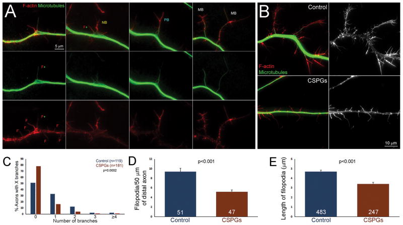
We first sought to determine the effects of a mixture of CSPGs, purified from embryonic day 14 chicken brains (see Methods), on the branching of embryonic day 7 chicken sensory neurons cultured in the presence of the branch inducing factor NGF (20 ng/mL). In all experiments throughout the study the control condition involved culturing the neurons on glass substrata coated with polylysine and laminin, and the experimental substrata were further coated with CSPGs as described in the Methods. Axon branches were analyzed in samples which underwent combined fixation and extraction to specifically retain the polymerized cytoskeleton without interference from soluble cytoskeletal proteins (Gallo and Letourneau, 1998; Gallo and Letourneau, 1999). Samples were subsequently stained with anti-α-tubulin and phalloidin to stain microtubules and actin filaments, respectively. Filopodia are defined as linear protrusions from the axon shaft characterized by a uniform bundle of actin filaments (Figure 1A; indicated by F). An early step in branch formation involves the targeting of a microtubule into a filopodium (Figure 1A; indicated by F+). As in our previous work, branches were defined as axonal protrusions containing one or more microtubules and a heterogeneous actin cytoskeleton (Figure 1A; indicated by PB, NB, MB at differing stages of formation), reflecting the maturation of filopodium into a branch (Figure 1A; Spillane et al., 2012; Spillane et al., 2013; Ketschek et al., 2015)). Analysis of the number of axon branches along the distal 100 μm of axons revealed that on CSPGs axons exhibited fewer branches per unit length of axon than controls (Figure 1B,C). Analysis of the number of axonal filopodia along the axons also revealed a 47% decrease in the number of filopodia on CSPGs (Figure 1D). The length of axonal filopodia on CSPGs was also decreased by 22% relative to the control substratum (Figure 1E). In contrast, the length of axon branches that formed on CSPGs was not different from that of branches that formed on the control substratum (p=0.52, Mann-Whitney test, n=24 and 26, respectively). These data are consistent with previous in vivo data demonstrating that CSPGs suppress the initiation of collateral axon branching, prompting us to investigate the mechanisms behind this suppression of filopodia and branching (Tom et al., 2009; Lee et al., 2010; Starkey et al., 2012; Lee et al., 2014; Lemarchant et al., 2014; Lang et al., 2015).
Figure 1.
CSPGs decrease axon branching. (A) Examples of filopodia and branches at different stages of branch formation. F refers to filopodia containing only actin filaments and exhibiting the characteristic linear morphology and actin filament bundles. F+ refers to a filopodium containing a microtubule. NB refers to a nascent branch. Note that the filopodial morphology has changed and the actin filaments are no longer organized in a uniform bundle (compare to examples of F). PB refers to a branchlet which has acquired a polarized distribution of actin filaments, greatest distally. MB refers to mature branches containing a microtubule core and a more prominent distal accumulation of actin filaments resembling a small growth cone. (B) Examples of the cytoskeletal organization of axons on control and CSPG coated substrata. The control example exhibits more axonal filopodia and branches than the axon on CSPGs. (C) Graph showing the distribution of axons exhibiting 0 to ≥4 branches/distal 100 μm on control and CSPG coated substrata. (D) Graph of the number of filopodia along distal axons. (E) Graph of the length of filopodia along distal axons. n = axons in C and D, and filopodia in E.
CSPGs partially depolarize the membrane potential of axonal mitochondria
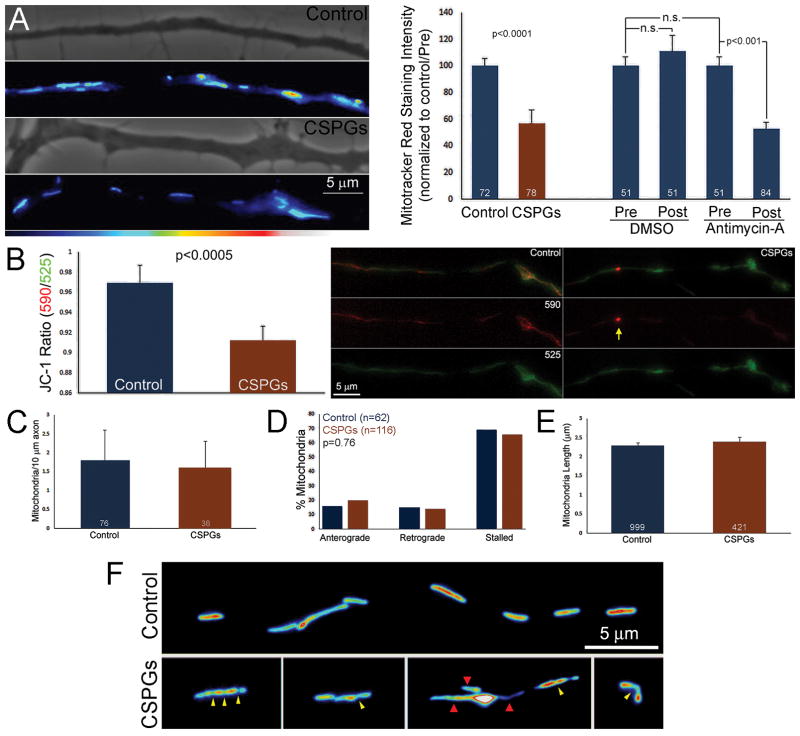
Mitochondria and their respiration have recently been determined to be important aspects of the mechanism of both spontaneous and NGF-induced axon branching (Courchet et al., 2013; Spillane et al., 2013; Tao et al., 2014). The mitochondrial membrane potential, which is established by the electron transport chain, is a major positive determinant of oxidative phosphorylation derived ATP production. In order to determine if mitochondrial membrane potential might be affected by CSPGs mitochondria were labeled with two dyes which are sensitive to mitochondrial membrane potential. Mitotracker Red incorporates into mitochondria as a function of mitochondrial membrane potential (Poot et al., 1996; Gilmore and Wilson, 1999; Krohn et al., 1999; Buckman et al., 2001; Pendergrass et al., 2004). Depolarized membrane potential result in decreased accumulation of the dye. Comparison of the intensity of mitotracker red in mitochondria using epifluorescence along the axons of control and CSPG coated cultures revealed a 43% decrease in mitotracker red staining intensity along axons on CSPGs (Figure 2A). Addition of antimycin A, an inhibitor of cytochrome C reductase that results in the disruption of the proton gradient, depolarization of the mitochondrial membrane potential and decreased ATP production, reproduced the effects of CSPGs by reducing mitotracker red staining intensity. Cultures labeled with mitotracker red were treated with varying concentrations of antimycin A and a 2.5 μM treatment resulted in a 53% decrease in labeling (Figure 2A; Figure S1).
Figure 2.
CSPGs depolarize mitochondrial membrane potential and affect mitochondrial morphology. (A) Examples of mitotracker red labeling intensity of axonal mitochondria on control and CSPG coated substrata. The graph shows the quantification of the mean staining intensity of mitotracker red in axonal mitochondria as a function of substratum (leftmost two bars). The graph also shows the effects of a 40 min treatment with 2.5 μM antimycin-A on mitotracker red labeling of axonal mitochondria (see Figure S1 for an example of the decline in mitotracker red staining intensity following antimycin-A treatment). Mitochondria were labeled and imaged before treatment with antimycin-A or the vehicle DMSO (pretreatment), and again 40 min later (post treatment). n = axons, 3 mitochondria sampled/axon. (B) Quantitative analysis and examples of the ratio of JC-1 emission at 590/525 on control and CSPG substrata. Although on CSPGs mitochondria exhibited mean decreased 590/525 ratios, and 590 signal, occasionally high potential mitochondria (yellow arrow) were observed in both the CSPGs and control groups. n=66 mitochondria/group from 22 axons/group. (C) Measurement of the density of axonal mitochondria. n = axons. (D) Duty cycles of axonal mitochondria as reflected by the percentage of mitochondria undergoing anterograde or retrograde transport or remaining stalled during a 5 min imaging sequence (6 sec interframe intervals). n = mitochondria. (E) Analysis of the lengths, end to end, of axonal mitochondria. n = mitochondria. (F) Examples of abnormal mitochondrial morphologies observed on CSPG coated substrata. The yellow arrowheads denote apparent constrictions along the mitochondrion. The red arrowheads denote elongated profiles from non-uniform mitochondria.
As an alternative approach to measure the relative membrane potential of mitochondria we labeled neurons with JC-1, a mitochondrially targeted dye that exhibits aggregation and change in emission wavelength as a function of membrane potential (Overly et al., 1996; Dedov and Roufogalis, 1999; Miller and Sheetz, 2004; Lee and Peng, 2006; Lee and Peng, 2008; Steketee et al., 2012). JC-1 emits at ~525 nm in monomer form and switches to emitting at ~590 when aggregated. Thus, a decrease in ratio of emission at 590/525 is indicative of membrane depolarization. The axonal mitochondria of neurons cultured on CSPGs exhibited lower values of the 590/525 emission ratio relative to neurons on the control substratum, demonstrating a reduction in mitochondria membrane potential by a second independent means (Figure 2B).
In contrast to reducing the mitochondria membrane potential CSPGs did not affect the number of mitochondria per unit length of the distal 20–70 μm of the axon (excluding the distal 20 micron reflective of the growth cones) (Figure 2C), the proportion of mitochondria exhibiting anterograde/retrograde movements or remaining stalled (Figure 2D) or the length of mitochondria (Figure 2E). However, axonal mitochondria morphology was affected on CSPGs. While under control conditions mitochondria exhibited the expected tubular morphology (Figure 2F), on CSPGs approximately 33% of mitochondria (n=258) exhibited a variety of aberrant morphologies (Figure 2F), in contrast to 6% on the control substratum (n=194)(p<0.001). On CSPGs mitochondria were observed to exhibit heterogeneous widths and/or apparent constrictions along their lengths (Figure 2F). Mitochondria exhibiting heterogeneous widths or apparent constrictions accounted for 71% and 26%, respectively, and 3% exhibited both malformations. The data indicate that CSPGs impact aspects of mitochondria function and morphology, but do not overtly affect their transport and density in axons.
Promotion of mitochondrial respiration increases axon branching on CSPGs
Given the relationship between mitochondrial localization and respiration in the initiation of axon collateral branches (Courchet et al., 2013; Spillane et al., 2013; Tao et al., 2014) and the observed deficits in mitochondrial function on CSPGs, we sought to experimentally manipulate mitochondrial respiration to elucidate this relationship. Acetyl-L-carnitine (ALC) is a natural metabolite which promotes mitochondrial oxidative phosphorylation (reviewed in Rosca et al., 2009; Onofrj et al., 2013). ALC promotes the sprouting of serotonergic fibers following central nervous system spinal cord injury (Karalija et al., 2014), regeneration of sensory axons following peripheral nerve injury (Fernandez et al., 1990; McKay Hart et al., 2002; Kokkalis et al., 2009; Wilson et al., 2010; Farahpour and Ghayour, 2014) and has tissue sparing effects in the spinal cord and other central nervous system injury models (Patel et al., 2010; Karalija et al., 2012; Patel et al., 2012; Karalija et al., 2014).
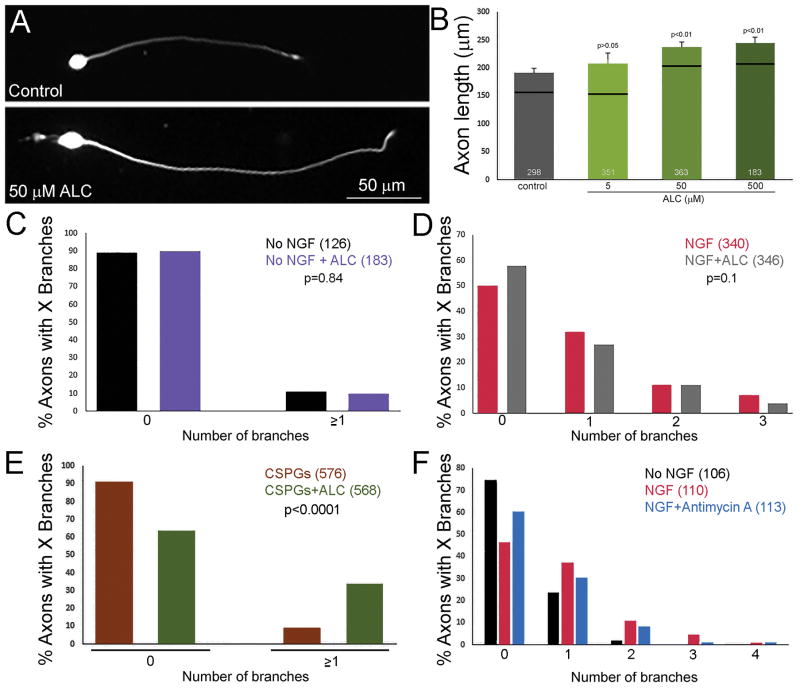
The effects of ALC on sensory axon morphogenesis have not previously been examined in vitro, thus we sought to determine whether ALC treatment would impact the growth and branching of sensory axons under control conditions in the presence and absence of NGF. Dissociated embryonic day 7 chicken sensory neurons were cultured in the absence of neurotrophins on laminin coated substrates. Under these conditions the NGF responsive population preferentially extends axons due to differential expression of integrin receptors between the sensory neuron populations in the ganglion (Guan et al., 2003; Roche et al., 2009; Ketschek and Gallo, 2010; Marsick et al., 2012; Spillane et al., 2012; Ketschek et al., 2015). Neurons were cultured overnight in medium with or without ALC and the lengths of the axons were measured from fixed samples stained with anti-α-tubulin antibodies. ALC dose dependently increased axon length over a range of 5–500 μM (Figure 3A,B). The effect of ALC becomes evident at 50 μM. At 50 and 500 μM ALC median axon lengths were increased by 26% and 30%, respectively. The effective concentrations of ALC observed in this study are in the same range as determined by other in vitro studies using primary neurons and neuron-like cell lines and for the remainder of the study 500 μM ALC was used (Forloni et al., 1994; Pisano et al., 2003; Picconi et al., 2006; Bagetta et al., 2008; Epis et al., 2008; Selvatici et al., 2009; Zhang et al., 2012).
Figure 3.
Effects of treatment with ALC on axon development on control and CSPG coated substrata. (A) Examples of dissociated neurons cultured overnight on control substrata in the absence of NGF either treated with 500 μM ALC or control medium. As reflected in the quantification in panel (B), ALC treatment increased axon lengths. (B) Dose dependent effects of ALC treatment on axon lengths on control substrata in the absence of NGF. Black lines represent medians and the number of neurons measured is shown in the bars. n = axons. (C) Graph showing the percentage of axons with 0 or ≥1 branches along their distal 100 μm cultured on control substrata in the absence of NGF ± treatment with 500 μM ALC. n = axons. (D) Graph showing the percentage of axons with 0 to 3 branches along their distal 100 μm cultured on control substrata in the presence of NGF ± treatment with 500 μM ALC. n = axons. (E) Graph showing the percentage of axons with 0 or ≥1 branches along their distal 100 μm cultured on CSPG coated substrata in the presence of NGF ± treatment with 500 μM ALC. n = axons. (F) Graph showing the percentage of axons with 0 to 3 branches along their distal 100 μm cultured overnight on control substrata in the absence of NGF, then treated with NGF for 40 min ± treatment with 2.5 μM antimycin A. NGF induces branching which is attenuated by antimycin A, as reflected by a leftward shit in the distribution. However, the effects of antimycin A was not statistically significant. n = axons.
To address whether ALC would also increase the length of axons when NGF was present, neurons were cultured for 6 hrs ± ALC in medium containing NGF. For this determination we used a 6 hr culturing period to avoid axons forming overlapping and fasciculated meshworks due to NGF-mediated promotion of axon growth. In the presence of NGF treatment with ALC increased median axon length by 14% (p=0.03, Mann-Whitney test, n=137 and 141 for NGF and NGF+ALC groups, respectively). These data indicate that ALC treatment increases the length of axons and that the effect is approximately double when NGF is not present. In contrast, on CSPGs + NGF treatment with ALC had no effect on axon lengths (p=0.32, n=576, 568).
Although ALC treatment increased axon lengths under control conditions with and without NGF, treatment with ALC did not alter the number of branches formed along axons regardless of the presence of NGF (Figure 3C–D). However, ALC treatment increased the proportion of axons that formed branches when cultured on CSPGs (Figure 3E). Collectively, these data indicate that promotion of mitochondrial function can partially reverse the effects of CSPGs on the suppression of axon branching but not the inhibition of axon extension, underscoring that while the two processes of axon extension and axon branching share much in common the mechanisms which control them are different (Gallo, 2011).
Conversely, we sought to determine if decreasing mitochondrial membrane potential to a similar extent as observed on CSPGs, using antimycin A (Figure 2B), can impair the branching of axons in response to acute treatment with NGF on control substrata. As in our previous work addressing the acute induction of axon branching by NGF (Ketschek and Gallo, 2010; Ketschek et al., 2015) E7 DRG explants were cultured on laminin coated substrata in the absence of NGF for 24 hours. Cultures were then treated with DMSO, NGF+DMSO or NGF+2.5 μM antimycin A for 40 min prior to fixation and staining. As expected, NGF induced branching relative to no NGF controls (Figure 3F; p<0.0001, Fisher’s test). Cotreatment with 2.5 μM antimycin A did not block NGF-induced branching (Figure 3F). Although there was a trend toward less branching it was not statistically significant (p=0.12, Fisher’s test). These data indicate that the suppression of mitochondrial membrane potential to a similar extent as that observed on CSPGs is not sufficient to block branching in the context of branching induced by acute treatment with NGF.
CSPGs impair cytoskeletal actin dynamics underlying the formation of axonal filopodia
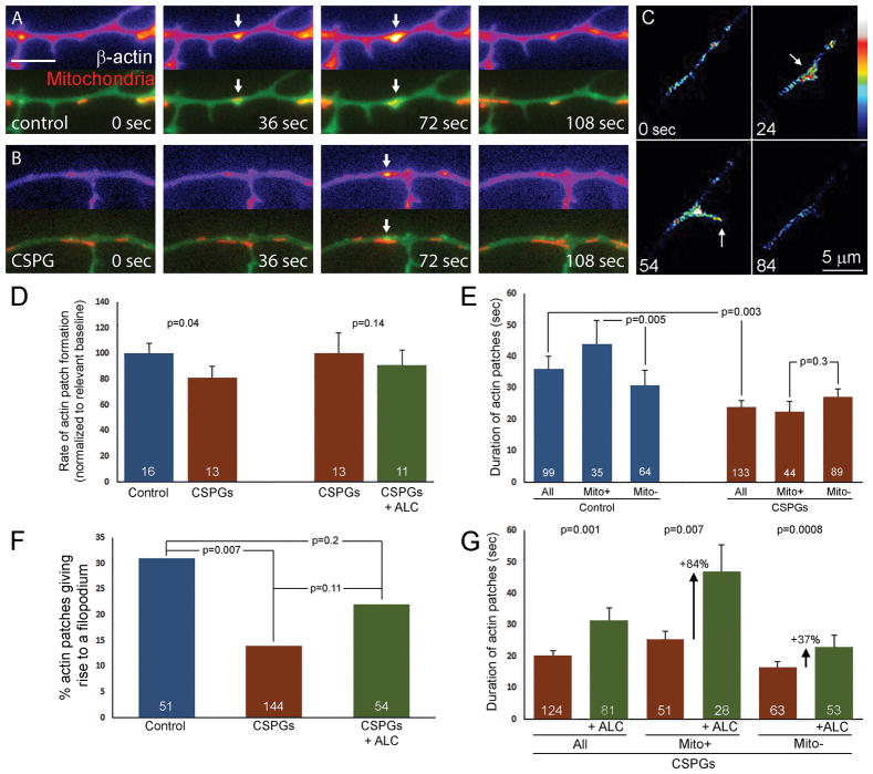
Axonal filopodia arise from precursor structures consisting of meshworks of actin filaments termed axonal actin patches (Figure 4A–C; Ketschek and Gallo, 2010; Gallo, 2011; Spillane et al., 2011; Gallo, 2013). As revealed by timelapse imaging of eYFP- β-actin patches are dynamic structures arising de novo along the axon, elaborating to their maximal size and subsequently diminishing in size until fully dissipated (Figure 4A–C). Axonal filopodia arise from actin patches (Figure 4C), but only a subset of the actin patches formed along the axon give rise to filopodia (Ketschek and Gallo, 2010; Gallo, 2011; Gallo, 2013). Actin patches are initiated in part through the Arp2/3 actin nucleation complex and its regulatory proteins (e.g., cortactin and WAVE1; Spillane et al., 2011; Spillane et al., 2012; Arnold and Gallo, 2013) and in the presence of NGF axonal actin patches and filopodia preferentially form at axonal sites populated by mitochondria (Ketschek and Gallo, 2010; Spillane et al., 2013).
Figure 4.
Effects of CSPGs and ALC treatment on axonal actin dynamics. (A) and (B) Examples of actin patches (imaged through expression of eYFP-β-actin; green in bottom images and false colored in top images showing only actin) in axons also expressing mitochondrially targeted DsRed (red). Panel (A) shows an axon on the control substratum, panel (B) shows an axon on a CSPGs coated substratum. The arrows denote actin patches colocalizing with mitochondria. Note that on the control substratum the patch has a greater duration and attains a greater size. Bar in panel A represents 5 μm and applies to A and B. (C) Example of an axonal actin patch giving rise to a filopodium (time in seconds; false coloring). The actin patch elaborates between 0–24 seconds, gives rise to a filopodium (54 sec) and by 84 seconds the filopodium has retracted and the patch fully dissipated. Images were contrast enhanced to resolve the fine filopodium which is otherwise dimmer than the actin patch. (D) Actin patch formation rates on control and CSPGs substrates (mean control rate of 7.9 patches/50 μm/6 min). The two data sets were acquired independently, with control and CSPG substrata acting as the relevant baselines for each comparison (control vs CSPGs and CSPGs vs CSPGs+ALC). n=axons. (E) Effects of CSPG substrata on the duration of actin patches and the relationship between patch duration and mitochondrial colocalization. In the control group patches associated with mitochondria exhibited longer durations than those not associated with mitochondria. On CSPGs patches exhibited shorter durations than in controls and the durations of patches did no correlate with mitochondria colocalization. n=patches. (F) The percentage of actin patches that give rise to filopodia is decreased by CSPGs relative to controls. Treatment with ALC restores the percentage of patches giving rise to filopodia by 47%. n=patches. (G) Treatment of neurons cultured on CSPG substrata with ALC increases actin patch duration regardless of mitochondrial colocalization. However, the relative effect of ALC on patch duration is most pronounced for patches associated with mitochondria. n=patches.
In order to determine if CSPGs control axonal actin dynamics and alter the role of mitochondria in this process we imaged actin dynamics through the expression of eYFP-β-actin in neurons cotransfected with mitochondrially targeted DsRed (mtDsRed) as in prior work (Spillane et al., 2013). On CSPGs the population of all actin patches regardless of their association with mitochondria formed patches at nearly the same frequency per unit length/unit time as axons on the control substratum (Figure 4A,B,D). On CSPGs there was a ~20% decrease in the number of patches formed per unit length of axon. However, the mean duration of all actin patches regardless of colocalization with mitochondria (i.e., time from first detection to full dissipation) was decreased by 33% on CSPGs relative to controls (Figure 4E). The slight decrease in the number of detectable patches formed along axons (Figure 4D) may thus reflect the decrease in the duration of patches, placing some patches below our ability to detect them. Analysis of actin patch duration as a function of their colocalization with mitochondria revealed that actin patches associated with mitochondria were more greatly affected by CSPGs (47% decrease in duration) than those not associated with mitochondria (Figure 4E), which was not significantly different between control and CSPGs conditions. Similar trends with respect to mitochondrial colocalization were also detected by measuring the area of individual patches when they reached maximal elaboration demonstrating that mitochondrially associated actin patches were more greatly affected by CSPGs (Figure S2). Additionally, the duration of patches under control conditions as a function of mitochondrial colocalization also revealed that actin patches associated with mitochondria exhibit longer durations than those not associated with mitochondria (Figure 4E). This observation is consistent with the mitochondrion being a positive regulator of actin patch dynamics. Finally, consistent with the observed decrease in axonal filopodia (Figure 1D) CSPGs decreased the percentage of actin patches which give rise to filopodia (Figure 4F).
Since the effects of CSPGs were most pronounced at mitochondria we next considered whether treatment with ALC altered the effects of CSPGs on actin patches. Compared to CSPGs alone and considering the entire population of actin patches treatment with ALC increased the duration of actin patches (Figure 4G). However, the effect of ALC on patch duration was proportionally much greater for actin patches associated with mitochondria (Figure 4G). ALC increased the mean duration of actin patches by 84% for patches associated with mitochondria and by 37% for those not associated with mitochondria. ALC treatment did not affect the rate of actin patch formation relative to CSPGs alone (Figure 4D), but partially restored the percentage of actin patches that gave rise to a filopodium on CSPGs coated substrata (Figure 4F). The increase in the percentage of patches giving rise to filopodia in the ALC treated group restored 47% of the decrease observed on CSPGs relative to the control, and statistically the ALC treated group was not different from either the control of the CSPG groups. These data demonstrate that CSPGs inhibit filopodia formation by regulating the actin dynamics of precursor actin patches, and this inhibition can in part be overcome locally at the mitochondrion by increasing mitochondrial metabolism through ALC, which is associated with an increase in ATP (Nishida et al., 1989; Aureli et al., 1994).
CSPGs decrease the duration of axonal filopodia associated with mitochondria
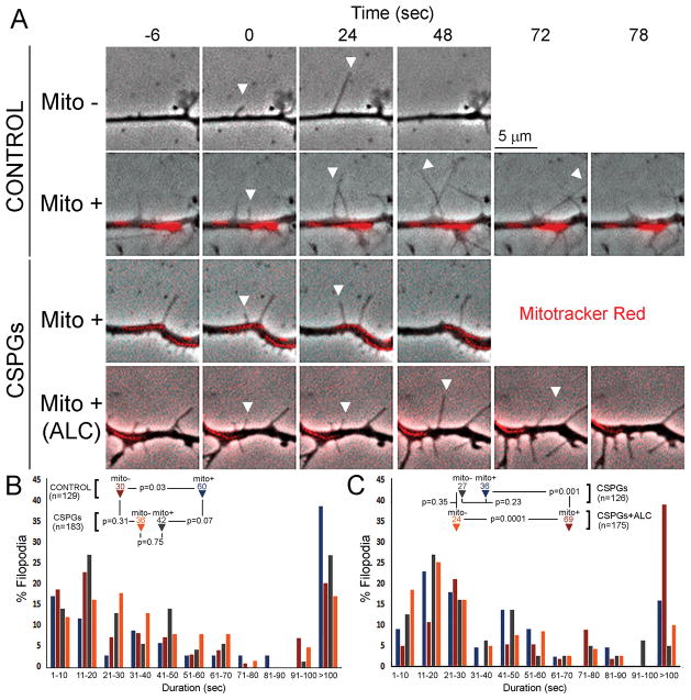
Filopodia elongate through the polymerization of the barbed ends of the actin filaments localized to the tip of the filopodium (reviewed in Gallo, 2013). To analyze filopodial dynamics in relation to axonal mitochondria, we labeled neurons with mitotracker red and imaged filopodia using phase contrast imaging. The duration of newly formed filopodia was analyzed on control substrata and CSPGs as a function of localization with mitochondria. Examples of filopodia emerging associated with or without mitochondria and their duration are shown in in Figure 5A. On control substrata, filopodia which formed in association with mitochondria exhibited approximately 100% greater median durations (denoted by arrowheads) compared to those formed in the absence of mitochondria (Figure 5A,B). In contrast, on CSPGs filopodia duration was independent of the association with mitochondria (Figure 5A,B) and overall all filopodia exhibited durations similar to those of filopodia not associated with mitochondria on the control substratum (Figure 5B). However, on CSPGs filopodia that formed in association with mitochondria exhibited a strong trend toward decreased durations relative to those on the control substratum (Figure 5B; although p=0.07). Furthermore, on CSPGs there was a 32% decrease in the percentage of mitochondria associated filopodia with durations greater than 100 seconds (Figure 5B; p=0.04, Fischer’s exact test).
Figure 5.
CSPGs decrease the duration of filopodia associated with mitochondria which is restored by ALC treatment. (A) Examples of filopodia emerging from axons and their durations. The full panel shows examples of filopodia emerging associated with mitochondria (Mito +) and not associated with mitochondria (Mito −) for the control substratum, and examples of filopodia associated with mitochondria on CSPGs ± ALC treatment. The tip of the filopodium is tracked over time by the white arrowheads. The mitotracker red signal was digitally manipulated to show the mitochondria overlaid onto the phase dark axons. (B) Histogram of the non-normal distributions of the duration of filopodia in 10 seconds bins from the respective color coded groups (control of CSPG substrata, association of filopodium with mitochondria +/−). The inset text shows the medians (arrowheads) for each color coded group and the outcomes of statistical comparisons. n=number of filopodia. (C) Same representation of filopodial duration rates as in panel A but all data was obtained from neurons cultured on CSPG substrata ± ALC treatment. n=number of filopodia.
In a separate experiment, we considered whether treatment with ALC would affect the duration of filopodia on CSPGs substrata (Figure 5C). Consistent with the prior results, we found no relationship between mitochondria colocalization and filopodia duration on CSPGs. Treatment with ALC increased filopodia duration for filopodia which formed associated with mitochondria, but did not affect the durations of filopodia formed independent of mitochondria (Figure 5C). ALC treatment also more than doubled the percentage of filopodia with durations greater than 100 sec (Figure 5C). Examples of filopodia emerging in association with mitochondria on CSPG ± ALC treatment are shown in Figure 5A. These data indicate that as with other aspects of actin filament dynamics on CSPGs ALC treatment results in effects which are specific to actin based structures (e.g., filopodia) associated with mitochondria.
The intra-axonal translation of cortactin is impaired on CSPGs
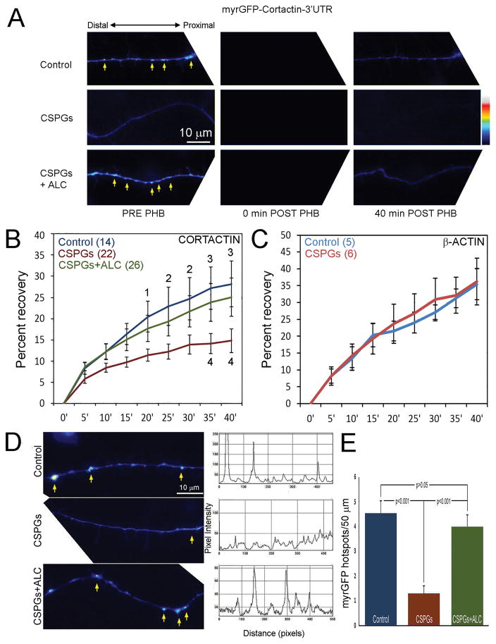
Cortactin is a regulator of the Arp2/3 actin nucleation complex that positively regulates the emergence of filopodia from actin patches and the duration of actin patches (Spillane et al., 2012), which are both decreased by CSPGs. In our culturing system the formation of axon branches in the presence of NGF requires NGF-induced intra-axonal translation of actin regulatory proteins including cortactin (Spillane et al., 2012; Spillane et al., 2013). The intra-axonal translation of cortactin is dependent on mitochondrial respiration (Spillane et al., 2013). mRNAs are targeted into axons through their association with ribonucleoprotein particles that undergo transport, and the association of mRNAs with the ribonucleoprotein particles is mediated by the 3′UTRs of mRNA in most but not all cases (reviewed in Gomes et al., 2014). The mRNAs for β-actin and cortactin are both localized to sensory axons through their 3′UTRs (Zhang et al., 2001; Willis et al., 2007; Spillane et al., 2012; Spillane et al., 2013). We have previously characterized reporter constructs consisting of the myristoylated-GFP (myrGFP) open reading frame with the 3′UTR of β-actin or cortactin to live image axonal translation (termed myrGFP-cortactin/β-actin-3′UTR as in prior work; Spillane et al., 2012, 2013). The cotranslational myristoylation of GFP confers membrane targeting and greatly decreases the diffusion of GFP providing increased spatial resolution over time when addressing the localization of the translation of the myrGFP reporter (Aakalu et al., 2001; Yudin et al., 2008). Expression of these constructs results in axonal myrGFP signal. The recovery of the myrGFP signal following photobleaching is reflective of local intra-axonal translation. Fluorescence recovery after photobleaching (FRAP) analysis of the myrGFP-cortactin-3′UTR and myrGFP-β-actin-3′UTR signals in distal axons revealed a suppression of cortactin translation on CSPGs (Figure 6A,B), but no effect on the translation of β-actin (Figure 6C). Treatment with ALC restored the FRAP of myrGFP-cortactin-3′UTR to levels similar to those observed in control conditions (Figure 6A,B).
Figure 6.
CSPGs suppress the axonal translation of cortactin mRNA. (A) Examples of axons expressing myrGFP-cortactin-3′UTR in control, CSPGs and CSPGs+ALC groups. The PRE images show the distribution of myrGFP before photobleaching (PHB). Examples of hotspots of myrGFP are denoted by arrows. The post PHB panels show the bleaching of the myrGF signal (0 min) and the recovery at 40 min post PHB. Uneven cropping is due to rotation of image to represent axons in horizontal position. (B) Quantification of the recovery from PHB. Anova with Bonferroni post-hoc tests was performed using time-matched comparisons. Post-hoc test p values are presented in the graph as follows: control vs CSPGs, 1 = p<0.05, 2 = p<0.01, 3 = p<0.001; CSPGs vs CSPGs+ALC, 4 = p<0.01. n = axons. (C) FRAP analysis of myrGFP-β-actin-3′UTR. (D) Additional examples of the distribution of myrGFP along axons, as in panel (A) and accompanying line scans of the intensity of myrGFP along the axon showing hotspots. (E) Quantification of the number of myrGFP hotspots. Sample sizes are the same as in panel B as this is a different analysis of the same population.
Axonal mitochondria determine sites of preferential translation along sensory axons, which when using the myrGFP translational reporters are reflected by focal accumulations of myrGFP termed myrGFP hotspots (Spillane et al., 2013). On the control substratum, axons exhibited the expected hotspots (denoted by arrows in Figure 6A, D). In contrast, few hotspots were visible along axons cultured on CSPGs (Figure 6A, D), and when elevations in the myrGFP signal were present on CSPGs they were not as focal (Figure 6D, CSPGs panel arrow). However, axons on CSPGs that were treated with ALC exhibited hotspots (Figure 6A, D). Quantification of the number of hotspots per unit length of axon showed that CSPGs decreased the number of hotspots while ALC treatment restored the number of hotspots to near control levels (Figure 6D).
Analysis of effects of CSPGs on axonal microtubules
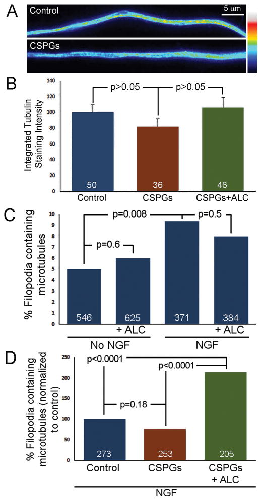
To investigate the effects of CSPGs and ALC treatment on axonal microtubules we used an established combined fixation and extraction protocol in cytoskeleton preserving buffer to retain the polymeric cytoskeleton (e.g., microtubules) while removing the soluble forms of the cytoskeletal proteins (e.g., tubulin) (Gallo and Letourneau, 1998; Gallo and Letourneau, 1999). The total integrated intensity of α-tubulin staining reflective of microtubules was then measured in fixed lengths of distal axons, as in prior work (e.g., Spillane et al., 2012). The total content of microtubules in distal axons was not affected by CSPGs, and ALC treatment did not increase microtubule content on CSPGs (Figure 7A,B). However, in the absence of NGF on control substrata treatment with ALC increased the microtubule content of axons by 63% (n=55 and 56 axons, p=0.004) but not in the presence of NGF (n=43 and 47 axons, p=0.23), as was also the case for neurons cultured on CSPGs substrata which are in the presence of NGF. Since microtubules are a major component of the mechanism of axon elongation, these observations are generally consistent with the more pronounced effect of ALC on increasing axon length in the absence of NGF (Figure 3).
Figure 7.
CSPGs have minimal effects on the axonal microtubule cytoskeleton. (A) False colored examples of the microtubule content of distal axons on control and CSPG substrata. (B) Quantification of the total integrated intensity in distal 50 microns of axons on control, CSPG substrata and CSPG substrata with ALC treatment all in the presence of NGF. No statistical differences amongst the groups were detected through a non-parametric ANOVA with Dunn’s post-hoc tests. Similarly, even when only the control and CSPG groups are considered alone through Mann-Whitney test no difference was detected (p=0.36). n = axons. (C) Graph of the percentage of axonal filopodia containing microtubules on control substrata ± NGF ± ALC treatment. As expected from prior investigations NGF increases the targeting of microtubules into axonal filopodia, but ALC treatment has no additional effect. n = filopodia. (D) Graph of the percentage of axonal filopodia containing microtubules on control substrata, CSPG and CSPG + ALC treatment. NGF is present in all groups. CSPGs did not affect the targeting of microtubules in filopodia, but ALC increased the percentage of filopodia containing microtubules on CSPG substrata. n = filopodia.
The localization of axonal microtubules into filopodia is a required step in order to form an axonal branch (Figure 1A). In both the absence and presence of NGF on control substrata treatment with ALC did not increase the percentage of axonal filopodia containing microtubules (Figure 7C). Consistent with prior observations (Spillane et al., 2012) the presence of NGF approximately doubled the percentage of filopodia containing microtubules relative to the absence of NGF (Figure 7C). The percentage of filopodia containing microtubules was not affected by CSPGs (Figure 7D). Unexpectedly, although ALC did not alter the microtubule content of axons cultured on CSPGs (Figure 7B) ALC approximately doubled the percentage of filopodia containing microtubules on CSPGs (Figure 7D).
DISCUSSION
This study demonstrates that CSPGs suppress the actin dynamics underlying early steps in the process of branching. The effects of CSPGs on the actin cytoskeleton are likely due to, at least in part, the downregulation of signaling pathways by CSPGs. CSPGs partially suppress PI3K-Akt signaling (Fisher et al., 2011; Silver et al., 2014) and restoration of PI3K signaling counters the effects of CSPGs on the inhibition of axon extension (Silver et al., 2014). PI3K is a major regulator of sensory neuron axon branching and drives both actin cytoskeletal dynamics (Gallo, 2011) and the intra-axonal protein synthesis required for sensory axons to mount a branching response to NGF (Spillane et al., 2012). This study also details effects of CSPGs on axonal mitochondria reflected in a depolarization of the mitochondrial membrane potential. The effects on membrane potential may also be due to impaired PI3K signaling on CSPGs (Fisher et al., 2011; Silver et al., 2014). PI3K-Akt signaling positively promotes mitochondrial respiration through regulation of the mitochondrial membrane potential (Huang et al., 2005; Miyamoto et al., 2008; Bassino et al., 2015) and other aspects of mitochondria biology (Parcellier et al., 2008). PI3K signaling is also required for NGF to hyperpolarize the mitochondrial membrane potential (Verburg and Hollenbeck, 2008). While the specific mechanism of the regulation of mitochondria membrane potential by CSPGs remains to be directly addressed, the above considerations suggest that CSPGs could depolarize mitochondria through the partial decrease in PI3K-Akt activity mediated by the activation of the LAR receptor by CSPGs (Fisher et al., 2011). A full understanding of the CSPG receptors that may be transducing signals to impact mitochondria will be a fruitful venue of future investigation.
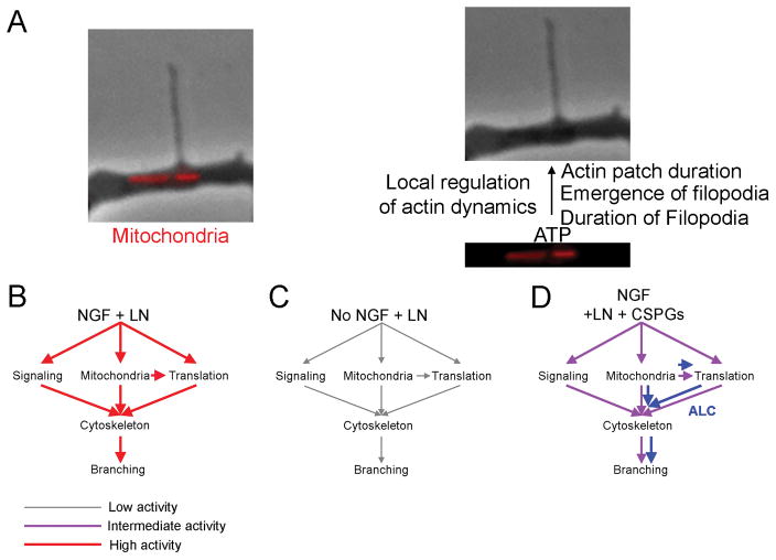
The role of the mitochondrion in determining branching sites along the axon has been established by multiple recent studies (Courchet et al., 2013; Spillane et al., 2013; Tao et al., 2014). Actin is an ATPase and in neurons the turnover of actin filaments is a strenuous metabolic demand that consumes approximately 50% of cellular ATP (Bernstein and Bamburg, 2003). We find that axonal actin patches and filopodia associated with mitochondria are more greatly affected by CSPGs than those not associated with mitochondria. This observation likely reflects the decreased respiration by axonal mitochondria on CSPGs as reflected in the partial depolarization of the mitochondrial membrane potential. Consistently, treatment with ALC, with the aim of promoting mitochondrial respiration, on CSPGs restored aspects of the dynamics of axonal actin patches and the duration of filopodia associated with mitochondria. Furthermore, in control conditions both actin patches and filopodia formed in association with mitochondria exhibited longer durations than those not associated with mitochondria. Collectively, these observations provide additional evidence for the local regulation of axonal actin dynamics by mitochondria (Figure 8A; Ketschek and Gallo, 2010).
Figure 8.
Diagram showing the effects of CSPGs on components of the mechanism of axon branching and the effects of promoting mitochondrial function with ALC. (A) Summary schematic of the contribution of axonal mitochondria to the regulation of axonal cytoskeletal and filopodial dynamics. (B–D) In each panel the relative “strength” of the activation of components of the mechanism of axon branching is denoted by arrows which depict relative strength by their color and size (shown in the bottom left of the figure). (B) NGF strongly drives signaling pathways (e.g., PI3K, Erk), promotes mitochondrial respiration (Verburg and Hollenbeck, 2008) and drives intra-axonal protein synthesis. The arrow between mitochondria and translation reflect the role of mitochondrial function in the promotion of axonal translation (Spillane et al., 2013). Ultimately, signaling, mitochondria function and axonal translation converge on the axonal cytoskeleton to promote branch formation. A connection between signaling pathways and the regulation of mitochondria function and translation is not shown, for clarity, but implied as both of these components of the mechanism of branching are also under regulation by signaling pathways (see Discussion). (C) In the absence of NGF activation of the components of branching is minimal. In this context promotion of mitochondrial function with ALC is unable to further drive branching. (D) In the presence of NGF but on CSPG coated substrata the activation of components of the branching mechanism is attenuated, but persists at moderate levels (Fisher et al., 2011; Silver et al., 2014). In this background, promotion of mitochondrial function with ALC is able to promote branching by working cooperatively with the available signaling and translational mechanisms while promoting the mitochondria respiration dependent aspects of the broader mechanism (blue).
Treatment with ALC on CSPGs promotes axon branching. However, ALC did not affect branching on the control substratum regardless of the presence of NGF. The absence of an effect of ALC on branching in the presence of NGF may reflect that the branching system is already at maximal capacity in the presence of NGF (Figure 8B). Conversely, in the absence of the activation of relevant signaling pathways by NGF the promotion of mitochondrial function is not sufficient to promote branching (Figure 8C), although it increases axon length. When axons are on CSPGs and in the presence of NGF, CSPGs suppress but do not abolish the activation of signaling pathways which drive axon branching (e.g., PI3K signaling; Fisher et al., 2011; Silver et al., 2014). Thus, in the context of CSPGs the partial activation of relevant signaling pathways may allow for production of branches when ALC promotes mitochondrial function and downstream associated actin filament dynamics (Figure 8D).
The mitochondrial respiration dependent intra-axonal translation of cortactin (Spillane et al., 2013) is negatively regulated by CSPGs, and this effect of CSPGs is reversed by treatment with ALC. The positive regulation of actin patch duration and filopodia emergence from patches are previously established functions of cortactin (Spillane et al., 2012), suggesting that the decrease in the axonal translation of cortactin on CSPGs is likely responsible, at least in part, for the decrease in the rate of emergence of filopodia from actin patches and decreased duration of actin patches. Translational hotspots reflect sites of mitochondria-supported translation (Spillane et al., 2013). The decrease in hotspots observed on CSPGs is reversed by treatment with ALC. This reversal is consistent with a role for altered mitochondrial function as contributing to the suppression of the translation of cortactin.
We did not find an effect of CSPGs on the axonal translation of β-actin. In the same experimental system used in the current study, NGF did not promote the axonal translation of β-actin (Roche et al., 2009). However, β-actin mRNA is present and translated in these axons (Spillane et al., 2013; Figure 5C). In contrast to the suppression of the axonal translation of cortactin, CSPGs promote the axonal translation of RhoA (Walker et al., 2012), a negative regulator of axon extension, branching and axonal filopodia formation (Loudon et al., 2006; Spillane and Gallo, 2014). CSPGs also control the axonal proteome through micro-RNAs that can target RhoA mRNA (Zhang et al., 2015). Collectively, these studies indicate that CSPGs do not have a unidirectional effect on axonal translation. The suppression of the axonal translation of cortactin by CSPGs may be reflective of the suppression of the translation of a set of functionally linked mRNA species (e.g., mRNAs coding for proteins involved in axon branching and positively regulated by NGF; Spillane et al., 2012, 2013). This notion is consistent with previous studies that found that extracellular signals differentially regulate the transport and translation of specific mRNAs in axons (Willis et al., 2005; Willis et al., 2007). It will be of interest to determine if CSPGs similarly inhibit the translation of other mRNA species positively regulated by NGF during NGF-induced axon branching (e.g., Arp2 and WAVE1; Spillane et al., 2012).
This study did not find overt effects of CSPGs on the total microtubule content of axons or the targeting of microtubules into axonal filopodia. However, a prior study found that NGF promoted and CSPGs decreased microtubule plus tip polymerization in the growth cones of PC12 cells (Kelly et al., 2010). It will be of interest to determine if CSPGs may differentially regulate microtubule plus tip polymerization in the growth cone relative to the axon shaft or between PC12 cells and primary neurons. Microtubules along the axon shaft are more stable than the highly dynamic microtubules in the growth cone. It is possible that growth cone microtubules may be more readily affected by CSPGs than those along the axon shaft. While we cannot exclude fine regulation of axonal microtubules by CSPGs the current data do not support a major role of CSPGs in the regulation of microtubules in the axon shaft. Furthermore, the failure to detect effects on microtubules in distal axons may be due to the relative concentrations of CSPGs and laminin used in coating substrata in the current study. This culturing environment generates partial suppression of axon extension, which is required in order to investigate branching from axons. However, microtubules may be affected when growth cones encounter either sharp borders between a permissive and CSPG coated environment or an escalating gradient of CSPGs that prevent further axon extension. Interestingly, axons have been noted to extend through in vivo environments enriched in CSPGs in both the central and peripheral nervous system (Bicknese et al., 1994; Gause et al., 2014). However, in these environments the growing axons do not exhibit branching. The suppression of branching by CSPGs in environments also containing additional growth promoting or permissive extracellular cues (e.g., laminin in peripheral nerves) may represent a greater sensitivity of the mechanism of branching to CSPGs than that of the extension of the growth cone.
ALC treatment did not affect the total microtubule content of axons on CSPGs. However, it unexpectedly increased the targeting of microtubules into axonal filopodia. The promotion of microtubule targeting into filopodia likely contributed to the ALC-induced increase in axon branching on CSPGs. The increase in microtubule targeting may be due to the effects of ALC on actin dynamics and filopodia duration. Alternatively, ALC may modulate the retention of microtubules in filopodia following entry. The mechanism through which ALC promotes microtubule targeting into filopodia will require subsequent analysis. It may also reflect a modulation of septin-7 and drebrin based mechanisms that target microtubules into filopodia (Hu et al., 2012; Ketschek et al., 2016).
A role for CSPGs in suppressing the branching of axons in the context of injury scenarios is well appreciated (see Introduction). CSPG expression increases during the developmental period of the refinement of axonal projections and the closing of critical periods for endogenous activity-mediated plasticity (Pizzorusso et al., 2002), which are in part reflective of changes in axon branching patterns. During the early stages of limb bud development the axons of sensory and motoneurons invade the limb bud mesenchyme and grow as fasciculated axons with minimal branching. The expression of CSPGs, including core and link proteins, in the limb bud is relatively diffuse during these early stages but then becomes restricted to the developing vasculature and cartilage at later stages of development when the axons segregate into different branches of nerves and innervate their target tissues (Kimata et al., 1986; Shinomura et al., 1990; Tsonis and Walker, 1991; Rayan et al., 2000). CSPGs also surround the path of developing axons from the spinal cord region into the limb bud (Oakley and Tosney, 1991). This distribution of CSPGs likely keeps the axons on the correct path and also suppresses branching along the way. In contrast, minimal CSPGs are detected within the chicken spinal cord during embryogenesis when sensory axons undergo extensive collateral branching (Hennig et al., 1992). However, CSPGs are detected in the chicken brain with levels peaking at later stages of maturation (Henning et al., 1992) suggesting a role in the consolidation of established patterns of connectivity and suppressing further plasticity (Pizzorusso et al., 2002). Future in vivo studies will be required to address the role of CSPGs in preventing the inappropriate branching of axons during development throughout the nervous system.
In conclusion, this study reports that the membrane potential of axonal mitochondria of embryonic sensory neurons is negatively impacted by CSPGs. The effects of CSPGs on mitochondria contribute to the impairment of axonal actin cytoskeletal dynamics, a key step underlying the formation of axonal filopodia and branches. The effects on mitochondria, at least in part, also contribute to the suppression of the axonal translation of cortactin. It remains to be determined whether these observations also apply to adult neurons. Finally, in both the normal and injured nervous system CSPGs decrease endogenous circuitry plasticity characterized by axonal branching (Pizzorusso et al., 2002; Massey et al., 2006; Harris et al., 2010; Alilain et al., 2011; Harris et al., 2013) and CSPGs may exert these effects through the combined regulation of the actin cytoskeleton and mitochondrial function.
Supplementary Material
Acknowledgments
This work was supported by the National Institutes of Health [NS078030 to G.G.], an award from the Morton Cure Paralysis Found [to G.G.] and a Shriners Hospitals Fellowship [to R.S.].
Footnotes
The authors have no conflicts of interest to disclose.
Referenced Literature
- Aakalu G, Smith WB, Nguyen N, Jiang C, Schuman EM. Dynamic visualization of local protein synthesis in hippocampal neurons. Neuron. 2001;30:489–502. doi: 10.1016/s0896-6273(01)00295-1. [DOI] [PubMed] [Google Scholar]
- Alilain WJ, Horn KP, Hu H, Dick TE, Silver J. Functional regeneration of respiratory pathways after spinal cord injury. Nature. 2011;475:196–200. doi: 10.1038/nature10199. [DOI] [PMC free article] [PubMed] [Google Scholar]
- Arnold DB, Gallo G. Structure meets function: actin filaments and myosin motors in the axon. J Neurochem. 2013 doi: 10.1111/jnc.12503. [DOI] [PMC free article] [PubMed] [Google Scholar]
- Aureli T, Miccheli A, Di Cocco ME, Ghirardi O, Giuliani A, Ramacci MT, Conti F. Effect of acetyl-L-carnitine on recovery of brain phosphorus metabolites and lactic acid level during reperfusion after cerebral ischemia in the rat--study by 13P- and 1H-NMR spectroscopy. Brain Res. 1994;643:92–99. doi: 10.1016/0006-8993(94)90013-2. [DOI] [PubMed] [Google Scholar]
- Bagetta V, Barone I, Ghiglieri V, Di Filippo M, Sgobio C, Bernardi G, Calabresi P, Picconi B. Acetyl-L-Carnitine selectively prevents post-ischemic LTP via a possible action on mitochondrial energy metabolism. Neuropharmacology. 2008;55:223–229. doi: 10.1016/j.neuropharm.2008.05.015. [DOI] [PubMed] [Google Scholar]
- Barritt AW, Davies M, Marchand F, Hartley R, Grist J, Yip P, McMahon SB, Bradbury EJ. Chondroitinase ABC promotes sprouting of intact and injured spinal systems after spinal cord injury. The Journal of neuroscience: the official journal of the Society for Neuroscience. 2006;26:10856–10867. doi: 10.1523/JNEUROSCI.2980-06.2006. [DOI] [PMC free article] [PubMed] [Google Scholar]
- Bartus K, James ND, Bosch KD, Bradbury EJ. Chondroitin sulphate proteoglycans: key modulators of spinal cord and brain plasticity. Exp Neurol. 2012;235:5–17. doi: 10.1016/j.expneurol.2011.08.008. [DOI] [PubMed] [Google Scholar]
- Bassino E, Fornero S, Gallo MP, Gallina C, Femmino S, Levi R, Tota B, Alloatti G. Catestatin exerts direct protective effects on rat cardiomyocytes undergoing ischemia/reperfusion by stimulating PI3K-Akt-GSK3beta pathway and preserving mitochondrial membrane potential. PloS one. 2015;10:e0119790. doi: 10.1371/journal.pone.0119790. [DOI] [PMC free article] [PubMed] [Google Scholar]
- Bernstein BW, Bamburg JR. Actin-ATP hydrolysis is a major energy drain for neurons. J Neurosci. 2003;23:1–6. doi: 10.1523/JNEUROSCI.23-01-00002.2003. [DOI] [PMC free article] [PubMed] [Google Scholar]
- Bicknese AR, Sheppard AM, O’Leary DD, Pearlman AL. Thalamocortical axons extend along a chondroitin sulfate proteoglycan-enriched pathway coincident with the neocortical subplate and distinct from the efferent path. J Neurosci. 1994;14:3500–3510. doi: 10.1523/JNEUROSCI.14-06-03500.1994. [DOI] [PMC free article] [PubMed] [Google Scholar]
- Buckman JF, Hernandez H, Kress GJ, Votyakova TV, Pal S, Reynolds IJ. MitoTracker labeling in primary neuronal and astrocytic cultures: influence of mitochondrial membrane potential and oxidants. J Neurosci Methods. 2001;104:165–176. doi: 10.1016/s0165-0270(00)00340-x. [DOI] [PubMed] [Google Scholar]
- Courchet J, Lewis TL, Jr, Lee S, Courchet V, Liou DY, Aizawa S, Polleux F. Terminal axon branching is regulated by the LKB1-NUAK1 kinase pathway via presynaptic mitochondrial capture. Cell. 2013;153:1510–1525. doi: 10.1016/j.cell.2013.05.021. [DOI] [PMC free article] [PubMed] [Google Scholar]
- Dedov VN, Roufogalis BD. Organisation of mitochondria in living sensory neurons. FEBS Lett. 1999;456:171–174. doi: 10.1016/s0014-5793(99)00951-5. [DOI] [PubMed] [Google Scholar]
- Dietz V, Fouad K. Restoration of sensorimotor functions after spinal cord injury. Brain. 2014;137:654–667. doi: 10.1093/brain/awt262. [DOI] [PubMed] [Google Scholar]
- Dyck SM, Karimi-Abdolrezaee S. Chondroitin sulfate proteoglycans: Key modulators in the developing and pathologic central nervous system. Exp Neurol. 2015;269:169–187. doi: 10.1016/j.expneurol.2015.04.006. [DOI] [PubMed] [Google Scholar]
- Epis R, Marcello E, Gardoni F, Longhi A, Calvani M, Iannuccelli M, Cattabeni F, Canonico PL, Di Luca M. Modulatory effect of acetyl-L-carnitine on amyloid precursor protein metabolism in hippocampal neurons. Eur J Pharmacol. 2008;597:51–56. doi: 10.1016/j.ejphar.2008.09.001. [DOI] [PubMed] [Google Scholar]
- Farahpour MR, Ghayour SJ. Effect of in situ delivery of acetyl-L-carnitine on peripheral nerve regeneration and functional recovery in transected sciatic nerve in rat. Int J Surg. 2014;12:1409–1415. doi: 10.1016/j.ijsu.2014.10.023. [DOI] [PubMed] [Google Scholar]
- Fernandez E, Pallini R, Gangitano C, Del Fa A, Olivieri-Sangiacomo C, Sbriccoli A, Ricoy J, Rossi GF. Studies on the degenerative and regenerative phenomena occurring after transection and repair of the sciatic nerve in rats: effects of acetyl-L-carnitine. Int J Clin Pharmacol Res. 1990;10:85–99. [PubMed] [Google Scholar]
- Fisher D, Xing B, Dill J, Li H, Hoang HH, Zhao Z, Yang X-L, Bachoo R, Cannon S, Longo FM, Sheng M, Silver J, Li S. Leukocyte common antigen-related phosphatase is a functional receptor for chondroitin sulfate proteoglycan axon growth inhibitors. The Journal of neuroscience: the official journal of the Society for Neuroscience. 2011;31:14051–14066. doi: 10.1523/JNEUROSCI.1737-11.2011. [DOI] [PMC free article] [PubMed] [Google Scholar]
- Forloni G, Angeretti N, Smiroldo S. Neuroprotective activity of acetyl-L-carnitine: studies in vitro. J Neurosci Res. 1994;37:92–96. doi: 10.1002/jnr.490370112. [DOI] [PubMed] [Google Scholar]
- Gallo G. The cytoskeletal and signaling mechanisms of axon collateral branching. Dev Neurobiol. 2011;71:201–220. doi: 10.1002/dneu.20852. [DOI] [PubMed] [Google Scholar]
- Gallo G. Mechanisms underlying the initiation and dynamics of neuronal filopodia: from neurite formation to synaptogenesis. Int Rev Cell Mol Biol. 2013;301:95–156. doi: 10.1016/B978-0-12-407704-1.00003-8. [DOI] [PubMed] [Google Scholar]
- Gallo G, Letourneau PC. Localized sources of neurotrophins initiate axon collateral sprouting. J Neurosci. 1998;18:5403–5414. doi: 10.1523/JNEUROSCI.18-14-05403.1998. [DOI] [PMC free article] [PubMed] [Google Scholar]
- Gallo G, Letourneau PC. Different contributions of microtubule dynamics and transport to the growth of axons and collateral sprouts. J Neurosci. 1999;19:3860–3873. doi: 10.1523/JNEUROSCI.19-10-03860.1999. [DOI] [PMC free article] [PubMed] [Google Scholar]
- Gause TM, 2nd, Sivak WN, Marra KG. The role of chondroitinase as an adjuvant to peripheral nerve repair. Cells Tissues Organs. 2014;200:59–68. doi: 10.1159/000369449. [DOI] [PubMed] [Google Scholar]
- Gilmore K, Wilson M. The use of chloromethyl-X-rosamine (Mitotracker red) to measure loss of mitochondrial membrane potential in apoptotic cells is incompatible with cell fixation. Cytometry. 1999;36:355–358. doi: 10.1002/(sici)1097-0320(19990801)36:4<355::aid-cyto11>3.0.co;2-9. [DOI] [PubMed] [Google Scholar]
- Gomes C, Merianda TT, Lee SJ, Yoo S, Twiss JL. Molecular determinants of the axonal mRNA transcriptome. Dev Neurobiol. 2014;74:218–232. doi: 10.1002/dneu.22123. [DOI] [PMC free article] [PubMed] [Google Scholar]
- Guan W, Puthenveedu MA, Condic ML. Sensory neuron subtypes have unique substratum preference and receptor expression before target innervation. J Neurosci. 2003;23:1781–1791. doi: 10.1523/JNEUROSCI.23-05-01781.2003. [DOI] [PMC free article] [PubMed] [Google Scholar]
- Harris NG, Mironova YA, Hovda DA, Sutton RL. Pericontusion axon sprouting is spatially and temporally consistent with a growth-permissive environment after traumatic brain injury. Journal of neuropathology and experimental neurology. 2010;69:139–154. doi: 10.1097/NEN.0b013e3181cb5bee. [DOI] [PMC free article] [PubMed] [Google Scholar]
- Harris NG, Nogueira MSM, Verley DR, Sutton RL. Chondroitinase enhances cortical map plasticity and increases functionally active sprouting axons after brain injury. Journal of neurotrauma. 2013;30:1257–1269. doi: 10.1089/neu.2012.2737. [DOI] [PMC free article] [PubMed] [Google Scholar]
- Hennig A, Krueger R, Mangoura D, Schwartz NB. Chondroitin sulfate proteoglycan expression during neuronal development. Cellular and molecular biology (Noisy-le-Grand, France) 1992;38:585–593. [PubMed] [Google Scholar]
- Hu J, Bai X, Bowen JR, Dolat L, Korobova F, Yu W, Baas PW, Svitkina T, Gallo G, Spiliotis ET. Septin-driven coordination of actin and microtubule remodeling regulates the collateral branching of axons. Curr Biol. 2012;22:1109–1115. doi: 10.1016/j.cub.2012.04.019. [DOI] [PMC free article] [PubMed] [Google Scholar]
- Huang T-J, Verkhratsky A, Fernyhough P. Insulin enhances mitochondrial inner membrane potential and increases ATP levels through phosphoinositide 3-kinase in adult sensory neurons. Molecular and cellular neurosciences. 2005;28:42–54. doi: 10.1016/j.mcn.2004.08.009. [DOI] [PubMed] [Google Scholar]
- Kadomatsu K, Sakamoto K. Sulfated glycans in network rewiring and plasticity after neuronal injuries. Neurosci Res. 2014;78:50–54. doi: 10.1016/j.neures.2013.10.005. [DOI] [PubMed] [Google Scholar]
- Kalil K, Dent EW. Branch management: mechanisms of axon branching in the developing vertebrate CNS. Nat Rev Neurosci. 2014;15:7–18. doi: 10.1038/nrn3650. [DOI] [PMC free article] [PubMed] [Google Scholar]
- Karalija A, Novikova LN, Kingham PJ, Wiberg M, Novikov LN. Neuroprotective effects of N-acetyl-cysteine and acetyl-L-carnitine after spinal cord injury in adult rats. PLoS One. 2012;7:e41086. doi: 10.1371/journal.pone.0041086. [DOI] [PMC free article] [PubMed] [Google Scholar]
- Karalija A, Novikova LN, Kingham PJ, Wiberg M, Novikov LN. The effects of N-acetyl-cysteine and acetyl-L-carnitine on neural survival, neuroinflammation and regeneration following spinal cord injury. Neuroscience. 2014;269:143–151. doi: 10.1016/j.neuroscience.2014.03.042. [DOI] [PubMed] [Google Scholar]
- Kelly T-AN, Katagiri Y, Vartanian KB, Kumar P, Chen I-I, Rosoff WJ, Urbach JS, Geller HM. Localized alteration of microtubule polymerization in response to guidance cues. Journal of neuroscience research. 2010;88:3024–3033. doi: 10.1002/jnr.22478. [DOI] [PMC free article] [PubMed] [Google Scholar]
- Ketschek A, Gallo G. Nerve growth factor induces axonal filopodia through localized microdomains of phosphoinositide 3-kinase activity that drive the formation of cytoskeletal precursors to filopodia. J Neurosci. 2010;30:12185–12197. doi: 10.1523/JNEUROSCI.1740-10.2010. [DOI] [PMC free article] [PubMed] [Google Scholar]
- Ketschek A, Jones S, Spillane M, Korobova F, Svitkina T, Gallo G. Nerve Growth Factor Promotes Reorganization of the Axonal Microtubule Array at Sites of Axon Collateral Branching. Developmental Neurobiology. 2015;75:1441–1461. doi: 10.1002/dneu.22294. [DOI] [PMC free article] [PubMed] [Google Scholar]
- Ketschek A, Spillane M, Dun XP, Hardy H, Chilton J, Gallo G. Drebrin Coordinates the Actin and Microtubule Cytoskeleton During the Initiation of Axon Collateral Branches. Dev Neurobiol. 2016 doi: 10.1002/dneu.22377. [DOI] [PMC free article] [PubMed] [Google Scholar]
- Kimata K, Oike Y, Tani K, Shinomura T, Yamagata M, Uritani M, Suzuki S. A large chondroitin sulfate proteoglycan (PG-M) synthesized before chondrogenesis in the limb bud of chick embryo. J Biol Chem. 1986;261:13517–13525. [PubMed] [Google Scholar]
- Kokkalis ZT, Soucacos PN, Terzis JK. Effect of acetyl-L-carnitine on axonal sprouting following donor nerve injury distal to an end-to-side neurorrhaphy model. J Reconstr Microsurg. 2009;25:483–495. doi: 10.1055/s-0029-1234027. [DOI] [PubMed] [Google Scholar]
- Krohn AJ, Wahlbrink T, Prehn JH. Mitochondrial depolarization is not required for neuronal apoptosis. J Neurosci. 1999;19:7394–7404. doi: 10.1523/JNEUROSCI.19-17-07394.1999. [DOI] [PMC free article] [PubMed] [Google Scholar]
- Lang BT, Cregg JM, DePaul MA, Tran AP, Xu K, Dyck SM, Madalena KM, Brown BP, Weng YL, Li S, Karimi-Abdolrezaee S, Busch SA, Shen Y, Silver J. Modulation of the proteoglycan receptor PTPsigma promotes recovery after spinal cord injury. Nature. 2015;518:404–408. doi: 10.1038/nature13974. [DOI] [PMC free article] [PubMed] [Google Scholar]
- Lee CW, Peng HB. Mitochondrial clustering at the vertebrate neuromuscular junction during presynaptic differentiation. J Neurobiol. 2006;66:522–536. doi: 10.1002/neu.20245. [DOI] [PubMed] [Google Scholar]
- Lee CW, Peng HB. The function of mitochondria in presynaptic development at the neuromuscular junction. Mol Biol Cell. 2008;19:150–158. doi: 10.1091/mbc.E07-05-0515. [DOI] [PMC free article] [PubMed] [Google Scholar]
- Lee DH, Luo X, Yungher BJ, Bray E, Lee JK, Park KK. Mammalian target of rapamycin’s distinct roles and effectiveness in promoting compensatory axonal sprouting in the injured CNS. J Neurosci. 2014;34:15347–15355. doi: 10.1523/JNEUROSCI.1935-14.2014. [DOI] [PMC free article] [PubMed] [Google Scholar]
- Lee H, McKeon RJ, Bellamkonda RV. Sustained delivery of thermostabilized chABC enhances axonal sprouting and functional recovery after spinal cord injury. Proc Natl Acad Sci U S A. 2010;107:3340–3345. doi: 10.1073/pnas.0905437106. [DOI] [PMC free article] [PubMed] [Google Scholar]
- Lelkes PIUBR, Saporta S, Cameron DF, Gallo G. Culture of neuroendocrine and neuronal cells for tissue engineering. Chapter 14 Wiley-Liss; 2006. [Google Scholar]
- Lemarchant S, Pruvost M, Hebert M, Gauberti M, Hommet Y, Briens A, Maubert E, Gueye Y, Feron F, Petite D, Mersel M, do Rego JC, Vaudry H, Koistinaho J, Ali C, Agin V, Emery E, Vivien D. tPA promotes ADAMTS-4-induced CSPG degradation, thereby enhancing neuroplasticity following spinal cord injury. Neurobiol Dis. 2014;66:28–42. doi: 10.1016/j.nbd.2014.02.005. [DOI] [PubMed] [Google Scholar]
- Loudon RP, Silver LD, Yee HF, Jr, Gallo G. RhoA-kinase and myosin II are required for the maintenance of growth cone polarity and guidance by nerve growth factor. J Neurobiol. 2006;66:847–867. doi: 10.1002/neu.20258. [DOI] [PMC free article] [PubMed] [Google Scholar]
- Marsick BM, Roche FK, Letourneau PC. Repulsive axon guidance cues ephrin-A2 and slit3 stop protrusion of the growth cone leading margin concurrently with inhibition of ADF/cofilin and ERM proteins. Cytoskeleton (Hoboken) 2012;69:496–505. doi: 10.1002/cm.21016. [DOI] [PMC free article] [PubMed] [Google Scholar]
- Massey JM, Hubscher CH, Wagoner MR, Decker JA, Amps J, Silver J, Onifer SM. Chondroitinase ABC digestion of the perineuronal net promotes functional collateral sprouting in the cuneate nucleus after cervical spinal cord injury. The Journal of neuroscience: the official journal of the Society for Neuroscience. 2006;26:4406–4414. doi: 10.1523/JNEUROSCI.5467-05.2006. [DOI] [PMC free article] [PubMed] [Google Scholar]
- McKay Hart A, Wiberg M, Terenghi G. Pharmacological enhancement of peripheral nerve regeneration in the rat by systemic acetyl-L-carnitine treatment. Neurosci Lett. 2002;334:181–185. doi: 10.1016/s0304-3940(02)00982-5. [DOI] [PubMed] [Google Scholar]
- Miao QL, Ye Q, Zhang XH. Perineuronal net, CSPG receptor and their regulation of neural plasticity. Sheng Li Xue Bao. 2014;66:387–397. [PubMed] [Google Scholar]
- Miller KE, Sheetz MP. Axonal mitochondrial transport and potential are correlated. J Cell Sci. 2004;117:2791–2804. doi: 10.1242/jcs.01130. [DOI] [PubMed] [Google Scholar]
- Miyamoto S, Murphy AN, Brown JH. Akt mediates mitochondrial protection in cardiomyocytes through phosphorylation of mitochondrial hexokinase-II. Cell death and differentiation. 2008;15:521–529. doi: 10.1038/sj.cdd.4402285. [DOI] [PubMed] [Google Scholar]
- Nishida N, Sugimoto T, Takeuchi T, Kobayashi Y. Effect of L-carnitine on glycogen synthesis and ATP production in cultured hepatocytes of the newborn rat. J Nutr. 1989;119:1705–1708. doi: 10.1093/jn/119.11.1705. [DOI] [PubMed] [Google Scholar]
- Oakley RA, Tosney KW. Peanut agglutinin and chondroitin-6-sulfate are molecular markers for tissues that act as barriers to axon advance in the avian embryo. Developmental biology. 1991;147:187–206. doi: 10.1016/s0012-1606(05)80017-x. [DOI] [PubMed] [Google Scholar]
- Ohtake Y, Li S. Molecular mechanisms of scar-sourced axon growth inhibitors. Brain Res. 2015;1619:22–35. doi: 10.1016/j.brainres.2014.08.064. [DOI] [PMC free article] [PubMed] [Google Scholar]
- Onifer SM, Smith GM, Fouad K. Plasticity after spinal cord injury: relevance to recovery and approaches to facilitate it. Neurotherapeutics. 2011;8:283–293. doi: 10.1007/s13311-011-0034-4. [DOI] [PMC free article] [PubMed] [Google Scholar]
- Onofrj M, Ciccocioppo F, Varanese S, di Muzio A, Calvani M, Chiechio S, Osio M, Thomas A. AcetylL-carnitine: from a biological curiosity to a drug for the peripheral nervous system and beyond. Expert Review of Neurotherapeutics. 2013;13:925–936. doi: 10.1586/14737175.2013.814930. [DOI] [PubMed] [Google Scholar]
- Overly CC, Rieff HI, Hollenbeck PJ. Organelle motility and metabolism in axons vs dendrites of cultured hippocampal neurons. J Cell Sci. 1996;109( Pt 5):971–980. doi: 10.1242/jcs.109.5.971. [DOI] [PubMed] [Google Scholar]
- Parcellier A, Tintignac LA, Zhuravleva E, Hemmings BA. PKB and the mitochondria: AKTing on apoptosis. Cellular signalling. 2008;20:21–30. doi: 10.1016/j.cellsig.2007.07.010. [DOI] [PubMed] [Google Scholar]
- Patel SP, Sullivan PG, Lyttle TS, Magnuson DS, Rabchevsky AG. Acetyl-L-carnitine treatment following spinal cord injury improves mitochondrial function correlated with remarkable tissue sparing and functional recovery. Neuroscience. 2012;210:296–307. doi: 10.1016/j.neuroscience.2012.03.006. [DOI] [PMC free article] [PubMed] [Google Scholar]
- Patel SP, Sullivan PG, Lyttle TS, Rabchevsky AG. Acetyl-L-carnitine ameliorates mitochondrial dysfunction following contusion spinal cord injury. J Neurochem. 2010;114:291–301. doi: 10.1111/j.1471-4159.2010.06764.x. [DOI] [PMC free article] [PubMed] [Google Scholar]
- Pendergrass W, Wolf N, Poot M. Efficacy of MitoTracker Green and CMXrosamine to measure changes in mitochondrial membrane potentials in living cells and tissues. Cytometry A. 2004;61:162–169. doi: 10.1002/cyto.a.20033. [DOI] [PubMed] [Google Scholar]
- Picconi B, Barone I, Pisani A, Nicolai R, Benatti P, Bernardi G, Calvani M, Calabresi P. Acetyl-L-carnitine protects striatal neurons against in vitro ischemia: the role of endogenous acetylcholine. Neuropharmacology. 2006;50:917–923. doi: 10.1016/j.neuropharm.2006.01.002. [DOI] [PubMed] [Google Scholar]
- Pisano C, Pratesi G, Laccabue D, Zunino F, Lo Giudice P, Bellucci A, Pacifici L, Camerini B, Vesci L, Castorina M, Cicuzza S, Tredici G, Marmiroli P, Nicolini G, Galbiati S, Calvani M, Carminati P, Cavaletti G. Paclitaxel and Cisplatin-induced neurotoxicity: a protective role of acetyl-L-carnitine. Clin Cancer Res. 2003;9:5756–5767. [PubMed] [Google Scholar]
- Pizzorusso T, Medini P, Berardi N, Chierzi S, Fawcett JW, Maffei L. Reactivation of ocular dominance plasticity in the adult visual cortex. Science (New York, N Y ) 2002;298:1248–1251. doi: 10.1126/science.1072699. [DOI] [PubMed] [Google Scholar]
- Pollard TD, Cooper JA. Actin, a central player in cell shape and movement. Science. 2009;326:1208–1212. doi: 10.1126/science.1175862. [DOI] [PMC free article] [PubMed] [Google Scholar]
- Poot M, Zhang YZ, Kramer JA, Wells KS, Jones LJ, Hanzel DK, Lugade AG, Singer VL, Haugland RP. Analysis of mitochondrial morphology and function with novel fixable fluorescent stains. J Histochem Cytochem. 1996;44:1363–1372. doi: 10.1177/44.12.8985128. [DOI] [PubMed] [Google Scholar]
- Rayan GM, Haaksma CJ, Tomasek JJ, McCarthy KJ. Basement membrane chondroitin sulfate proteoglycan and vascularization of the developing mammalian limb bud. The Journal of hand surgery. 2000;25:150–158. doi: 10.1053/jhsu.2000.jhsu025a0150. [DOI] [PubMed] [Google Scholar]
- Roche FK, Marsick BM, Letourneau PC. Protein Synthesis in Distal Axons Is Not Required for Growth Cone Responses to Guidance Cues. Journal of Neuroscience. 2009;29:638–652. doi: 10.1523/JNEUROSCI.3845-08.2009. [DOI] [PMC free article] [PubMed] [Google Scholar]
- Roche FK, Marsick BM, Letourneau PC. Protein synthesis in distal axons is not required for growth cone responses to guidance cues. The Journal of neuroscience: the official journal of the Society for Neuroscience. 2009;29:638–652. doi: 10.1523/JNEUROSCI.3845-08.2009. [DOI] [PMC free article] [PubMed] [Google Scholar]
- Rosca MG, Lemieux H, Hoppel CL. Mitochondria in the elderly: Is acetylcarnitine a rejuvenator? Advanced Drug Delivery Reviews. 2009;61:1332–1342. doi: 10.1016/j.addr.2009.06.009. [DOI] [PMC free article] [PubMed] [Google Scholar]
- Selvatici R, Previati M, Marino S, Marani L, Falzarano S, Lanzoni I, Siniscalchi A. Sodium azide induced neuronal damage in vitro: evidence for non-apoptotic cell death. Neurochem Res. 2009;34:909–916. doi: 10.1007/s11064-008-9852-0. [DOI] [PubMed] [Google Scholar]
- Sharma K, Selzer ME, Li S. Scar-mediated inhibition and CSPG receptors in the CNS. Exp Neurol. 2012;237:370–378. doi: 10.1016/j.expneurol.2012.07.009. [DOI] [PMC free article] [PubMed] [Google Scholar]
- Shinomura T, Jensen KL, Yamagata M, Kimata K, Solursh M. The distribution of mesenchyme proteoglycan (PG-M) during wing bud outgrowth. Anat Embryol (Berl) 1990;181:227–233. doi: 10.1007/BF00174617. [DOI] [PubMed] [Google Scholar]
- Silver DJ, Silver J. Contributions of chondroitin sulfate proteoglycans to neurodevelopment, injury, and cancer. Curr Opin Neurobiol. 2014;27:171–178. doi: 10.1016/j.conb.2014.03.016. [DOI] [PMC free article] [PubMed] [Google Scholar]
- Silver L, Michael JV, Goldfinger LE, Gallo G. Activation of PI3K and R-Ras signaling promotes the extension of sensory axons on inhibitory chondroitin sulfate proteoglycans. Developmental neurobiology. 2014;74:918–933. doi: 10.1002/dneu.22174. [DOI] [PMC free article] [PubMed] [Google Scholar]
- Spillane M, Gallo G. Involvement of Rho-family GTPases in axon branching. Small GTPases. 2014;5:e27974. doi: 10.4161/sgtp.27974. [DOI] [PMC free article] [PubMed] [Google Scholar]
- Spillane M, Ketschek A, Donnelly CJ, Pacheco A, Twiss JL, Gallo G. Nerve growth factor-induced formation of axonal filopodia and collateral branches involves the intra-axonal synthesis of regulators of the actin-nucleating Arp2/3 complex. J Neurosci. 2012;32:17671–17689. doi: 10.1523/JNEUROSCI.1079-12.2012. [DOI] [PMC free article] [PubMed] [Google Scholar]
- Spillane M, Ketschek A, Jones SL, Korobova F, Marsick B, Lanier L, Svitkina T, Gallo G. The actin nucleating Arp2/3 complex contributes to the formation of axonal filopodia and branches through the regulation of actin patch precursors to filopodia. Dev Neurobiol. 2011;71:747–758. doi: 10.1002/dneu.20907. [DOI] [PMC free article] [PubMed] [Google Scholar]
- Spillane M, Ketschek A, Merianda TT, Twiss JL, Gallo G. Mitochondria coordinate sites of axon branching through localized intra-axonal protein synthesis. Cell Rep. 2013;5:1564–1575. doi: 10.1016/j.celrep.2013.11.022. [DOI] [PMC free article] [PubMed] [Google Scholar]
- Starkey ML, Bartus K, Barritt AW, Bradbury EJ. Chondroitinase ABC promotes compensatory sprouting of the intact corticospinal tract and recovery of forelimb function following unilateral pyramidotomy in adult mice. Eur J Neurosci. 2012;36:3665–3678. doi: 10.1111/ejn.12017. [DOI] [PMC free article] [PubMed] [Google Scholar]
- Steketee MB, Moysidis SN, Weinstein JE, Kreymerman A, Silva JP, Iqbal S, Goldberg JL. Mitochondrial dynamics regulate growth cone motility, guidance, and neurite growth rate in perinatal retinal ganglion cells in vitro. Invest Ophthalmol Vis Sci. 2012;53:7402–7411. doi: 10.1167/iovs.12-10298. [DOI] [PMC free article] [PubMed] [Google Scholar]
- Tao K, Matsuki N, Koyama R. AMP-activated protein kinase mediates activity-dependent axon branching by recruiting mitochondria to axon. Dev Neurobiol. 2014;74:557–573. doi: 10.1002/dneu.22149. [DOI] [PubMed] [Google Scholar]
- Tom VJ, Kadakia R, Santi L, Houle JD. Administration of chondroitinase ABC rostral or caudal to a spinal cord injury site promotes anatomical but not functional plasticity. J Neurotrauma. 2009;26:2323–2333. doi: 10.1089/neu.2009.1047. [DOI] [PMC free article] [PubMed] [Google Scholar]
- Tsonis PA, Walker E. Cell populations synthesizing cartilage proteoglycan core protein in the early chick limb bud. Biochem Biophys Res Commun. 1991;174:688–695. doi: 10.1016/0006-291x(91)91472-o. [DOI] [PubMed] [Google Scholar]
- Verburg J, Hollenbeck PJ. Mitochondrial membrane potential in axons increases with local nerve growth factor or semaphorin signaling. The Journal of neuroscience: the official journal of the Society for Neuroscience. 2008;28:8306–8315. doi: 10.1523/JNEUROSCI.2614-08.2008. [DOI] [PMC free article] [PubMed] [Google Scholar]
- Walker BA, Ji SJ, Jaffrey SR. Intra-axonal translation of RhoA promotes axon growth inhibition by CSPG. J Neurosci. 2012;32:14442–14447. doi: 10.1523/JNEUROSCI.0176-12.2012. [DOI] [PMC free article] [PubMed] [Google Scholar]
- Wang D, Sun T. Neural plasticity and functional recovery of human central nervous system with special reference to spinal cord injury. Spinal Cord. 2011;49:486–492. doi: 10.1038/sc.2010.124. [DOI] [PubMed] [Google Scholar]
- Willis D, Li KW, Zheng J-Q, Chang JH, Smit AB, Smit A, Kelly T, Merianda TT, Sylvester J, van Minnen J, Twiss JL. Differential transport and local translation of cytoskeletal, injury-response, and neurodegeneration protein mRNAs in axons. The Journal of neuroscience: the official journal of the Society for Neuroscience. 2005;25:778–791. doi: 10.1523/JNEUROSCI.4235-04.2005. [DOI] [PMC free article] [PubMed] [Google Scholar]
- Willis DE, van Niekerk EA, Sasaki Y, Mesngon M, Merianda TI, Williams GG, Kendall M, Smith DS, Bassell GJ, Twiss JL. Extracellular stimuli specifically regulate localized levels of individual neuronal mRNAs. Journal of Cell Biology. 2007;178:965–980. doi: 10.1083/jcb.200703209. [DOI] [PMC free article] [PubMed] [Google Scholar]
- Willis DE, van Niekerk EA, Sasaki Y, Mesngon M, Merianda TT, Williams GG, Kendall M, Smith DS, Bassell GJ, Twiss JL. Extracellular stimuli specifically regulate localized levels of individual neuronal mRNAs. The Journal of cell biology. 2007;178:965–980. doi: 10.1083/jcb.200703209. [DOI] [PMC free article] [PubMed] [Google Scholar]
- Wilson AD, Hart A, Wiberg M, Terenghi G. Acetyl-l-carnitine increases nerve regeneration and target organ reinnervation - a morphological study. J Plast Reconstr Aesthet Surg. 2010;63:1186–1195. doi: 10.1016/j.bjps.2009.05.039. [DOI] [PubMed] [Google Scholar]
- Yudin D, Hanz S, Yoo S, Iavnilovitch E, Willis D, Gradus T, Vuppalanchi D, Segal-Ruder Y, Ben-Yaakov K, Hieda M, Yoneda Y, Twiss JL, Fainzilber M. Localized regulation of axonal RanGTPase controls retrograde injury signaling in peripheral nerve. Neuron. 2008;59:241–252. doi: 10.1016/j.neuron.2008.05.029. [DOI] [PMC free article] [PubMed] [Google Scholar]
- Zhang HL, Eom T, Oleynikov Y, Shenoy SM, Liebelt DA, Dictenberg JB, Singer RH, Bassell GJ. Neurotrophin-induced transport of a beta-actin mRNP complex increases beta-actin levels and stimulates growth cone motility. Neuron. 2001;31:261–275. doi: 10.1016/s0896-6273(01)00357-9. [DOI] [PubMed] [Google Scholar]
- Zhang R, Zhang H, Zhang Z, Wang T, Niu J, Cui D, Xu S. Neuroprotective effects of pre-treatment with l-carnitine and acetyl-L-carnitine on ischemic injury in vivo and in vitro. Int J Mol Sci. 2012;13:2078–2090. doi: 10.3390/ijms13022078. [DOI] [PMC free article] [PubMed] [Google Scholar]
- Zhang Y, Chopp M, Liu XS, Kassis H, Wang X, Li C, An G, Zhang ZG. MicroRNAs in the axon locally mediate the effects of chondroitin sulfate proteoglycans and cGMP on axonal growth. Developmental neurobiology. 2015;75:1402–1419. doi: 10.1002/dneu.22292. [DOI] [PMC free article] [PubMed] [Google Scholar]
Associated Data
This section collects any data citations, data availability statements, or supplementary materials included in this article.


