Abstract
Background:
It is recognized that patients who undergo endoscopic sinus surgery (ESS) do not always achieve control of their disease. The causes are multifactorial; variations in surgical practice have been identified as possible factors in refractory disease.
Objective:
To reflect on the frequent anatomic findings of patients with chronic rhinosinusitis (CRS) who require revision ESS.
Methods:
A retrospective review of patients who required revision ESS at a tertiary institution over a 3-year period. Patients for whom maximal medical therapy failed for CRS underwent computed tomography of the paranasal sinuses and image-guided surgery. Surgical records of anatomic findings were reviewed and analyzed.
Results:
Over 3 years, a total of 75 patients underwent revision procedures, 28% of all ESS performed in the unit. The most frequent finding was a residual uncinate process in 64% of the patients (n = 48); other findings included a maxillary antrostomy not based on the natural ostium of the maxillary sinus in 47% (n = 35), an oversized antrostomy in 29% (n = 22), resected middle turbinates in 35% (n = 26), middle meatal stenosis in 15% (n = 11), synechiae in 29% (n = 22), and osteitic bone that required drilling in 13% (n = 10).
Conclusion:
Surgical technique can give rise to anatomic variations that may prevent adequate mucociliary clearance and medication delivery, which leads to failure in ESS in patients with CRS. This study demonstrated the surgical findings encountered in revision ESS that should be highlighted in the training of Ear, Nose and Throat surgeons to help prevent primary failure and reduce health care costs.
Keywords: Anatomic, endoscopic sinus surgery, frontal sinusotomy, rhinosinusitis, SNOT-22, surgical revision, uncinectomy
Revision endoscopic sinus surgery (ESS) presents a significant burden to health care systems. The U.K. national sinonasal audit demonstrated that ∼20% of patients who underwent ESS required revision within 5 years, which presents a burden to both the patient and the wider health care system.1 A recent large U.K. epidemiologic study (Chronic Rhinosinusitis Epidemiology Study) reported rates of previous surgery in patients with chronic rhinosinusitis (CRS) of 43%; subgroup analysis showed rates to be higher in those with CRS and nasal polyposis (CRSwNP) and allergic fungal rhinosinusitis (AFRS) compared with those with CRS and without nasal polyps.2 In the CRSwNP group, 50% had undergone previous surgery, with a mean of 3.3 nasal polypectomies per patient. This level of revision surgery could cost the National Health Service (NHS) as much as £15 million (19.64 US dollar) and, therefore, investigating surgical technique applied during primary or subsequent surgery may help establish whether the multiple revision cases seen are due to disease burden or the surgery itself.
Functional ESS (FESS) was pioneered in the 1970s by Messerklinger, with the aid of the Hopkins rod telescope, the aim being to preserve nasal mucosa.3,4 Kennedy and Adappa5 modified this further by reintroducing the use of maxillary antrostomy through the middle meatus. Both developments proved a major milestone in sinus surgery, which led to the expectation that a full uncinectomy, adequate antrostomy, and anterior ethmoidectomy are routinely performed as part of FESS. A further evolution of this concept now involves extended ESS for all diseased paranasal sinus cavities as appropriate.1 Complications of ESS include bleeding, cerebrospinal fluid leak, and orbital damage that leads to visual impairment. These risks are exacerbated in revision surgery in which the usual anatomic landmarks may be distorted or absent.6
Previous reports from North America5,7 indicated that multiple factors, both anatomic and systemic, may predispose to failure of ESS. These include scarring at the middle meatal antrostomy, failure to incorporate the natural maxillary sinus ostium in the antrostomy, oversized (nonphysiologic) antrostomy, residual uncinate process, scarring at the frontal recess, and recurrent polyps. With the newly published Chronic Rhinosinusitis Epidemiology Study2 data in mind, we aimed to identify the anatomic findings of patients with CRS who underwent revision ESS at a U.K. tertiary center to shed light on the variability of surgical practice.
METHODS
This retrospective study received approval from the local clinical research and audit governance committee (James Paget University Hospital). Between January 2011 and December 2013, 75 patients who required revision ESS were identified by the senior author (C.M.P.) from a total of 272 cases of ESS performed. One patient was from the center's own cohort, the rest were from external, nationwide referrals. Demographic variables and preoperative computed tomographic findings were noted for all the patients. The Sino-Nasal Outcome Test version 22 (SNOT-22) was completed by patients before and after surgery (between 3 and 6 months), along with allergy testing (skin-prick testing for local patients, the radioallergosorbent test for distant patients unable to attend many short visits).
The operation notes were examined for the following factors: recurrent polyposis, synechiae formation (between the septum and lateral wall and/or turbinates, or between the middle turbinate and lateral wall), incorrectly performed antrostomy (i.e., based on an accessory ostium with or without recirculation of mucus), large antrostomy (more than four times the natural ostium diameter), retained uncinate process, middle meatal antrostomy stenosis, osteitic bone (defined as the need to drill sclerotic bone to access a sinus as opposed to simple dissection with curette and/or bone cutting forceps), lack of frontal sinus dissection when indicated, and the presence of AFRS (Bent and Kuhn criteria8) and/or eosinophilic mucinous rhinosinusitis9 (EMRS) and/or eosinophilic fungal rhinosinusitis (EFRS).10 All data were analyzed by using IBM SPSS for Windows version 20.0 (SPSS Inc., Chicago, IL), and the paired Student's t-test was used to compare pre- and postoperative SNOT-22 scores.
RESULTS
A total of 272 patients had undergone ESS, of whom 27.6% (n = 75) underwent revision ESS by using image guidance (Table 1). The mean (standard deviation) age was 59 ± 12.7 years; there was a male predominance (57.3%). The number of previous ESS procedures ranged from 1 to 20, with a mean of 2.26. The mean time to recurrence of disease symptomatic enough to require surgery was 107 months (range, 11–360 months). A number of patients had concurrent systemic illnesses, including asthma in 58.2% and aspirin sensitivity in 26%. Proven atopy was present in 68% of patients tested. With the exception of one patient who had undergone primary ESS in our center, the remainder had undergone primary surgery before 2010 (when the senior author (C.M.P.) joined this center) or the surgery was performed at another site. The patient in the one primary case performed at this center had excessive bleeding throughout the primary procedure, which prevented frontal sinusotomies; this was addressed in the revision surgery.
Table 1.
Subgroup analysis of preoperative details and paired Student's t-test analysis of pre- and postoperative SNOT-22 scores
SNOT-22 = 22-Item Sino-Nasal Outcome Test; SD = standard deviation; CRSwNP = chronic rhinosinusitis with nasal polyp; CRSsNP = chronic rhinosinusitis without nasal polyp; AFRS = allergic fungal rhinosinusitis; EMRS = eosinophilic mucinous rhinosinusitis; EFRS = eosinophilic fungal rhinosinusitis.
A greater proportion of patients had CRSwNP (65.3%) and AFRS, EMRS, EFRS (26.7%) than CRS and without nasal polyps. Clinical outcomes by using the SNOT-22 scores were compared for all three subgroups and displayed a statically significant improvement in pre- and post-SNOT-22 scores in the CRSwNP and the AFRS, EMRS, EFRS subgroups (Table 1). Long-term follow-up SNOT-22 scores (mean, 22 month; range, 3–59 months) worsened, although they were still with significant improvement overall from preoperative scores. The number of procedures was also significantly different among the AFRS, EMRS, EFRS subgroups and the other two subgroups. The mean preoperative Lund-Mackay score was 17.74 (range, 3–24); the score of 3 was reported in two patients, one with a frontal sinus mucocele and the other with localized CRS and without nasal polyps that did not respond to medical therapy due to recirculation of mucus associated with a residual uncinate process and incorrectly placed antrostomy.
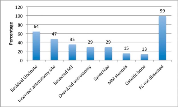
Preoperative radiologic assessment and endoscopic findings included a residual uncinate process in 64%, misplaced antrostomy (not based on the natural ostium of the maxillary sinus) in 47%, full or partial resection of the middle turbinate in 35%, synechiae in 29%, an oversized antrostomy in 29%, middle meatal stenosis in 15%, osteitic bone in 13%, and previously undiagnosed allergic fungal sinusitis in 14% (Fig. 1). With regard to the osteitic cases, significant revision surgery was required in two patients: the first patient in whom restenosis of the right frontal and maxillary sinuses occurred; the second procedure involved the placement of stents and the use of topical mitomycin C. Restenosis of the maxillary ostia occurred in a second case, in which multiple inferior antrostomies (on three occasions) had been performed previously with no removal of the uncinate processes. Frontal sinusotomy had been performed in one patient previously.
Figure 1.
Chart that shows operative anatomic findings. The results are described as percentages. MT = Middle turbinate; MM = middle meatus; FS = frontal sinus.
DISCUSSION
ESS has evolved rapidly since its introduction in the mid-20th century. In part, this is due to technological advances, but equally important has been our gain in the anatomic and physiologic understanding of the sinonasal cavities over the past 4 to 5 decades, which has led to improved outcomes as surgery aims to restore overall function while respecting key landmarks and structures. This study demonstrated that there was a wide variation with regard to the practice and extent of sinus surgery. Subsequently, patients developed recurrent sinonasal disease that was difficult to treat medically. It is important that surgeons who perform ESS are aware of the common surgical errors that can contribute to failure, especially when disease recurs and perhaps when compliance with medical treatment falls. This attention to technique will help to improve outcomes and reduce revision rates. The most-frequent variations denoted above are discussed.
Incomplete Uncinectomy
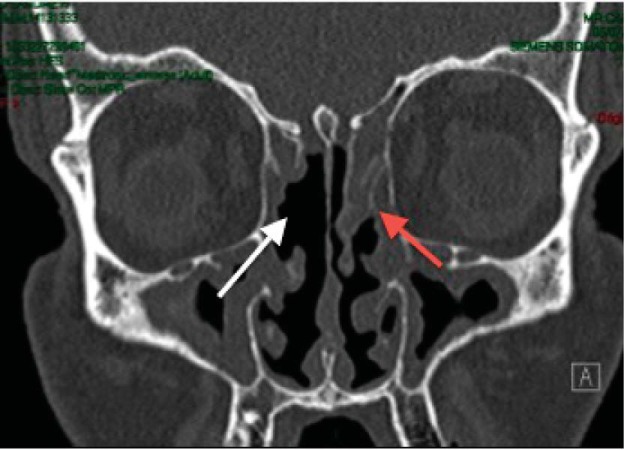
Dissection and removal of the entire uncinate process allows visualization of the natural maxillary ostium, especially with an angled endoscope. In addition, both the mucosa and bone of the uncinate are commonly diseased, and residual tissue may result in persistent symptoms and prevent adequate medication delivery to the osteomeatal complex (Fig. 2). The variations in uncinate anatomy should be considered by using radiologic examination before resection, with particular reference to its superior attachment and its relationship to the orbit.11 Sixty-four percent of patients in this study had residual uncinate; although the residual components varied, this echoed radiologic findings in a previous revision ESS series.12
Figure 2.
An incomplete uncinectomy. Coronal computed tomography, showing a residual left uncinate process (red arrow) and resected right middle turbinate (white arrow).
One patient in our cohort had previous visual loss after intraorbital hemorrhage; on this occasion, the uncinate was found laterally fractured into the maxillary sinus. Techniques vary, but we advocate a back-to-front approach by using a backbiter (pediatric if required) to divide the uncinate at the junction of the inferior third and superior two thirds. The superior component is then dissected out with a pediatric 90° forceps, and the inferior component is removed with a down-biting antral punch. Without performing this step correctly, ESS outcomes may well be inadequate because the uncinate is often considered the doorway to the sinuses.
Incorrectly Placed Antrostomy
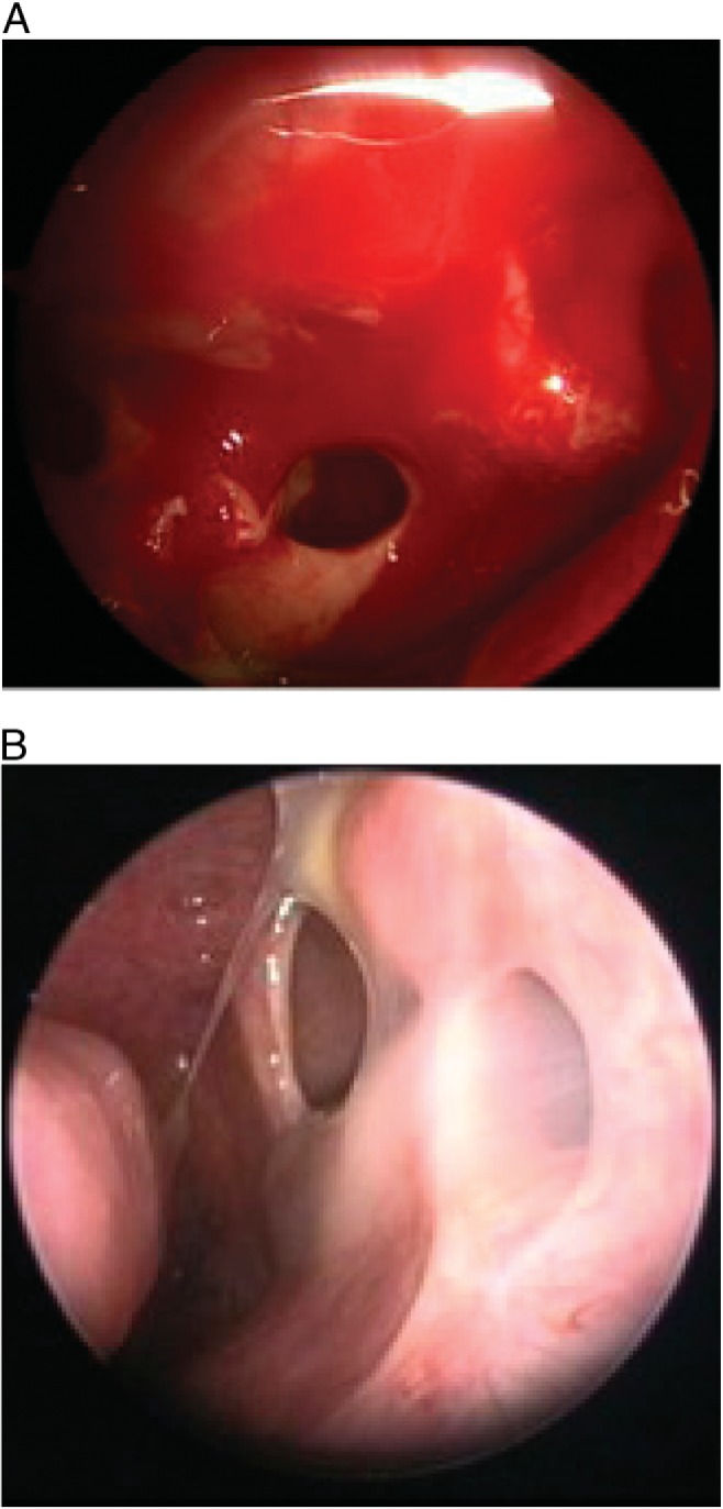
Locating the natural maxillary ostium is paramount in avoiding false surgical ostium formation and goes hand in hand with the uncinectomy because failure to adequately perform the latter will prevent adequate visualization of the ostium. In our series, 47% of the patients were found to have an incorrectly placed antrostomy, which risks mucociliary recirculation as a result of mucus flowing out of the natural ostium and in through the surgical ostium (Fig. 3).3 An angled endoscope can be very helpful during an uncinectomy, which allows the surgeon to clearly visualize the natural ostium once the uncinate is properly removed. Our standard practice is to use a 30° endoscope to remove the lower uncinate and visualize the natural ostium; on occasions, a 70° scope may even be necessary. The wider adoption of the use of angled endoscopes would help to avoid this problem.
Figure 3.

(A, B) Accessory ostium with recirculation. The use of an angled endoscope is key to visualizing the natural maxillary ostium. Incorrect antrostomy that does not incorporate the natural sinus ostium (A) may lead to recirculation of mucus (B).
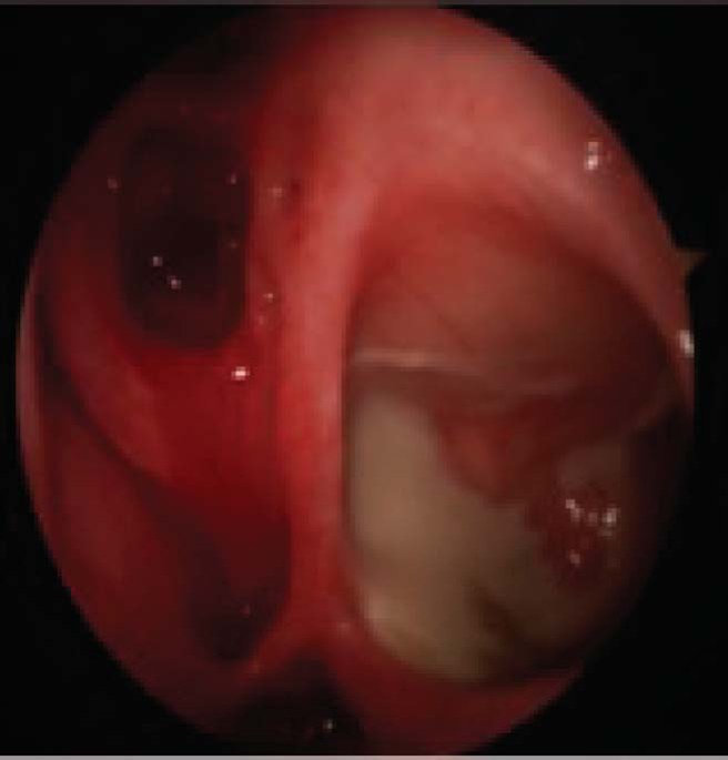
There is debate regarding posterior enlargement of the maxillary antrostomy, the main concern being the drying effect of nasal airflow on the sinus mucosa that may, in turn, predispose to biofilm formation.13 The other major concern is the loss of protective nitric oxide concentration within the maxillary sinus. The posterior aspect of the ostium and mucosa in the ethmoid infundibulum posteriorly should also be preserved to avoid impairment of mucociliary clearance. An oversized antrostomy was demonstrated in 29% of patients, in whom it accounted for >50% of the medial maxillary wall (Fig. 4).14 It is well documented that there are polar opinions on this matter; Cho and Hwang15 reported their successful series of “mega-antrostomies” in recalcitrant maxillary sinusitis, although their series contained many patients with cystic fibrosis and previous Caldwell-Luc procedures, which was not the case in our series. In select cases, as dictated by pathology or underlying systemic disease, there is certainly a case for making a larger antrostomy, but adherence to the principles mentioned above for the patients with uncomplicated CRS should help to reduce the chances of failure.
Figure 4.

An oversized antrostomy. A large antrostomy (>4 times the size of the natural sinus ostium) may predispose to biofilm formation.
Middle Turbinate Resection
The middle turbinate is a key landmark as well as being important in the physiologic function of the nose. A recent study showed that, although lateralization of the middle turbinate is not associated with increased symptoms, it is associated with the need for revision ESS.16 Valdes et al.17 also found it to be a common finding (48% of revision cases) in patients undergoing revision frontal sinus procedures. In 35% of patients in our study, the middle turbinate had been resected. We hypothesized that this was to prevent adhesion to the lateral wall and/or obstruction of the osteomeatal complex or perhaps was removed among a dense clutch of polyps. The former issue is important to avoid, but, rather than resecting the middle turbinate (and hence altering the anatomic appearance), we would advocate the placement of a middle meatal “spacer” at the end of the procedure18,19
Frontal Sinusotomy
Despite several cases that had previous radiologic evidence of frontal sinus disease, the majority of patients in this cohort had not undergone frontal sinus dissection. We did not have sufficient data on our entire cohort regarding the presence of frontal sinus disease before the first episode of FESS, and it is possible that, in some patients, there was no involvement of this area and hence deemed not to require sinusotomy. One patient in our series had erosion of the posterior table of the frontal sinus, and two had previously had external approaches to a frontal sinus through Lynch-Howarth incisions due to proptosis from expansile disease; none of these had had an endoscopic approach. Operative intervention to this area requires good instrumentation, including angled instruments and endoscopes as well as a sound knowledge of the anatomic variations. The suggestion here may be that greater emphasis is needed on training or that certain patients require the input of a specialist rhinologist trained to dissect this area. With a goal of improving access for postoperative medications, failure to open diseased sinuses may ultimately lead to failure of medical therapy and subsequent uncontrolled disease.
Osteitis
Osteitis of the flat bones that compose the sinuses is well recognized, and, although its exact role in disease progression is not fully understood, post-FESS cases often have higher rates of osteitic bone by using both radiologic and histologic analyses.20,21 This is thought to be due to either surgical technique (mucosal stripping or residual bone fragments) and/or disease pathophysiology in patients who re-present after primary surgery (who by their very nature may have more-severe disease). Osteitic bone formation can obstruct sinus drainage and hence reverse the advantage of primary FESS.7 In our experience, this complication was more prevalent in the frontal recess, and the 13% rate reported in this study was of osteitis that prevented simple dissection, which often required drilling to remove the affected bone and stenting in four patients to prevent further stenosis. Adequate topical steroid therapy to prevent progressive inflammatory reaction and subsequent osteitis is important, and, hence, education of the patient in the need for long-term steroids, as well as nasal irrigation, is imperative.
Study Limitations
Information regarding the primary surgery was not always available for these cases, and we, therefore, are unable to draw conclusions regarding the extent of surgery performed at this time (in particular, with regard the frontal sinus). We appreciated that the current opinion at the time may have dictated the type of surgery performed. In addition, due to the retrospective nature of the audit, information on the advice and subsequent compliance of patients with topical medication is not available. It is possible that those with poor compliance are more likely to re-present, and, hence, this population of patients may have skewed our results. At the time of the analysis, only one primary case from our center had needed revision surgery. There were also three revisions in two secondary cases in which the first surgery predated the senior author (C.M.P.) joining the department. We acknowledged the short time span of this series. Lastly, and perhaps most importantly, there was no record of patients who had had successful primary surgery and did not require revision. One could speculate that some of the common anatomic variations are seen in these patients also but why they did not develop uncontrolled disease is not known.
ESS in the Age of Austerity
A recent study indicated that surgery is more effective (than continued medical therapy) for patients with refractory disease who deteriorate when on medical treatment22 and also more cost effective, although the optimal timing of intervention is not yet clear.23 However, ESS outcomes are under much scrutiny. Within the United Kingdom, procedures of potential limited clinical effectiveness are under threat in a NHS under significant financial strain.24 ESS has been considered in this bracket,25 partly due to high rates of recurrent surgery as demonstrated by the long-term follow-up to a U.K. national sinonasal audit.26,27 Hospital Episode Statistics data for 2011–2012 indicate that ∼40,000 nose and/or sinus operations are performed each year in England and Wales, in addition to an estimated 75,000 outpatient consultations.28 Based on Hospital Episode Statistics data regarding admission for sinus surgery for CRSwNP, a 20% revision rate, and, when considering NHS reference costs, the total cost of revision ESS (with outpatient activity included) to the NHS is likely to be more than £30 million (39.28 million US dollars) per year (NHS England Health resource group code costs for intermediate, major, and complex nasal surgery totaled £86 million (112.61 million US dollars) in 2012–2013).29
The cohort in our study included patients referred from multiple locations around the United Kingdom with persistent sinonasal disease uncontrolled on medical therapy despite having undergone ESS. We believe that the study provided a realistic depiction of such referrals to tertiary centers and echoed radiologic findings of U.K. revision ESS cases identified by Kahlil et al.30 Of these, the highest rates of failure demonstrated here related to those steps that may be considered to be the simplest to perform, e.g., uncinectomy and antrostomy. It is likely that commissioners will pay closer scrutiny to outcomes at individual units, and a careful audit of ESS practice will increasingly be required. It, therefore, is crucial that surgeons who perform such procedures are aware of the common anatomic findings that may contribute to uncontrolled CRS so that these can be avoided in the future.
CONCLUSION
This study demonstrated a number of frequent anatomic and subsequent pathophysiologic findings encountered at revision surgery. It shed light on some of the issues raised in the recent Chronic Rhinosinusitis Epidemiology Study2, viz., that different surgical strategies are used by surgeons who undertake such work and indicates that further trials are required to assess whether these contribute to poor disease control. Those who perform ESS should be aware of the common pitfalls that require correction during revision surgery.
ACKNOWLEDGMENTS
We thank Jane Woods for her help with the Rhinology Database.
Footnotes
No external funding sources reported
C. Philpott is a consultant for Acclarent, Aerin Medical, Entellus. The remaining authors have no conflicts of interest pertaining to this article
REFERENCES
- 1. Philpott CM, Thamboo A, Lai L, et al. Endoscopic frontal sinusotomy—Preventing recurrence or a route to revision? Laryngoscope 120:1682–1686, 2010. [DOI] [PubMed] [Google Scholar]
- 2. Philpott C, Hopkins C, Erskine S, et al. The burden of revision sinonasal surgery in the UK—Data from the Chronic Rhinosinusitis Epidemiology Study (CRES): A cross-sectional study. BMJ Open 5:e006680, 2015. [DOI] [PMC free article] [PubMed] [Google Scholar]
- 3. Stammberger H. Endoscopic endonasal surgery—Concepts in treatment of recurring rhinosinusitis: Part I. Anatomic and pathophysiologic considerations. Otolaryngol Head Neck Surg 94:143–147, 1986. [DOI] [PubMed] [Google Scholar]
- 4. Stammberger H. Endoscopic endonasal surgery—Concepts in treatment of recurring rhinosinusitis: Part II. Surgical technique. Otolaryngol Head Neck Surg 94:147–156, 1986. [DOI] [PubMed] [Google Scholar]
- 5. Kennedy DW, Adappa ND. Endoscopic maxillary antrostomy: Not just a simple procedure. Laryngoscope 121:2142–2145, 2011. [DOI] [PubMed] [Google Scholar]
- 6. Jiang RS, Hsu CY. Revision functional endoscopic sinus surgery. Ann Otol Rhinol Laryngol 111:155–159, 2002. [DOI] [PubMed] [Google Scholar]
- 7. Mechor B, Javer AR. Revision endoscopic sinus surgery: The St. Paul's Sinus Centre experience. J Otolaryngol Head Neck Surg 37:676–680, 2008. [PubMed] [Google Scholar]
- 8. Bent JP, III, Kuhn FA. Diagnosis of allergic fungal sinusitis. Otolaryngol Head Neck Surg 111:580–588, 1994. [DOI] [PubMed] [Google Scholar]
- 9. Ferguson BJ. Eosinophilic mucin rhinosinusitis: A distinct clinicopathological entity. Laryngoscope 110(pt. 1):799–813, 2000. [DOI] [PubMed] [Google Scholar]
- 10. Aeumjaturapat S, Saengpanich S, Isipradit P, Keelawat S. Eosinophilic mucin rhinosinusitis: Terminology and clinicopathological presentation. J Med Assoc Thai 86:420–424, 2003. [PubMed] [Google Scholar]
- 11. Awad Z, Bhattacharyya M, Jayaraj SM. Anatomical margins of uncinectomy in endoscopic sinus surgery. Int J Surg 11:188–190, 2013. [DOI] [PubMed] [Google Scholar]
- 12. Gore MR, Ebert CS, Jr, Zanation AM, Senior BA. Beyond the “central sinus”: Radiographic findings in patients undergoing revision functional endoscopic sinus surgery. Int Forum Allergy Rhinol 3:139–146, 2013. [DOI] [PubMed] [Google Scholar]
- 13. Phillips PS, Sacks R, Marcells GN, et al. Nasal nitric oxide and sinonasal disease: A systematic review of published evidence. Otolaryngol Head Neck Surg 144:159–169, 2011. [DOI] [PubMed] [Google Scholar]
- 14. Kirihene RK, Rees G, Wormald PJ. The influence of the size of the maxillary sinus ostium on the nasal and sinus nitric oxide levels. Am J Rhinol 16:261–264 2002. [PubMed] [Google Scholar]
- 15. Cho DY, Hwang PH. Results of endoscopic maxillary mega-antrostomy in recalcitrant maxillary sinusitis. Am J Rhinol 22:658–662, 2008. [DOI] [PubMed] [Google Scholar]
- 16. Bassiouni A, Chen PG, Naidoo Y, Wormald PJ. Clinical significance of middle turbinate lateralization after endoscopic sinus surgery. Laryngoscope 125:36–41, 2015. [DOI] [PubMed] [Google Scholar]
- 17. Valdes CJ, Bogado M, Samaha M. Causes of failure in endoscopic frontal sinus surgery in chronic rhinosinusitis patients. Int Forum Allergy Rhinol 4:502–506, 2014. [DOI] [PubMed] [Google Scholar]
- 18. Akbari E, Philpott CM, Ostry AJ, et al. A double-blind randomised controlled trial of gloved versus ungloved merocel middle meatal spacers for endoscopic sinus surgery. Rhinology 50:306–310, 2012. [DOI] [PubMed] [Google Scholar]
- 19. Hobson CE, Choby GW, Wang EW, et al. Systematic review and metaanalysis of middle meatal packing after endoscopic sinus surgery. Am J Rhinol Allergy 29:135–140, 2015. [DOI] [PubMed] [Google Scholar]
- 20. Georgalas C, Videler W, Freling N, Fokkens W. Global Osteitis Scoring Scale and chronic rhinosinusitis: A marker of revision surgery. Clin Otolaryngol 35:455–461, 2010. [DOI] [PubMed] [Google Scholar]
- 21. Kim HY, Dhong HJ, Chung SK, et al. Clinical characteristics of chronic rhinosinusitis with asthma. Auris Nasus Larynx 33:403–408, 2006. [DOI] [PubMed] [Google Scholar]
- 22. Smith KA, Smith TL, Mace JC, Rudmik L. Endoscopic sinus surgery compared to continued medical therapy for patients with refractory chronic rhinosinusitis. Int Forum Allergy Rhinol 4:823–827, 2014. [DOI] [PMC free article] [PubMed] [Google Scholar]
- 23. Bernic A, Dessouky O, Philpott C, et al. Cost-Effective Surgical Intervention in Chronic Rhinosinusitis. Current Otorhinolaryngology Reports 1–7. Available online at http://link.springer.com/article/10.1007/s40136-015-0077-x; accessed June 5, 2014.
- 24. London CSf. Procedures of Limited Clinical Effectiveness 2011. Available online at http://www.londonhp.nhs.uk/wp-content/uploads/2011/03/PoLCE-original-evidence-summary.doc; accessed June 5, 2014.
- 25. Soni-Jaiswal A, Philpott C, Hopkins C. The impact of commissioning for rhinosinusitis in England. Clin Otolaryngol 40:639–645, 2015. [DOI] [PubMed] [Google Scholar]
- 26. Hopkins C, Browne JP, Slack R, et al. The national comparative audit of surgery for nasal polyposis and chronic rhinosinusitis. Clin Otolaryngol 31:390–398, 2006. [DOI] [PubMed] [Google Scholar]
- 27. Hopkins C, Slack R, Lund V, et al. Long-term outcomes from the English national comparative audit of surgery for nasal polyposis and chronic rhinosinusitis. Laryngoscope 119:2459–2465, 2009. [DOI] [PubMed] [Google Scholar]
- 28. Care TICfHaS. Hospital Episode Statistics: Main Operations 2011 to 2012. Available online at http://www.hscic.gov.uk/hes; accessed June 5, 2014.
- 29. Department of Health UK. Reference costs guidance 2013–14. Available online at http://www.gov.uk/government/collections/nhs-reference-costs; accessed June 5, 2014.
- 30. Khalil HS, Eweiss AZ, Clifton N. Radiological findings in patients undergoing revision endoscopic sinus surgery: A retrospective case series study. BMC Ear Nose Throat Disord 11:4, 2011. [DOI] [PMC free article] [PubMed] [Google Scholar]


