Case Report
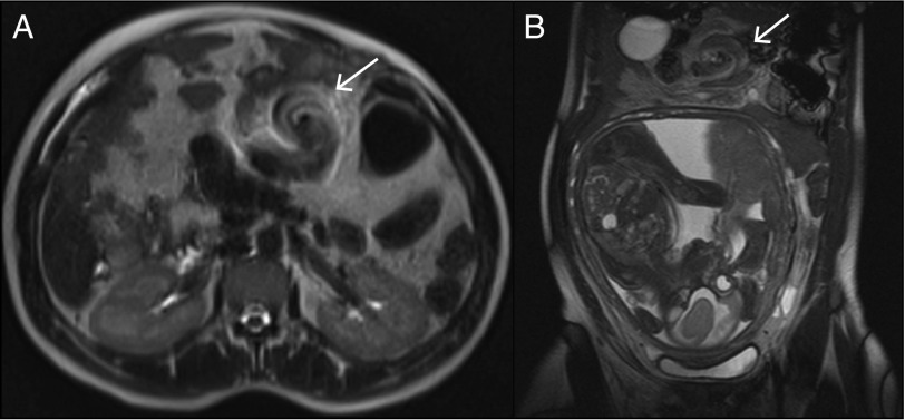
A 38-year-old woman in the 27th week of gestation was admitted for sudden onset of epigastric pain, vomiting, and nausea, which worsened after food ingestion. The patient described 1 year of self-limited episodes of abdominal pain that improved with defecation and were associated with a change in frequency and consistency of stools that were interpreted as irritable bowel syndrome. On physical examination, she had stable vital signs, a distended abdomen, and tenderness in both upper quadrants. Blood tests showed leukocytosis 21.8 x 10⁹/L and C-reactive protein 10 mg/L. Abdominal x-ray was normal, and abdominal ultrasonography revealed a small amount of anechogenic fluid between intestinal loops and in the hepatorenal recess. Obstetric ultrasonography showed fetal well-being. Upper endoscopy was inconclusive because of abundant gastric residual fluid. Contrast-enhanced magnetic resonance imaging revealed features of malrotation (the large bowel was predominantly located on the left side and the small bowel predominantly on the right side) and a whirlpool image in the proximal small bowel (Figure 1). A diagnosis of a small bowel volvulus and midgut malrotation was made. Due to the risk of miscarriage, the patient refused surgery, and a conservative management with antibiotics, intravenous fluids, and parenteral nutrition was started. One week later, although the patient presented with normal vital signs, the abdominal pain and vomiting worsened and there was an increase in C-reactive protein to 102 mg/L. The patient accepted surgery, which confirmed the midgut malrotation associated with small bowel volvulus, and a Ladd’s procedure was done (Figure 2). Treitz ligament was absent. No complications were described postoperatively, and she gave birth to a healthy newborn with no apparent malformations at 38 weeks gestation.
Figure 1.
Axial T1-weighted abdominal magnetic resonance imaging slices showing the whirlpool sign or the clockwise rotation of the duodenum and jejunum around the axis of the superior mesentery artery and vein.
Figure 2.
Surgical intervention confirming the clinical and radiological diagnosis.
Midgut malrotation is a common congenital anomaly, resulting from a complete nonrotation or an incomplete counterclockwise rotation of the primitive intestinal loop around the superior mesenteric artery during fetal development. It is rare for malrotation to present in adulthood, and its presentation in pregnancy is described only in case reports.1–5 The standard treatment is a Ladd’s procedure, although there are reports of successful detorsion by endoscopy (nevertheless, the risk of recurrence persists).5 Surgery consists in a counterclockwise detorsion of bowel, division of anomalous peritoneal fibrous bands (Ladd’s bands), broadening of mesenteric base, appendectomy, and repositioning of small bowel and caecum to the right and the large bowel to the left of the abdominal cavity.
Disclosures
Author contributions: AS Gião Antunes wrote the manuscript and is the article guarantor. B. Peixe designed the study and aquired the data. H. Guerreiro drafted the manuscript.
Financial disclosure: None to report.
Informed consent was obtained for this case report.
References
- 1.Gilbert HW, Armstrong CP, Thompson MH. The presentation of malrotation of the intestine in adults. Ann R Coll Surg Engl. 1990; 72(4):239–42. [PMC free article] [PubMed] [Google Scholar]
- 2.Vassiliou I, Tympa A, Derpapas M, Kottis G, Vlahos N. Small bowel ischemia due to jejunum volvulus in pregnancy: A case report. Case Rep Obstet Gynecol. 2012; 2012:485863.. [DOI] [PMC free article] [PubMed] [Google Scholar]
- 3.Connoly MM, Unti JA, Nora PF. Bowel obstruction in pregnancy. Surg Clin North Am. 1995; 75(1):101–13. [DOI] [PubMed] [Google Scholar]
- 4.Zheng ZH, Huang JL, Wei HB, Liu JP, Huang Y. Adult congenital intestinal malrotation accompanied by midgut volvulus: Report of eight cases. Int J Clin Exp Med. 2014; 7(6):1614–8. [PMC free article] [PubMed] [Google Scholar]
- 5.Siwatch S, Noor MT, Dutta U, Kochhar R, Behera A, Singh K. Endoscopic management of a pregnant lady with duodenal obstruction due to malrotation with midgut volvulus. Trop Gastroenterol. 2011; 32(4):339–41. [PubMed] [Google Scholar]


