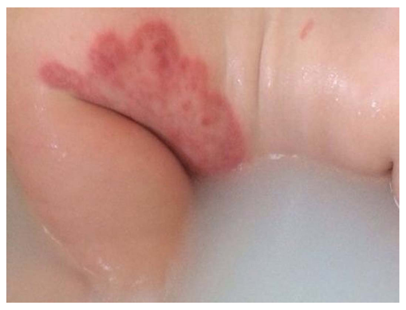
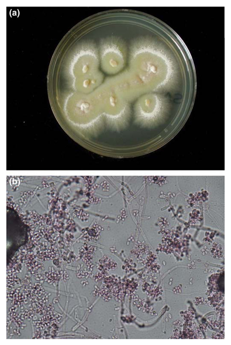
A two year old white Caucasian girl presented with a two month history of small, red itchy patches on the right flank. Over this period the lesions grew in size. They improved temporarily with trials of topical mometasone and betamethasone, but her symptoms worsened when these were stopped. Her dermatological history included mild atopic eczema controlled with emollients. On examination well demarcated, erythematous, annular plaques on the right flank were noted, with central clearing and scaly edges (Figure 1). Similar satellite lesions were noted on the lower abdomen and back. Culture of scrapings yielded pure colonies of a mould which superficially resembled Microsporum canis colonially, but which developed concentric zones of heavy conidiation upon prolonged incubation (Figure 2a). Microscopic examination of the culture revealed numerous, small oval to clavate aleuriospores, consistent with isolates of Arthroderma benhamiae (Figure 2b). Identification was confirmed by PCR amplification and sequencing of the Internal transcribed Spacer Region1 (ITS1) of genomic DNA extracted and amplified exactly as described previously 1.The resulting sequence shared 100% identity over the full 310 nucleotide amplicon with confirmed isolates of A. benhamiae in the public synchronised EMBL databases (EMBL accession number LT223563). The current isolate has been conserved as viable material in the National Collection of Pathogenic Fungi, Mycology Reference Laboratory, Bristol, under the unique identifier NCPF 5137.
Figure 1.
Well demarcated, erythematous plaque with central clearing on the right flank.
Figure 2.
Colonial and microscopic appearance of A. benhamiae, NCPF 5137.
Panel (a): Colonies on Sabouraud’s agar after 10 days culture at 30°C.
Panel (b): Microscopic appearance of needle tease mounts prepared in lactofuschin.
Scale bar = 10 microns.
Further history revealed that she had, until recently, had a guinea pig at home, but the family had not noticed hair loss or claw changes. Her skin lesions were treated with topical terbinafine cream used twice daily for two weeks.
Arthroderma benhamiae is the teleomorphic (sexual) state of a dermatophyte in the Trichophyton mentagrophytes complex. It is a zoophilic dermatophyte that can result in a severe inflammatory keratinocytic cytokine pattern response in humans, where a superficial mycosis involving the keratinized layer can become difficult to treat.
Arthroderma benhamiae was first isolated as a cause of tinea in a human in Japan in 2002, and more recently has been recognised as a cause of tinea in Europe and America.2,3Trichophyton species of Arthroderma benhamiae can cause tinea capitis, tinea corporis, tinea manuum and tinea faciei, with severe inflammation in children/adolescents and immunosuppressed patients.4
Guinea pigs are the most common source of Arthroderma benhamiae, but it has also been isolated from rabbits, cats and dogs.3,5,6 In infected guinea pigs it may manifest as circumscribed areas of alopecia with erythema, scaling and crusting and onychomycosis with brittle, irregular claws that lack lustre and are unusually coloured.6
Diagnosis can be made by conventional identification of the dermatophyte by microscopic examination of isolates obtained from clinical material. Amplification and sequencing of the ribosomal DNA internal transcribed spacer region and matrix-assisted laser desorption/ionization time-of-flight mass spectrometry (MALDI TOF MS) can be used to confirm Arthroderma benhamiae in cultures growing Trichophyton species.5
Widespread dermatophytosis due to Trichophyton species of Arthroderma benhamiae, in particular tinea capitis, requires oral antimycotics. Terbinafine has proven to be effective, with fluconazole and itraconazole as alternatives.
It is only in recent years that Arthroderma benhamiae, the teleomorphic state of many well characterised Trichophyton species, has gained recognition as a zoophilic dermatophyte causing tinea in humans. Whilst uncommon, it is increasing in prevalence. To date there are very few reports in the dermatological literature and little recognition of the potential for guinea pigs to be a source of dermatophytosis. It is important to consider pets as a potential source of infection and to adequately treat both humans and pets to prevent new and re-occurring infections.
Multiple choice questions.
Question 1.Which of the following animals is considered the most common source of Arthroderma benhamiae?
-
a)
Cats
-
b)
Dogs
-
c)
Birds
-
d)
Horses
-
e)
Guinea pigs
Answers to question 1
-
a)
Incorrect. Cats are not considered the most common source of Arthroderma benhamiae.
-
b)
Incorrect. Dogs are not considered the most common source of Arthroderma benhamiae.
-
c)
Incorrect. Birds are not considered the most common source of Arthroderma benhamiae.
-
d)
Incorrect. Horses are not considered the most common source of Arthroderma benhamiae.
-
e)
Correct. Guinea pigs are considered the most common source of Arthroderma benhamiae.
Question 2. Arthroderma benhamiae is the teleomorphic (sexual) state of which dermatophyte
-
a)
Trichophyton mentagrophytes
-
b)
Trichophyton equinum
-
c)
Trichophyton simii
-
d)
Trichophyton verrucosum
-
e)
Trichophyton redellii
Answers to question 2
-
a)
Correct. Arthroderma benhamiae is the teleomorphic (sexual) state of Trichophyton mentagrophytes.
-
b)
Incorrect. Arthroderma benhamiae is the not teleomorphic (sexual) state of Trichophyton equinum.
-
c)
Incorrect. Arthroderma benhamiae is not the teleomorphic (sexual) state of Trichophyton simii.
-
d)
Incorrect. Arthroderma benhamiae is not the teleomorphic (sexual) state of Trichophyton verrucosum.
-
e)
Incorrect. Arthroderma benhamiae is not the teleomorphic (sexual) state of Trichophyton redellii.
Footnotes
No conflict of interest
References
- 1.Borman AM, Fraser M, Linton CJ, et al. An improved method for the preparation of total genomic DNA from isolates of yeast and mould using Whatman FTA filter papers. Mycopathologia. 2010;169:445–449. doi: 10.1007/s11046-010-9284-7. [DOI] [PubMed] [Google Scholar]
- 2.Mochizuki T, Kawasaki M, Ishizaki H, et al. Molecular epidemiology of Arthroderma benhamiae an emerging pathogen of dermatophytoses in Japan, by polymorphisms of the non-transcribed spacer region of the ribosomal DNA. J Dermatol Sci. 2001;27:14–20. doi: 10.1016/s0923-1811(01)00101-3. [DOI] [PubMed] [Google Scholar]
- 3.Nakamura Y, Kano R, Nakamura E, et al. Case Report. First report on human ringworm caused by Arthroderma benhamiae in Japan transmitted from a rabbit. Mycoses. 2002;45:129–131. doi: 10.1046/j.1439-0507.2002.00732.x. [DOI] [PubMed] [Google Scholar]
- 4.Budihardja D, Freund V, Mayser P. Widespread erosive tinea corporis by Arthroderma benhamiae in a renal transplant recipient: casereport. Mycoses. 2010;53:530–532. doi: 10.1111/j.1439-0507.2009.01736.x. [DOI] [PubMed] [Google Scholar]
- 5.Nenoff P, Krüger C, Schaller J, et al. Mycology - an update part 2: dermatomycoses: clinical picture and diagnostics. J Dtsch Dermatol Ges. 2014;12:749–77. doi: 10.1111/ddg.12420. [DOI] [PubMed] [Google Scholar]
- 6.Drouot S, Mignon B, Fratti M, et al. Pets as the main source of two zoonotic species of the Trichophyton mentagrophytes complex in Switzerland, Arthroderma vanbreuseghemii and Arthroderma benhamiae. Veterinary Dermatology. 2009;20:13–18. doi: 10.1111/j.1365-3164.2008.00691.x. [DOI] [PubMed] [Google Scholar]


