Abstract
Objectives:
Masking ability of a restorative material plays a role in hiding colored substructures; however, the masking ability of zirconia ceramic (ZRC) has not yet been clearly understood in zirconia-based restorations. This study evaluated the effect of three different core materials on masking ability of a ZRC.
Materials and Methods:
Ten zirconia disc samples, 0.5mm in thickness and 10mm in diameter, were fabricated. A white (W) substrate (control) and three substrates of nickel-chromium alloy (NCA), non-precious gold alloy (NPGA), and ZRC were prepared. The zirconia discs were placed on the four types of substrates for spectrophotometry. The L*, a*, and b* values of the specimens were measured by a spectrophotometer and color change (ΔE) values were calculated to determine color differences between the test and control groups and were then compared with the perceptual threshold. Randomized block ANOVA and Bonferroni test analyzed the data. A significance level of 0.05 was considered.
Results:
The mean and standard deviation values of ΔE for NCA, NPGA, and ZRC groups were 10.26±2.43, 9.45±1.74, and 6.70±1.91 units, respectively. Significant differences were found in the ΔE values between ZRC and the other two experimental groups (NCA and NPGA; P<0.0001 and P=0.001, respectively). The ΔE values for the groups were more than the predetermined perceptual threshold.
Conclusions:
Within the limitations of this study, it was concluded that the tested ZRC could not well mask the examined core materials.
Keywords: Color, Spectrophotometry, Visual Perception, Yttria Stabilized Tetragonal Zirconia
INTRODUCTION
Metal-ceramic restorations have been successfully applied in restorative dentistry due to high fracture strength [1,2]. However, achieving a natural translucency is more difficult with metal-ceramic restorations than all-ceramic restorations because metal substructures prevent light transmission in metal-ceramic restorations [3,4]. This has led to an increase in use of all-ceramic restorations [5]. Among all-ceramic restorations, zirconia restorations have shown acceptable physical and mechanical properties [6,7]. However, high translucency of a restoration is not always an advantage, for instance in cases with discolored teeth, colored core materials and cast metal post and cores [8]. In these clinical conditions, a restorative material with optimal masking ability of the underlying substructures is rationally recommended to achieve acceptable esthetic results. The masking ability is defined as the ability to hide background color [5].
A method to evaluate the masking ability of restorative materials is to measure color differences in CIE Lab color system. In this color system, the L*, a* and b* color parameters and ΔE refer to lightness, red-green value, yellow-blue value, and color difference, respectively, which can be measured by a spectrophotometer. The ΔE color difference is calculated using this formula:
which is the most commonly used formula for ΔE [5]. This formula can detect even a small amount of color difference between two objects [9, 10]. As spectrophotometers can detect even the slightest amount of color difference not perceptible by the human eye, perceptual and clinically acceptable thresholds have been defined based on ΔE values [5]. It has been reported that the clinically acceptable threshold is more than the perceptual threshold, because of optical conditions of oral environment [11]. If the ΔE (color difference) between two objects is more than the perceptual threshold, a color mismatch will be detected by the human eye. The perceptual threshold has been reported from ΔE=1 to ΔE=5.5 in different studies [12–14]. If a restorative material has an ideal masking ability, ΔE will be equal to zero when placing the material on white and black substrates [8], namely that the material has a similar color on different substrates, and the substrates cannot affect the color of the material. From a clinical point of view, if ΔE of a material on different backgrounds is less than the threshold, it can thoroughly mask the background color. Several studies have surveyed the optical properties of different ceramic materials [8, 15–18]. Some studies have evaluated various factors affecting the final color of zirconia restorations including dental substrates [19–22], luting agents [21, 23], ceramic veneers [24–26], glaze [24], and firing process [27].
Suputtamongkol et al, [19] reported that the background color could affect the overall color of posterior zirconia restorations on a metal post and core or a prefabricated post and a composite core; however, color changes did not exceed the perceptual threshold. Choi and Razzoog [20] assessed the masking ability of a zirconia ceramic (ZRC) with and without the veneering porcelain and revealed that the zirconium oxide coping material alone had some degree of masking ability. Oh and Kim [22] disclosed that abutment shade, ceramic thickness and coping type affected the resulting color of zirconia restorations. Vichi et al, [21] evaluated the effects of different substrates with different colors on the final color of all-ceramic IPS-Empress glass-ceramic restorations, and advised to take the color of substrate into consideration in cases with ceramic thickness of less than 1mm [21].
Masking ability of a ZRC is related to its color coverage over underlying substructures including cement and dental substrates. It may not be feasible to mask a substrate with cements, because different shades do not exist for all cements. Furthermore, the availability of different cement shades does not allow major color corrections [21].
Thus, it seems that the color masking ability of a ZRC on different substrates may be an important factor affecting the esthetic results of zirconia-based restorations. On the other hand, posts and cores, which are widely applied in restorative dentistry to restore severely damaged teeth may be fabricated of different materials such as cast metal alloys or ceramics. The effects of these core materials (as substrates) on masking ability of ZRCs (as substructures in zirconia-based restorations) have to be clearly determined.
Therefore, this in vitro study aimed to evaluate the effect of three different core materials including a nickel-chromium alloy (NCA), a non-precious gold alloy (NPGA), and a ZRC on masking ability of a ZRC. The null hypothesis was that the ZRC would mask the three aforementioned core materials with no significant difference.
MATERIALS AND METHODS
Totally, 10 zirconia discs were fabricated. The discs were placed over four substrates including a white (W) substrate (as the control) [20], NCA, NPGA and ZRC. Spectrophotometric measurements were made on the specimens. The procedure was conducted as follows:
Specimen preparation: A computer-aided design/computer-aided manufacturing system (CORITEC 250i, imes-icore GmbH, Eiterfeld, Germany) milled zirconia blanks (Luminesse High Strength/Low Translucency, 98mm Discs #5113; Talladium, Valencia, CA, USA) to fabricate zirconia discs. The zirconia blanks had 37% translucency and were used to fabricate zirconia frameworks in zirconia-based restorations. The discs had 0.5mm thickness and 10mm diameter [20, 23]. All the zirconia discs were sintered at 1520°C for a 12-hour process in a sintering furnace (iSINT HT, imes-icore GmbH, Eiterfeld, Germany). A digital micrometer (293 MDC-MX Lite; Mitutoyo Corporation, Tokyo, Japan) with the accuracy of 0.002mm was employed to measure the thicknesses of the discs. The discs were adjusted to have a thickness of 0.5±0.01mm. An adjustment/polishing kit (BruxZir; Glidewell Direct, Irvine, CA, USA) was used to reduce the thicknesses according to the afore-mentioned acceptable range. In case of lack of acceptable thickness, the disc was excluded from the study. The zirconia discs were polished, cleaned in an ultrasonic bath (Elmasonic S-30; Dentec, North Shore, Australia) containing 98% ethanol for 15 minutes, and were finally dried. The zirconia disc specimens were prepared as such, and had natural white color of zirconia (Fig. 1).
- Substrate preparation: In order to fabricate the W substrate with the dimensions of 10mm in diameter and 10mm in height, a white Teflon material (PTFE, Omnia Plastica SPA, Busto Arsizio, Italy) was milled. Then, two cylindrical acrylic resin (Duralay, Reliance Dental Mgf Co., Worth, IL, USA) patterns were formed according to the afore-mentioned dimensions. The patterns were cast by a NCA (VeraBond V; Alba Dent, Fairfield, CA, USA) and a NPGA (Alba Dent, Fairfield, CA, USA) to fabricate NCA and NPGA substrates, respectively. An adjusting/polishing kit (NP Alloy Adjustment Kit; Shofu Inc., Kyoto, Japan) was employed to polish the alloy substrates. In order to fabricate the ZRC substrate, a cylindrical pattern was virtually designed by a software program (SOLIDWORKS 2015, Solidworks Corp., Dallas, TX, USA) according to the aforementioned dimensions. The same computer-aided design/computer-aided manufacturing system milled a zirconia blank (Luminesse High Strength 98mm Discs #5113; Talladium, Valencia, CA, USA) to fabricate a zirconia substrate based on the above-mentioned virtual design. The zirconia substrate was dipped in A2 shade color liquid (DD Bio ZX2 monolithic zero LZDD; Dental Direkt GmbH, Spenge, Germany) for 10 seconds. The zirconia substrate was sintered at 1520°C for a 12-hour process in the same sintering furnace, and was then polished by the same polishing kit. The four substrates of W, NCA, NPGA, and ZRC were prepared as such. All the substrates were cleaned in the same ultrasonic bath containing 98% ethanol for 15 minutes (Fig. 2) Color measurement: A spectrophotometer (SpectroShade Micro, MHT, Verona, Italy) was employed for spectrometric measurements [28]. A putty silicone material (Speedex; Coltene, Altstatten, Switzerland) was adapted to the mouth piece of the spectrophotometer to match the conditions of spectrophotometry for all specimens and to prevent external lights. The specimens were located at the center of this putty mold. Before each measurement, the spectro-photometer was calibrated by the white and green calibration plates, respectively. The discs were placed over the substrates with a water drop in-between to prevent the refraction of light [29]. Each disc was placed on each of the substrates, and the color measurements were made. All the color measurements were made at the center of specimens marked by a pen on the monitor screen of spectrophotometer. The color parameters of L*, a*, and b* were recorded for each specimen. Additionally, ΔE values were calculated to determine the ΔE of disc on the substrates. In order to compare the color of specimens on the NCA, NPGA, and ZRC substrates with their color on the W substrate, the ΔE values were measured in three situations including: W-NCA, W-NPGA, and W-ZRC. The ΔE W-NCA ΔE W-NPGA , and ΔE W-ZRC values were calculated using this formula:
Fig. 1:

Zirconia disc specimens
Fig. 2:

Tested substrates (from left to right: zirconia ceramic, non-precious gold alloy, nickel-chromium alloy and white substrate)
The perceptual threshold of ΔE=2.6 was considered in this study [12–14]. Normal distribution of the data was accepted in all groups by the Kolmogorov-Smirnov test (P>0.05). SPSS version 21 (SPSS Inc., Chicago, IL, USA) analyzed the data.
The values of L*, a*and b* for each disc in NPGA, NCA and ZRC groups were subtracted from L*white, a*white and b*white values, which were the corresponding values for W substrate. In order to compare the standard L*, a*, b* (from which, the values of W substrate were subtracted) and ΔE values among the groups, randomized block ANOVA was employed. Pairwise comparisons of the groups were performed by Bonferroni test. STATA software (StataCorp LP, Lakeway, TX, USA) compared the ΔE values with the predetermined perceptual threshold of ΔE=2.6 using One-sample t-test. All tests were carried out at 0.05 level of significance.
RESULTS
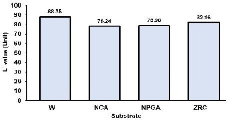
The results were explained according to the measured variables of L*, a*, b*, and ΔE in four sections. L* parameter (lightness): The mean and standard deviation of the L* values for the W, NCA, NPGA and ZRC groups were 88.35±1.46, 78.24±1.31, 78.98±1.26, and 82.16±1.16 units, respectively (Table 1, Fig. 3). Statistical analysis showed a significant difference among the groups in this regard (P<0.0001). Pairwise comparisons of the groups using Bonferroni test revealed significant differences between the NCA and ZRC (P<0.0001), and NPGA and ZRC (P<0.0001). No significant difference was found between NCA and NPGA (P=0.72). The L* values decreased in all the groups compared to the control. The decrease in L* value was the lowest in ZRC.
Table 1:
The color parameter values in the four groups of white substrate (W), nickel-chromium alloy (NCA), non-precious gold alloy (NPGA) and zirconia ceramic (ZRC)
| Substrate group | Color parameter | Mean | Standard deviation | Minimum | Maximum | %95 confidence interval |
|---|---|---|---|---|---|---|
| W | L * | 88.35 | 1.46 | 85.40 | 90.50 | (87.31, 89.39) |
| a * | −0.40 | 0.42 | −1.10 | 0.30 | (−0.70, −0.10) | |
| b * | 3.38 | 0.36 | 2.90 | 4.10 | (3.12, 3.64) | |
| NCA | L * | 78.24 | 1.31 | 76.10 | 80.00 | (77.31, 79.17) |
| a * | 0.04 | 0.52 | −0.40 | 1.40 | (−0.33, 0.41) | |
| b * | 1.92 | 1.01 | 0.70 | 4.00 | (1.20, 2.64) | |
| ΔE | 10.26 | 2.43 | 6.22 | 14.65 | (8.52, 12.00) | |
| NPGA | L * | 78.98 | 1.26 | 76.50 | 80.80 | (78.08, 79.88) |
| a * | 0.60 | 0.42 | 0.20 | 1.70 | (0.30, 0.90) | |
| b * | 3.86 | 0.49 | 3.30 | 4.90 | (3.51, 4.21) | |
| ΔE | 9.45 | 1.74 | 5.83 | 12.29 | (8.21, 10.70) | |
| ZRC | L * | 82.16 | 1.16 | 80.30 | 83.90 | (81.33, 82.99) |
| a * | 1.52 | 0.38 | 1.00 | 2.30 | (1.25, 1.79) | |
| b* | 4.88 | 0.57 | 4.30 | 6.20 | (4.48, 5.28) | |
| ΔE | 6.70 | 1.91 | 2.66 | 9.17 | (5.33, 8.07) |
Fig. 3:
The mean L* values of the groups
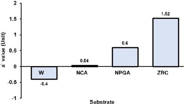
a* parameter (red-green value): The mean and standard deviation of the a* values for the W, NCA, NPGA and ZRC groups were −0.40±0.42, 0.04±0.52, 0.60±0.42, and 1.52±0.38 units, respectively (Table 1, Fig. 4). A significant difference was found among the groups in this respect (P<0.0001). Pairwise comparisons of the groups using Bonferroni test revealed significant differences between NCA and NPGA (P<0.0001), NCA and ZRC (P<0.0001) and NPGA and ZRC (P<0.0001). The a* values increased in all the groups compared to the control. The increase in the a* value was the highest in ZRC.
Fig. 4:
The mean a* values of the groups
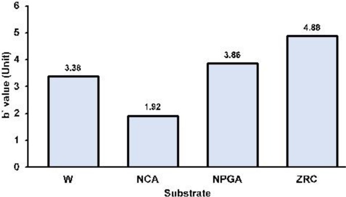
b* parameter (yellow-blue value): The mean and standard deviation of the b* values for the W, NCA, NPGA, and ZRC groups were 3.38±0.36, 1.92±1.01, 3.86±0.49, and 4.88±0.57 units, respectively (Table 1, Fig. 5). A significant difference was detected among the groups in this respect (P<0.0001). Pairwise comparisons of the groups using Bonferroni test showed significant differences between NCA and NPGA (P<0.0001), NCA and ZRC (P<0.0001), and NPGA and ZRC (P<0.001). ZRC had the highest b* value, and NCA had the lowest b* value among the groups.
Fig. 5:
The mean b* values of the groups
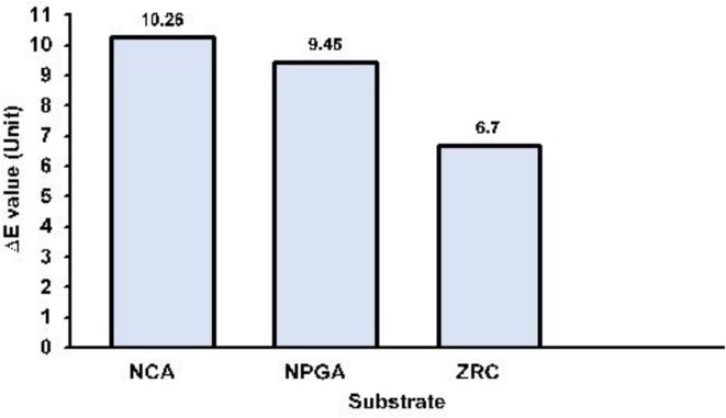
ΔE (color difference): The mean and standard deviation of the ΔE values for the NCA, NPGA and ZRC groups were 10.26±2.43, 9.45±1.74, and 6.70±1.91 units, respectively (Table 1, Fig. 6). A significant difference was found among the groups (P<0.0001). Pairwise comparisons of the groups using Bonferroni test showed significant differences between NCA and ZRC (P<0.0001), and NPGA and ZRC (P=0.001). The difference between NCA and NPGA was not statistically significant (P=0.65). In order to compare the ΔE values of the groups with the predetermined perceptual threshold of ΔE=2.6, one-sample t-test (one-sided) was employed. The null hypothesis of ΔE ≤ 2.6 was rejected for all the groups (P<0.0001).
Fig. 6:
The mean ΔE values of the groups
DISCUSSION
The present study evaluated the color parameters of L*, a*, b*, and ΔE for zirconia disc specimens on four different substrates including W, NCA, NPGA and ZRC. Statistical analysis indicated significant differences among the groups in the L*, a*, b* and ΔE values. The measured ΔE values were more than the predetermined perceptual threshold. The examined ZRC on the tested substrates showed perceptible color change namely that the examined ZRC could not thoroughly mask the tested substrates. Hence, the null hypothesis of the study was rejected.
The L* (lightness) values decreased in all the groups compared with the control. The decrease in L* value was the lowest in ZRC. The change in L* value shows the impact of the substrates on the zirconia specimens. This can be explained by the optical properties of ZRC, which is a semi-translucent material. According to the results, metal core materials such as NCAs and NPGAs can decrease the lightness of ZRC more than a zirconia core material. This may be due to less lightness of metals compared to ceramics.
The a* (red-green) values increased in all the groups compared to the control. The increase of a* value was the highest in ZRC. A reason for this may be the natural a* value of the ZRC as a substrate. The tested zirconia substrate (A2 shade) positively shifted the a* value towards red.
The b* (yellow-blue) values changed in the groups depending on the substrates. The nickel-chromium substrate decreased the b* value while the non-precious gold and zirconia substrates increased the b* value compared to the control. This may be due to the yellow color tendency of A2 shade zirconia substrate and NPGA, which impact on the color of ZRC and shift it towards yellow. On the contrary, NCA shifted the color of ZRC towards blue, and negatively affected the b* value.
The ΔE values of all groups were more than the predetermined perceptual threshold of ΔE=2.6. The color changes induced by the substrates were beyond the perceptual threshold. Consequently, the tested ZRC could not completely mask the tested core materials.
Assessment of the L*, a* and b* values indicated their changes in the substrate groups. Thus, color changes were derived from all three color parameters. The L*, a*, and b* values manifested significant differences between the substrate groups. This demonstrated that the quality of the color change was different depending on the substrate. The ΔE values manifested no significant difference between the metal substrates; however, significant differences existed between the zirconia substrate and the metal substrates, and the least color change belonged to the zirconia substrate. This demonstrated that the quantity of the color change was different depending on the substrate as well. Hence, both quality and quantity of the color of ZRC were altered by the substrates. The used metal substrates did not differ in terms of the quantity of color change, and both affected the quantity of the color change more than the zirconia substrate.
Suputtamongkol et al, [19] reported that the color of an underlying substructure could affect the overall color of posterior zirconia-based restorations on a metal post and core or a prefabricated post and a composite core, ranging from ΔE =1.2 to 3.1. Additionally, slight color changes of zirconia crowns were detected by measuring the ΔE [19]. Although some differences exist between the afore-mentioned study and the current research such as ZRC brands, thicknesses of ceramics, veneered versus non-veneered ZRCs, hypothesized perceptual thresholds and substrate types, both studies showed that ZRC could not thoroughly mask the underlying materials.
Oh and Kim [22] in an in vitro study of color masking ability assessed the effects of abutment shade, ceramic thickness and coping type on the final color of zirconia restorations. The abutments were fabricated of gold alloy, base metal (nickel-chromium) alloy, and four different shades of composite resins. In their study, the average ΔE value of Lava specimens between the A2 shade composite resin and gold alloy abutments was higher than that between the A2 shade composite resin and other abutments, and was close to ΔE=5.5. This means that the tested ZRC could not ideally mask the substrates.
A similar result was obtained in the current study, although the substrates and ZRCs used in the two studies were different. According to the study by Oh and Kim [22], gold alloy substrate yielded the highest ΔE among the tested substrates; however, the NPGA and the NCA yielded close ΔE values in the current study. This dissimilarity in the results may be caused by the color difference of the precious gold alloy used by Oh and Kim [22] and the NPGA used in the current study.
Choi and Razzoog [20] evaluated the masking ability of ZRC with and without a veneering porcelain. In their study, color differences caused by ZRC and the veneering porcelain were compared with substrates alone. They concluded that the non-veneered ZRC had some degree of masking ability over different tested substrates (white, black, gray, and A3 shade tooth-colored substrates). However, the current study compared substrate-induced color differences with a ZRC over W substrate and concluded that the tested ZRC could not mask the examined substrates. Choi and Razzoog [20] measured the ΔE values between the substrate alone (as control) and the ZRC over the substrate, while the current study measured the ΔE values between the ZRC over W substrate (as control) and ZRC over the tested substrates. The different results may be due to differences in the control groups.
Based on the results of our study, the three core materials including NCA, NPGA and ZRC can change the color of tested ZRC. The quality and quantity of color change depend on the type of core material. ZRC as a core material creates less color change than metal cores. It seems that the substrates shift the color of ZRC to their inherent colors. The color of ZRC may be closer to the final color of a restoration than the metal cores. Therefore, it is esthetically advised to preferably choose a ZRC post and core instead of NCA or NPGA post and cores in zirconia-based restorations and to reduce the core material-induced color change by further core reduction, applying sufficient thickness of the veneering porcelain, and use of proper luting agents in zirconia-based restorations. It should be noted that given that the physical and mechanical properties of zirconia post and core are confirmed in further studies, zirconia post and core should be clinically recommended. This needs future investigations.
Considering the optical properties of ZRC and the thickness of zirconia sub-structures, which is approximately 0.5mm in a normal case [30], light transmission through the zirconia structure can be expected in zirconia-based restorations. Thus, substrates including cements and core materials may affect the color. The present study evaluated the effects of three core materials in this respect. On the other hand, the zirconia core overlaying materials such as porcelain veneers and glaze may influence the final color of zirconia-based restorations, which were not assessed in this study. Therefore, evaluation of the effect of other factors in this respect is recommended in future studies. The present study had some limitations such as using a specific brand of ZRC, an uncolored ZRC, and a specific brand of composite resin. More studies on the mentioned subjects are suggested.
CONCLUSION
Within the limitations of this study, it was concluded that the tested ZRC could not thoroughly mask the three core materials of NCA, NPGA, and ZRC.
ACKNOWLEDGMENT
This paper has been entirely drawn up from a “M.D. Thesis” which was successfully completed by Dr Faeze Masoomi under the supervision of Dr. Farhad Tabatabaian.
REFERENCES
- 1-. Walton TR. An up to 15-year longitudinal study of 515 metal-ceramic FPDs: Part 1. Outcome. Int J Prosthodont. 2002. Sep-Oct; 15( 5): 439– 45. [PubMed] [Google Scholar]
- 2-. Näpänkangas R, Raustia A. Twenty-year follow-up of metal-ceramic single crowns: a retrospective study. Int J Prosthodont. 2008. Jul-Aug; 21 (4): 307– 11. [PubMed] [Google Scholar]
- 3-. Heffernan MJ, Aquilino SA, Diaz-Arnold AM, Haselton DR, Stanford CM, Vargas MA. Relative translucency of six all-ceramic systems. Part I: core materials. J Prosthet Dent. 2002. July; 88 (1): 4– 9. [PubMed] [Google Scholar]
- 4-. Heffernan MJ, Aquilino SA, Diaz-Arnold AM, Haselton DR, Stanford CM, Vargas MA. Relative translucency of six all-ceramic systems. Part II: core and veneer materials. J Prosthet Dent. 2002. July; 88 (1): 10– 5. [PubMed] [Google Scholar]
- 5-. Vichi A, Louca C, Corciolani G, Ferrari M. Color related to ceramic and zirconia restorations: a review. Dent Mater. 2011. January; 27 (1): 97– 108. [DOI] [PubMed] [Google Scholar]
- 6-. Raigrodski AJ. Contemporary materials and technologies for all-ceramic fixed partial dentures: a review of the literature. J Prosthet Dent. 2004. December; 92 (6): 557– 62. [DOI] [PubMed] [Google Scholar]
- 7-. Luthardt RG, Holzhüter M, Sandkuhl O, Herold V, Schnapp JD, Kuhlisch E, et al. Reliability and properties of ground Y-TZP-zirconia ceramics. J Dent Res. 2002. July; 81 (7): 487– 91. [DOI] [PubMed] [Google Scholar]
- 8-. Chu FC, Chow TW, Chai J. Contrast ratios and masking ability of three types of ceramic veneers. J Prosthet Dent. 2007. November; 98 (5): 359– 64. [DOI] [PubMed] [Google Scholar]
- 9-. Swain VL, Pesun IJ, Hodges JS. The effect of metal ceramic restoration framework design on tooth color. J Prosthet Dent. 2008. June; 99 (6): 468– 76. [DOI] [PubMed] [Google Scholar]
- 10-. Seghi RR, Hewlett ER, Kim J. Visual and instrumental colorimetric assessments of small color differences on translucent dental porcelain. J Dent Res. 1989. December; 68 (12): 1760– 4. [DOI] [PubMed] [Google Scholar]
- 11-. Douglas RD, Steinhauer TJ, Wee AG. Intraoral determination of the tolerance of dentists for perceptibility and acceptability of shade mismatch. J Prosthet Dent. 2007. April; 97 (4): 200– 8. [DOI] [PubMed] [Google Scholar]
- 12-. Lindsey DT, Wee AG. Perceptibility and acceptability of CIELAB color differences in computer-simulated teeth. J Dent. 2007. July; 35 (7): 593– 9. [DOI] [PMC free article] [PubMed] [Google Scholar]
- 13-. Ishikawa-Nagai S, Yoshida A, Sakai M, Kristiansen J, Da Silva JD. Clinical evaluation of perceptibility of color differences between natural teeth and all-ceramic crowns. J Dent. 2009; 37 Suppl 1: e57– 63. [DOI] [PubMed] [Google Scholar]
- 14-. Ghinea R, Pérez MM, Herrera LJ, Rivas MJ, Yebra A, Paravina RD. Color difference thresholds in dental ceramics. J Dent. 2010; 38 Suppl 2: e57– 64. [DOI] [PubMed] [Google Scholar]
- 15-. Fabbri G, Mancini R, Marinelli V, Ban G. Anterior discolored teeth restored with procera all-ceramic restorations: a clinical evaluation of the esthetic outcome based on the thickness of the core selected. Eur J Esthet Dent. 2011. Spring; 6 (1): 76– 86. [PubMed] [Google Scholar]
- 16-. Zhang F, Heydecke G, Razzoog ME. Double-layer porcelain veneers: effect of layering on resulting veneer color. J Prosthet Dent. 2000. October; 84 (4): 425– 31. [DOI] [PubMed] [Google Scholar]
- 17-. Chen YM, Smales RJ, Yip KH, Sung WJ. Translucency and biaxial flexural strength of four ceramic core materials. Dent Mater. 2008. November; 24 (11): 1506– 11. [DOI] [PubMed] [Google Scholar]
- 18-. Lee YK, Cha HS, Ahn JS. Layered color of all-ceramic core and veneer ceramics. J Prosthet Dent. 2007. May; 97 (5): 279– 86. [DOI] [PubMed] [Google Scholar]
- 19-. Suputtamongkol K, Tulapornchai C, Mamani J, Kamchatphai W, Thongpun N. Effect of the shades of background substructures on the overall color of zirconia-based all-ceramic crowns. J Adv Prosthodont. 2013. August; 5 (3): 319– 25. [DOI] [PMC free article] [PubMed] [Google Scholar]
- 20-. Choi YJ, Razzoog ME. Masking ability of zirconia with and without veneering porcelain. J Prosthodont. 2013. February; 22 (2): 98– 104. [DOI] [PubMed] [Google Scholar]
- 21-. Vichi A, Ferrari M, Davidson CL. Influence of ceramic and cement thickness on the masking of various types of opaque posts. J Prosthet Dent. 2000. April; 83 (4): 412– 7. [DOI] [PubMed] [Google Scholar]
- 22-. Oh SH, Kim SG. Effect of abutment shade, ceramic thickness, and coping type on the final shade of zirconia all-ceramic restorations: in vitro study of color masking ability. J Adv Prosthodont. 2015. October; 7 (5): 368– 74. [DOI] [PMC free article] [PubMed] [Google Scholar]
- 23-. Fazi G, Vichi A, Ferrari M. Influence of four different cements on the color of zirconia structures of varying ceramic thickness. Int Dent SA. 2006; 8: 20– 6. [Google Scholar]
- 24-. Sinmazisik G, Demirbas B, Tarcin B. Influence of dentin and core porcelain thickness on the color of fully sintered zirconia ceramic restorations. J Prosthet Dent. 2014. February; 111 (2): 142– 9. [DOI] [PubMed] [Google Scholar]
- 25-. Son H, Kim WC, Jun SH, Kim YS, Ju SW, Ahn JS. Influence of dentin porcelain thickness on layered all-ceramic restoration color. J Dent. 2010; 38 Suppl 2: e71– 7. [DOI] [PubMed] [Google Scholar]
- 26-. Barão VA, Gennari-Filho H, Goiato MC, dos Santos DM, Pesqueira AA. Factors to achieve aesthetics in all-ceramic restorations. J Craniofac Surg. 2010. November; 21 (6): 2007– 12. [DOI] [PubMed] [Google Scholar]
- 27-. Bachhav VC, Aras MA. The effect of ceramic thickness and number of firings on the color of a zirconium oxide based all ceramic system fabricated using CAD/CAM technology. J Adv Prosthodont. 2011. June; 3 (2): 57– 62. [DOI] [PMC free article] [PubMed] [Google Scholar]
- 28-. Llena C, Lozano E, Amegual J, Forner L. Reliability of two color selection devices in matching and measuring tooth color. J Contemp Dent Pract. 2011. January 1; 12 (1): 19– 23. [DOI] [PubMed] [Google Scholar]
- 29-. Chang J, Da Silva JD, Sakai M, Kristiansen J, Ishikawa-Nagai S. The optical effect of composite luting cement on all ceramic crowns. J Dent. 2009. December; 37 (12): 937– 43. [DOI] [PubMed] [Google Scholar]
- 30-. Denry I, Kelly JR. State of the art of zirconia for dental applications. Dent Mater. 2008. March; 24 (3): 299– 307. [DOI] [PubMed] [Google Scholar]


