Abstract
The successful use of Bacillus thuringiensis insecticidal toxins to control agricultural pests could be undermined by the evolution of insect resistance. Under selection pressure in the laboratory, a number of insects have gained resistance to the toxins, and several cases of resistance in the diamondback moth have been reported from the field. The use of protein engineering to develop novel toxins active against resistant insects could offer a solution to this problem. The display of proteins on the surface of phages has been shown to be a powerful technology to search for proteins with new characteristics from combinatorial libraries. However, this potential of phage display to develop Cry toxins with new binding properties and new target specificities has hitherto not been realized because of the failure of displayed Cry toxins to bind their natural receptors. In this work we describe the construction of a display system in which the Cry1Ac toxin is fused to the amino terminus of the capsid protein D of bacteriophage lambda. The resultant phage was viable and infectious, and the displayed toxin interacted successfully with its natural receptor.
The bacterium Bacillus thuringiensis produces insecticidal proteins during sporulation. These toxins are expressed as protoxins that are packaged into crystalline inclusions. When ingested by susceptible insect larvae, the protoxin is solubilized by the unique environment of the host gut and activated by proteolytic cleavage with gut enzymes. The activated toxins are able to bind to receptor molecules present on the insect gut epithelium and insert into the membrane (6). This membrane penetration results in the formation of pores which cause colloid osmotic lysis and eventual insect death (17, 32, 38).
Due to their receptor specificity, Cry proteins are only toxic to certain insects and are harmless to other organisms, including humans, other mammals, fish, and most beneficial insects (31). B. thuringiensis is one of the few microbes that have been successfully used in agricultural insect pest control. Liquid and powder formulation mixtures of spores and crystals from this bacterium have been used for more than 40 years on many crops. B. thuringiensis toxin genes have also been successfully expressed in plants, providing environmentally benign control of insect pests (11).
Although in the past it was hoped that insects would not develop resistance to these toxins, insect resistance to B. thuringiensis toxins has been detected in insect populations, mainly in the laboratory, but also in the field (8, 36).
One possible way of overcoming the problem of resistance is to generate novel toxins by genetic manipulation of B. thuringiensis genes. Several combinatorial methods are now available to create large libraries of mutant proteins that can be searched for toxins with higher potency and different specificities. However, the rate-limiting step is the selection of valuable proteins from the mutant pool.
One way to overcome this problem is to display libraries of peptides or proteins on the surface of bacteriophages (23, 28, 29, 34). This technique is particularly suitable for B. thuringiensis toxins due to the nature of Cry proteins. Because these proteins form insoluble crystals when expressed in B. thuringiensis or inclusion bodies when expressed in Escherichia coli, phage display of B. thuringiensis toxins could be a good way to produce soluble proteins without the need of a solubilization step.
Display of Cry toxins in filamentous phages has been attempted previously (13, 20), but the displayed proteins did not bind to their natural receptor in the midgut of insects. In this report we describe the successful display of a biologically functional Cry1Ac1 toxin with a different approach, based on λ phage. The Cry1Ac1 toxin was fused at its C terminus to the λ phage protein encoded by the gpD gene. D is a 109-amino-acid protein present in the head of λ phage, nonessential for head assembling but necessary for phage stability (12, 35, 42). The fusion between the toxin and the D protein was stable, and the chimera was successfully assembled into λ phages. Importantly, we have shown that the displayed toxin is functional and able to bind to its natural receptors.
MATERIALS AND METHODS
Bacterial and phage strains.
E. coli strain XL1-Blue (Stratagene) was used for cloning purposes, while E. coli BLR strain (Novagene) was used for expression studies.
EMBL3 BamHI arms from Stratagene were used for cloning and protein display. EMBL3 phage and its derivatives were propagated and titrated in E. coli LE392 grown at 37°C in LB medium (25) supplemented with 0.2% maltose and 10 mM MgSO4 until an optical density at 600 nm of 0.8 was reached.
Construction of the cry1Ac1::gpD fusion.
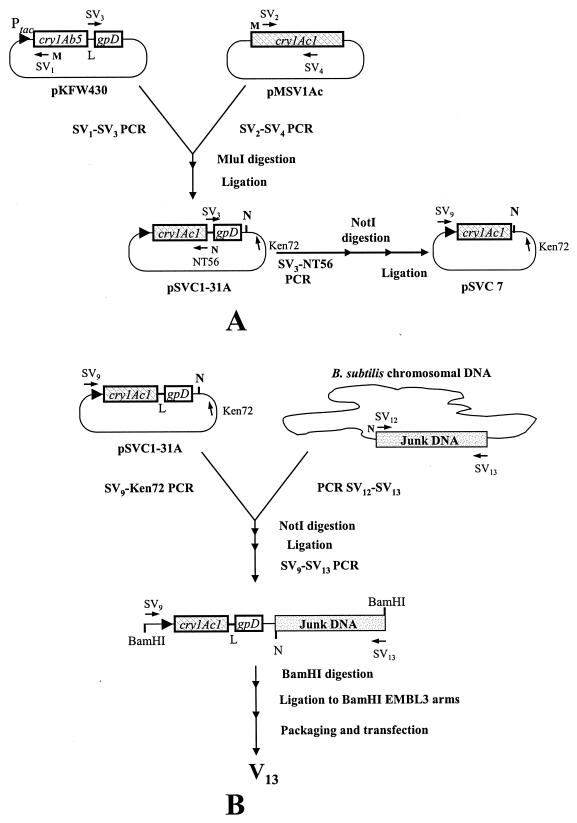
The transcriptional fusion between cry1Ac1 and gpD was carried out as shown in Fig. 1A. The sequences of the primers used in this strategy are shown in Table 1. Plasmid pKFW430 (constructed by K. F. Whelan), containing the transcriptional fusion between cry1Ab5 and gpD separated by a glycine-serine linker coding for the sequence SGGGGSGGGG, was used as a template in a PCR with Pfu polymerase. Primers SV1 (containing an MluI restriction site) and the phosphorylated SV3 were used for amplification of a fragment containing the linker (L), the gpD gene, the vector, and the first 228 nucleotides of the 5′ end of the cry1Ab5 gene (identical to cry1Ac1). To obtain the rest of the active core of the cry1Ac1 gene, plasmid pMSV1Ac (33) was used as the template in a PCR involving the SV2 and SV4 primers. SV1-SV3 and SV2-SV4 fragments were digested with MluI and ligated following the supplier's instructions. XL1-Blue competent cells were transformed with this ligation mix, and transformants were selected on LB plus carbenicillin (250 mM) plates. After that, transformants were screened by colony PCR with two pairs of primers, SV2-SV4 (to check for the presence of the cry1Ac1 gene) and SV3-Ken72 (to check for the presence of the linker and the gpD gene). A clone positive for both PCRs was selected and named pSVC1-31A.
FIG. 1.
Cloning strategy for the translational fusion cry1Ac1::gpD (A) and its cloning into EMBL3 phage (B). Primers are shown as horizontal arrows pointing in the 5′-3′ direction. Primers with MluI (M) and NotI (N) sites are indicated.
TABLE 1.
Primers
| Primer | Sequence | Restriction site underlined |
|---|---|---|
| SV1 | 5′-AAACGCGTCCCATTGAGAGG | MluI |
| SV2 | 5′-ATGGGACGCGTTTCTTGTAC | MluI |
| SV3 | 5′-AGTGGAGGTGGTGGATCAGGTGGAG GAGGT | |
| SV4 | 5′-CTCCAGATTATATATTCAGCC | |
| NT56 | 5′-CACCGCTGCGGCCGCTCTTTCCGATT ATATTC | NotI |
| Ken72 | 5′-CACACAAGCTGTGACCGTC | |
| SV9 | 5′-AAATGGATCCAGACGAAAGGG | BamHI |
| SV12 | 5′-TTAGAGCGGCCGCAACAATTATTAG AAACATTAGC | NotI |
| SV13 | 5′-GCTATGGATCCGCCTACTATTTTTG GTAGCATCCCC | BamHI |
A control plasmid bearing only the N-terminal end and core of the cry1Ac1 gene was constructed by PCR with pSVC1-31A as the template and following the strategy shown in Fig. 1A. For that pSVC1-31A was amplified by PCR with SV3 and NT56. The fragment obtained was digested with NotI, and the resulting larger NotI fragment was circularized by ligation. E. coli XL1-Blue competent cells were transformed with the ligation reaction, and transformants were screened by colony PCR with the SV9-Ken72 primers to check for the absence of gpD. A positive clone was selected, and the plasmid obtained was designated pSVC7.
Cloning of cry1Ac1::gpD into the EMBL3 vector.
EMBL3 is a λ replacement vector derived from lambda 1059 (Promega) that can be used to clone DNA fragments of between 9 and 14 kb. As the fragment containing the translational fusion cry1Ac1::gpD is 2.6 kb, an extra fragment of DNA was necessary for the cloning of this fusion into the EMBL3 arms. The strategy followed for the cloning is detailed in Fig. 1B. The translational fusion between cry1Ac1 and gpD was obtained by PCR from pSVC1-31A with the primers SV9 and Ken72. A DNA fragment of approximately 7.3 kb with no open reading frames (junk DNA) was obtained from Bacillus subtilis genomic DNA by PCR with primers SV12 and SV13 with a mixture of Taq and Pfu polymerases in a ratio of 16:1. The SV9-Ken72 and SV12-SV13 fragments were digested with NotI and ligated for 3 h at room temperature after NotI inactivation at 65°C for 20 min. With the SV9 and SV13 primers and the ligation reaction as the template, a 9.3-kb fragment containing the fusion cry1Ac1::gpD and the junk DNA was obtained. This fragment was digested with BamHI and ligated to EMBL3 BamHI arms under the supplier's instructions. The cloned DNA was packaged with the PhageMaker in vitro λ packaging system from Novagen. E. coli LE392, grown as described before, was transfected with serial dilutions of the packaging reaction. Plaques were screened by plaque PCR with the SV9 and SV4 primers. A positive clone was selected and named V13.
A control phage (Vc) containing only the cry1Ac1 gene and the junk DNA was constructed following the same strategy as V13, the only difference being that plasmid pSVC7 was used instead of pSVC1-31A.
Phage preparation.
Phage lysates were obtained with the method described by Blattner et al. (2) with minor modifications. Briefly, a single plaque was cored out with a Pasteur pipette and eluted in 0.5 ml of SM buffer (for 1 liter, 5.8 g of NaCl, 2 g of MgSO4 · 7H2O, 50 ml of 1 M Tris-HCl, pH 7.5, 5 ml of 2% [wt/vol] gelatin). After incubation for 2 h at room temperature to allow phage particles to diffuse from the agar, 10 μl of this suspension were incubated at 37°C for 30 min with 100 μl of an overnight culture of E. coli LE392 grown in LB supplemented with 0.2% (wt/vol) maltose and 10 mM MgSO4. A volume of 50 μl of this suspension was used to inoculate 50 ml of fresh LB. This culture was incubated with agitation at 37°C until lysis occurred. After that, 100 μl of chloroform was added to the lysate and incubated with agitation for 15 min and subsequently centrifuged. Cell-free lysates were obtained by filtration with 0.45-μm filters, and after phage titration, lysates were kept at 4°C. When higher concentrations of phages were needed, particles were precipitated from lysates following the method described by Santos (30).
Protein expression and solubilization conditions.
Protein expression was carried out in E. coli BLR. The strain, bearing the appropriate plasmid, was cultured at 37°C in 30 ml of LB containing 1% glucose and 250 mM carbenicillin until the culture reached an optical density at 600 nm of 0.5. Afterwards, isopropylthiogalactopyranoside (IPTG) was added to the culture to 1 mM final concentration, followed by additional incubation for 2.5 h under the same conditions. Cells were harvested by centrifugation, and the pellets were resuspended in 1.5 ml of TBS buffer (10 mM Tris-HCl, pH 7.5, 150 mM NaCl) plus 0.2 mM phenylmethylsulfonyl fluoride. The suspension was sonicated in ice with a Soniprep 150 ultrasonic disintegrator. Ten cycles of 10 s on and 10 s off on full power were used. Soluble and insoluble fractions were separated by centrifugation at maximal speed in a microcentrifuge. Pellet fractions containing Cry toxins were solubilized with 250 μl of 50 mM Na2HPO4 buffer, pH 12.
Brush border membrane vesicle preparation.
Brush border membrane vesicles were prepared from the midguts of fifth instar Manduca sexta larvae by differential centrifugation according to the method described by Carroll and Ellar (5). The M. sexta eggs were obtained from S. Reynolds (University of Bath, School of Biological Sciences, United Kingdom) and raised on an artificial diet (1).
Brush border membrane protein preparation.
Brush border membrane proteins were prepared from brush border membrane vesicles as follows. The supernatant was removed from a brush border membrane vesicle suspension by centrifugation. The pellet was resuspended in 20 mM Tris-HCl, pH 8.5-100 mM NaCl-5 mM EDTA-1 mM phenylmethylsulfonyl fluoride-1% (wt/vol) CHAPS (3-[(3-cholamidopropyl)dimethylammonio]-1-propanesulfonate), at a final concentration of 5 mg/ml (26). The suspension was incubated with gentle agitation at 4°C overnight. Insoluble material was removed by centrifugation at 12,000 × g for 10 min at 4°C. The supernatant was washed three times with TBS buffer with a 2-ml Vivaspin concentrator bearing a polyethylsulfone membrane with a 100,000-molecular-weight cutoff (Vivascience). Protein concentration was measured by the Bradford method (3) with bovine serum albumin as the standard.
Immunoblot analysis.
Proteins were separated by electrophoresis with glycine-sodium dodecyl sulfate-polyacrylamide gels followed by electroblotting onto nitrocellulose membranes (37). Fusion proteins were detected with polyclonal anti-Cry1Ac1 or anti-D antibodies as primary antibodies (15) followed by peroxidase-conjugated goat anti-rabbit immunoglobulin G antiserum (Sigma) as the secondary antibody.
Primary and secondary antibodies were diluted 1:1,000 in blocking buffer (5% nonfat dry milk, 5% glycerol, 0.1% Tween 20 in TBS buffer). Nitrocellulose membranes were incubated for 1 h and 20 min, respectively, with these antibody dilutions. Detection was performed with a fresh TBS solution containing 0.6 mg of 4-chloro 1-naphthol (dissolved in methanol) and 2 μl of H2O2 per ml.
Gut extract preparation.
Gut extract was prepared from fifth instar Pieris brassicae larvae by placing dissected whole guts in ice-cold 50 mM Na2CO3 (pH 10.5) plus 10 mM dithiothreitol (pH 9.5). Guts were chopped with scissors, homogenized on ice with a prechilled 5-ml-capacity Dounce homogenizer (30 strokes), and repeatedly centrifuged at 15,000 × g for 5 min at 4°C (resuspending in the above buffer) to remove debris and fat deposits. The final supernatant was sterile filtered through a 0.2-μm filter, and samples of 25 μl to 200 μl were aliquoted and stored at −80°C.
Ligand blotting.
For ligand blot assays, brush border membrane vesicles were prepared as described before and separated by electrophoresis in a sodium dodecyl sulfate-10% polyacrylamide gel (25). Brush border membrane vesicle proteins were transferred to a nitrocellulose membrane in transfer buffer (190 mM glycine, 25 mM Tris-HCl, pH 7.5, and 10% methanol). The membrane was blocked with blocking buffer (5% nonfat dry milk, 5% glycerol, 0.1% Tween 20 in TBS buffer) at room temperature with gentle agitation for 1 h. Membranes were incubated for 1 h with either activated toxins or nonactivated toxins added to a final concentration of 0.02 μg/ml in blocking buffer. Solubilized toxins were activated by incubation either with 2.0% (vol/vol) P. brassicae gut extract for 20 min at 37°C or with 1:1 (wt/wt) trypsin (Sigma)-toxin at 37°C for 60 min. After washing, the membrane was treated as described above in the immunoblot analysis section with anti-Cry1Ac1 antibody.
Manduca sexta bioassays.
Bioassays were performed as described previously (10) with modifications. With a circular punching device with the same area as the plate wells, approximately 500 μl of artificial diet was deposited in each well of a 48-well microtiter plate (Costar). Solubilized proteins were diluted to the appropriate concentration, and 20 μl was added to the surface of each food disk. Each protein was tested in eight independent wells. After drying for 1 h, one M. sexta neonate was placed on each well. The microtiter plate was covered in Saran Wrap in which single holes were pricked above each well with a 1.1-mm needle. The plates were incubated at 25°C, and larval weights were recorded after 5 days of incubation.
Biopanning.
Biopanning assays were carried out as described before (28) with minor modifications. Affinity selection assays were performed in microtiter plates (Nunc) coated overnight at 4°C with 150 μl of the appropriate protein diluted in coating buffer (50 mM NaHCO3, pH 9.6). The coated plate was washed five times with washing buffer (0.02% Tween 20 in PBS [8 g of NaCl, 0.2 g of KCl, 1.44 g of Na2HPO4 per liter]), blocked 1 h at room temperature with blocking buffer (5% nonfat dry milk, 0.5% Tween 20 in PBS), and washed again five times with washing buffer. Each well was incubated 1 h at room temperature with 150 μl of a phage suspension containing 106 PFU/μl in SM buffer. After that, the wells were thoroughly washed a further six times with the above washing buffer followed by 20 min of incubation at 37°C with 150 μl of an E. coli LE392 culture grown under the conditions described above. After infection, the cells were removed, and serial dilutions were plated for PFU determination.
RESULTS
Cry1Ac1-D fusion protein.
For the display of a Cry toxin on the surface of the head of λ phage, a translational fusion was designed between the C-terminal end of the active core (plus the N-terminal end) of the Cry1Ac1 toxin and the N terminus of protein D of λ phage. This fusion included the first 619 amino acids of Cry1Ac1 toxin (GenBank number U89872) and the complete protein D (GenBank number NC_001416), linked by a 10-amino-acid-long linker rich in glycine and serine residues. The construction of a plasmid bearing the translational fusion between cry1Ac1 and gpD and a control plasmid containing only cry1Ac1 is detailed in Fig. 1A and in Materials and Methods. The entire cry1Ac1::gpD fusion in pSVC1-31A and the cry1Ac1 gene in pSVC7 were verified by DNA sequencing.
Cry1Ac1-D fusion protein is stable and expressed well in E. coli.
To check the expression and stability of the fusion protein Cry1Ac1-D and its control Cry1Ac1, expression studies in E. coli BLR(pSVC1-31A) and BLR(pSVC7) were performed as described in the Materials and Methods section. The results are shown in Fig. 2. In a Coomassie-stained sodium dodecyl sulfate-polyacrylamide gel electrophoresis gel (Fig. 2A), two strong bands migrating at 65 kDa (lanes 2 and 4) and 81 kDa (lanes 6 and 8), corresponding to the Cry1Ac1 and Cry1Ac1-D proteins, respectively, were found in the pellet fraction of cultures under noninduced (lanes 2 and 6) and induced (lanes 4 and 8) conditions. The 81-kDa and 65-kDa proteins were detected by immunoblotting with anti-Cry1Ac1 antibody (Fig. 2B) in both supernatant (lanes 1, 3, 5, and 7) and pellet (lanes 2, 4, 6, and 8) fractions. As expected, only the 81-kDa protein (the fusion protein) was detected when anti-D antibody was used in the immunoblot (Fig. 2C). Along with the 81- and 65-kDa proteins, breakdown products of these proteins were detected.
FIG. 2.
Coomassie staining (A) and immunodetection (B and C) of Cry1Ac1 (lanes 1 to 4) and Cry1Ac1-D (lanes 5 to 8) expressed from plasmids pSVC7 and pSVC1-31A. Lanes 1, 3, 5, and 7 show the supernatant fraction of the cultures, and lanes 2, 4, 6, and 8 show the pellet fraction of cultures under noninduced conditions (lanes 1, 2, 5, and 6) and under induced conditions (lanes 3, 4, 7, and 8). Lanes M, molecular size markers. Panels B and C show immunodetection with anti-Cry1Ac1 and anti-D antibodies, respectively.
Cry1Ac1-D fusion protein recognizes the aminopeptidase N receptor and is toxic for M. sexta.
Ligand blot assays are frequently used to detect binding between Cry toxins and their natural receptors present in the midguts of insects (9, 39, 40). The 120-kDa aminopeptidase N protein has been previously reported as a receptor in M. sexta for Cry1A toxins (14, 21, 27).
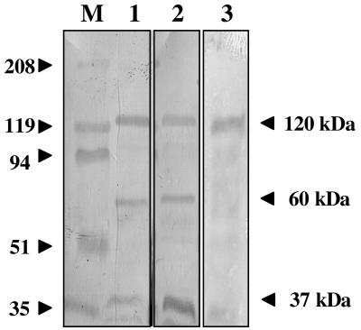
To test whether the fusion protein Cry1Ac1-D was able to recognize and bind receptors present in brush border membrane vesicles isolated from M. sexta midgut tissues, ligand blot assays were carried out (Fig. 3). For these, trypsin-treated (data not shown) and gut extract-treated Cry1Ac1 (lane 1) and Cry1Ac1-D proteins (lane 2) and nontreated Cry1Ac1-D protein (lane 3) were included in the assay. Three bands were observed migrating at 120, 60, and 37 kDa in all of the blots. Treated and nontreated Cry1Ac1-D protein produced the same pattern of binding to brush border membrane vesicle proteins as the treated Cry1Ac1 toxin control. The 120-kDa band was identified as the aminopeptidase N receptor (14), while the 60- and 37-kDa bands are frequently observed degradation products of aminopeptidase N that vary in intensity from preparation to preparation (D. J. Ellar, unpublished data). These results suggest that the fusion protein Cry1Ac1-D is able to recognize and bind to the aminopeptidase N receptor present in M. sexta midguts.
FIG. 3.
Cry1Ac1 and Cry1Ac1-D ligand blot of brush border membrane vesicles samples. Lane M, molecular size markers (in kilodaltons). Lane 1: overlay of gut extract-treated Cry1Ac1 protein over brush border membrane vesicles (40 μg). Lane 2: overlay of gut extract-treated Cry1Ac1-D fusion protein over brush border membrane vesicles (40 μg). Lane 3: overlay of nontreated Cry1Ac1-D fusion protein over brush border membrane vesicles (40 μg).
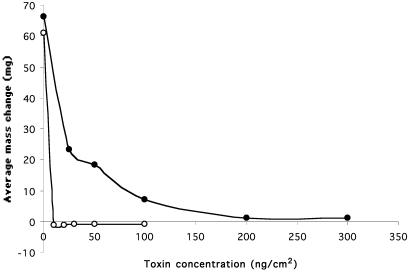
To determine the toxicity profile of the Cry1Ac1-D fusion protein, toxicity bioassays were carried out with M. sexta with solubilized Cry1Ac1 and Cry1Ac1-D proteins in a multiwell plate. The concentrations tested in the assay ranged from 0 to 100 ng/cm2 for Cry1Ac1 and 0 to 300 ng/cm2 for the fusion protein Cry1Ac1-D. Each concentration was tested with eight different M. sexta larvae. Figure 4 shows the average mass change of M. sexta larvae after 5 days. The observation that the wild-type Cry1Ac toxin was lethal starting at a concentration of 10 ng/cm2 is in accordance with previous reports (18, 19, 41). The fusion protein was lethal starting at a concentration of 200 ng/cm2, although a decrease in larval weight compared with the control was observed at a toxin concentration of 25 ng/cm2. These results showed that the fusion protein is toxic for M. sexta, although at least 20 times less toxic than the wild type.
FIG. 4.
Results of the diet surface contamination assay with M. sexta, comparing insecticidal activities of Cry1Ac1 (open circles) and Cry1Ac1-D (solid circles) proteins. The average mass change of eight larvae after 5 days of growth is represented as a function of the toxin concentration used in the assay.
Cry1Ac1 is expressed in λ phage.
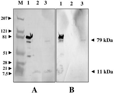
To confirm the expression of the fusion protein Cry1Ac1-D in cells infected by the V13 phage containing the cry1Ac1::gpD fusion, an immunoblot was performed. For this, lysates of the V13 and Vc phages were produced in E. coli LE392. Phage proteins present in the V13 and Vc lysates were analyzed by immunoblotting with anti-D (Fig. 5A) and anti-Cry1Ac1 (Fig. 5B) antibodies. As shown in Fig. 5, a 79-kDa protein band in V13 lysates (lanes 3) was detected with both anti-Cry1Ac1 and anti-D antibodies. This protein had the same size as the second band in lane 1, which contains the fusion protein expressed from the pSVC1-31A plasmid. The size of both these proteins corresponds to the fusion protein Cry1Ac1-D lacking approximately 29 N-terminal amino acids, a product normally generated during toxin activation by gut proteases. (The 81-kDa protein detected in lanes 1 is the nonactivated toxin.) The Cry1Ac1 protein was not detected in the Vc lysate (lanes 2).
FIG. 5.
Immunoblot detection of Cry1Ac1-D and Cry1Ac1-phage with anti-D (A) and anti-Cry1Ac1 (B) antibodies. Lane M: molecular size markers (in kilodaltons). Lanes 1: Cry1Ac1-D fusion protein expressed in E. coli BLR. Lanes 2: E. coli LE392 lysates obtained by infection with Vc phage. Lanes 3: E. coli LE392 lysates obtained by infection with V13 phage.
Protein bands migrating at 11 kDa were detected in lanes 2 and 3 in the anti-D immunoblot (Fig. 5A), corresponding to the wild-type D protein present in Vc and V13 heads.
Cry1Ac1 is displayed on the surface of λ phage.
To demonstrate that Cry1Ac1 toxin was displayed on the surface of the V13 phage, the technique termed biopanning (23) was used. This technique is based on the ability to capture phage physically with reagents that recognize peptides or proteins displayed on the phage surface. A plate coated overnight with 150 μl of a solution containing 0.5 mg of anti-Cry1Ac antibody per ml was incubated with V13 and Vc phage lysates under the conditions described in Materials and Methods. To determine if V13 phage was captured as a consequence of the binding between the displayed Cry1Ac1 and its antibody, 150 μl of an exponential culture of LE392 were placed in every well. After incubation, cells were removed and the number of PFU was determined. As shown in Table 2, the recovery of phage particles from the well incubated with V13 lysate was almost 300 times higher than the corresponding samples incubated with Vc lysate and those in noncoated wells. These results suggested that the expressed Cry1Ac1-D protein detected in Fig. 5 was assembled into the V13 head and displayed on the surface of the phage.
TABLE 2.
Recovery of bound phages in coated and noncoated platesa
| Phage | No. of bound phages (PFU)
|
|||
|---|---|---|---|---|
| Anti-CrylAc1 | BBMP | BBMP + GalNAc | Noncoated well | |
| V13 | 280 | 290 | 119 | 1 |
| Vc | 1 | 10 | 1 | 4 |
BBMP, brush border membrane proteins.
Displayed Cry1Ac1 is biologically functional.
To demonstrate the biological functionality of the displayed Cry1Ac1, several experiments were carried out. First we checked that the V13 phage was able to bind to receptor proteins found on the brush border of M. sexta larval midgut cells. For that, approximately 300 μg of total brush border membrane proteins prepared as described in Materials and Methods, were used in a biopanning experiment with phages V13 and Vc. As shown in Table 2, the number of bound V13 phage was approximately 30 times higher than those found in wells incubated with Vc phage and 300 times higher compared with noncoated wells.
To determine if this binding was specific between Cry1Ac1 protein and its receptors, a analogous biopanning experiment was carried out in the presence of the amino sugar N-acetylgalactosamine (GalNAc). GalNAc is known to bind to domain III of the Cry1Ac1 toxin (4, 7) and block binding to the aminopeptidase N receptor (15, 16, 21). As shown in Table 2, the addition of 250 mM GalNAc to the phage suspension reduced by almost threefold the binding between Cry1Ac1 phage and the brush border membrane proteins fixed on the microtiter plate.
These results suggested that the displayed Cry1Ac1 toxin is biologically functional and able to bind to its natural receptors present in insect midguts. Binding is partially blocked by GalNAc.
DISCUSSION
Previous attempts to display Cry toxins on filamentous phages in a fully functional form able to bind their natural receptors in vitro have not been successful (13, 20). In this work we have successfully displayed the Cry1Ac1 toxin on the surface of a λ phage in a form that can bind its receptor. One of the most critical issues in phage display with filamentous virus is that the fusion protein must necessarily be transported through the plasma membrane before assembly into viral particles. We selected bacteriophage λ for display because λ is assembled intracellularly prior to release of viral particles from the cell, thereby avoiding the need for secretion of the displayed protein across the bacterial membrane (34).
The 11.4-kDa D protein encoded by the gpD gene was selected for fusion with the Cry1Ac1 toxin. This protein appears to play a role in stabilizing the phage prohead as it fills with DNA (12, 35). Although D protein is necessary for phage stability, it is not essential for phage viability. Recent work with cryoelectron microscopy and image processing has shown that D protein is assembled as trimers on the capsid surface (42). The conditional requirement for D protein in viral assembly together with the accessibility of the protein on the phage surface make this protein the ideal choice for the display of proteins on the viral capsid. Previous reports show that N-terminal (22, 34) and C-terminal (22, 29) fusions to protein D have been successfully displayed.
One important reason for fusing the N terminus of the D protein to the C terminus of the Cry1Ac1 toxin in the display model prior to construction of a mutant library is that phages carrying mutations in the cry1Ac1 gene resulting in truncations or frameshifts will automatically be eliminated from the phage pool because they will not assemble into the λ head, thus reducing the background of undesirable proteins in the library.
In our fusion, a linker was inserted between the Cry1Ac1 and D proteins. It was hoped that this separation of the Cry toxin from the D protein would allow the individual folding of both proteins. Although it is claimed that polyglycine linkers maximize polypeptide conformational freedom, it has been shown that they do not result in optimal stability of the fusion protein (24). For this reason, a glycine-serine linker was selected for the fusion.
The Cry1Ac1-D fusion protein was successfully expressed in E. coli from the pSVC1-31A plasmid. The expressed fusion protein showed the expected size (81 kDa), was obtained in good quantity, and was stable under the usual manipulations of solubilization or protease treatment (activation). The Cry1Ac1 core plus the N-terminal end of the toxin expressed from pSVC7 showed similar behavior, although the size of the protein observed (65 kDa) was slightly smaller than expected (68 kDa).
Several results demonstrate the ability of the Cry1Ac1-D fusion protein to fold into a biologically active conformation. First, Cry1Ac1-D showed the same binding pattern as the wild-type toxin in ligand blot experiments. This shows that addition of the phage protein to the Cry toxin does not affect the latter's ability to recognize its aminopeptidase N receptor. Binding was observed not only with activated but also with nonactivated Cry1Ac1-D. Second, although the Cry1Ac1-D protein was 20 times less toxic than the wild type, it was lethal to M. sexta larvae in bioassay experiments. (This reduction in in vivo toxicity despite the normal receptor binding pattern of the displayed toxin is not unexpected. There is accumulated experimental evidence that formation of the lytic pore that kills the insect cells requires not only binding to the receptor and insertion of the Cry toxin into the membrane, but subsequent [or accompanying] toxin oligomerization. While receptor binding might reasonably be expected to be unaffected by attachment of even a relatively large fused protein, the same is unlikely to be true for the insertion and oligomerization steps.)
Both in vitro and in vivo data show that addition of the phage protein to the Cry1Ac1 toxin does not affect its biological function and the linker allows the correct folding of both proteins.
The results also demonstrate that Cry1Ac1 can be incorporated into viable phage particles. First, the fusion protein was detected in phage lysates. The size of the fusion protein detected by immunoblot in V13 lysates was lower than that expressed from the pSVC1-31A plasmid. The smaller size of the protein could be explained by proteolytic processing of the fusion protein by the proteases in the E. coli lysates. The missing fragment (2 kDa) might correspond to the N-terminal segment of the Cry1Ac1 toxin in the fusion, which is normally removed by gut proteases during activation of the protoxin.
Second, the biopanning results showed that the Cry toxin is successfully displayed on the lambda head and is accessible for interaction with the anti-Cry1Ac1 antibody. A 300-fold enrichment of the Cry phage (V13) was obtained compared with the control phage.
In order to check binding between V13 and the natural receptors present in the brush border membrane vesicles of M. sexta guts, a biopanning experiment with brush border membrane vesicles was performed. No differences in binding between V13 and Vc were obtained (data not shown). The high level of binding of Vc phage obtained in these experiments suggested that nonspecific binding between brush border membrane vesicles and phages might be taking place. We reasoned that this might be minimized with a partially purified preparation of brush border membrane vesicle proteins to coat the enzyme-linked immunosorbent assay plates. For this purpose we prepared a detergent-solubilized protein extract from brush border membranes. When this was used in biopanning experiments, V13 phage was found to be enriched 30-fold compared with Vc, confirming that the displayed Cry1Ac1 is able to recognize receptors present in the brush border membrane protein fraction from insect brush border membranes. Furthermore, we have shown that this binding is partially blocked by the presence of the amino sugar GalNAc on the receptor, since a threefold decrease in binding was observed when GalNAc was present in the assay.
To our knowledge, this is the first report which describes the binding of a phage-displayed Cry toxin to its natural receptors. This property of the display system is an essential requirement in further refinement of the system to select novel Cry toxins with desirable modifications from a mutant library.
Acknowledgments
We thank Kenneth Whelan and Johanna Rees for providing plasmid pKFW430 and insect gut extracts, respectively.
This work was funded by Bayer CropScience.
REFERENCES
- 1.Bell, R. A., and F. G. Joachim. 1976. Techniques for rearing laboratory colonies of tobacco hornworms and pink bollworms. Ann. Entomol. Soc. Am. 69:365-373. [Google Scholar]
- 2.Blattner, F. R., A. E. Blechl, K. Denniston-Thompson, H. E. Faber, J. E. Richards, J. L. Slightom, P. W. Tucker, and O. Smithies. 1978. Cloning human fetal gamma globin and mouse alpha-type globin DNA: preparation and screening of shotgun collections. Science 202:1279-1284. [DOI] [PubMed] [Google Scholar]
- 3.Bradford, M. M. 1976. A rapid and sensitive method for the quantitation of microgram quantities of protein utilizing the principle of protein-dye binding. Anal. Biochem. 72:248-254. [DOI] [PubMed] [Google Scholar]
- 4.Burton, S. L., D. J. Ellar, J. Li, and D. J. Derbyshire. 1999. N-acetylgalactosamine on the putative insect receptor aminopeptidase-N is recognised by a site on the domain III lectin-like fold of a Bacillus thuringiensis insecticidal toxin. J. Mol. Biol. 287:1011-1022. [DOI] [PubMed] [Google Scholar]
- 5.Carroll, J., and D. J. Ellar. 1993. An analysis of Bacillus thuringiensis δ-endotoxin action on insect midgut-membrane permeability using a light scattering assay. Eur. J. Biochem. 214:771-778. [DOI] [PubMed] [Google Scholar]
- 6.de Maagd, R. A., A. Bravo, and N. Crickmore. 2001. How Bacillus thuringiensis has evolved specific toxins to colonize the insect world. Trends Genet. 17:193-199. [DOI] [PubMed] [Google Scholar]
- 7.Derbyshire, D. J., D. J. Ellar, and J. Li. 2001. Crystallization of the Bacillus thuringiensis toxin Cry1Ac and its complex with the receptor ligand N-acetyl-D-galactosamine. Acta Crystallogr. Sect. D Biol. Crystallogr. 57:1938-1944. [DOI] [PubMed] [Google Scholar]
- 8.Ferré, J., and J. Van Rie. 2002. Biochemistry and genetics of insect resistance to Bacillus thuringiensis. Annu. Rev. Entomol. 47:501-533. [DOI] [PubMed] [Google Scholar]
- 9.Garczynski, S. F., J. V. Crim, and M. J. Adang. 1991. Identification of putative insect brush border membrane binding molecules specific to Bacillus thuringiensis δ-endotoxin by protein blot analysis. Appl. Environ. Microbiol. 57:2816-2820. [DOI] [PMC free article] [PubMed] [Google Scholar]
- 10.Gilliland, A., C. E. Chambers, E. J. Bone, and D. J. Ellar. 2002. Role of Bacillus thuringiensis Cry1 delta endotoxin binding in determining potency during Lepidopteran larval development. Appl. Environ. Microbiol. 68:1509-1515. [DOI] [PMC free article] [PubMed] [Google Scholar]
- 11.Gould, F. 1998. Sustainability of transgenic insecticidal cultivars: Integrating pest genetics and ecology. Annu. Rev. Entomol. 43:7017-7026. [DOI] [PubMed] [Google Scholar]
- 12.Imber, R., A. Tsugita, M. Wurtz, and T. Hohn. 1980. Outer surface protein of bacteriophage lambda. J. Mol. Biol. 139:277-295. [DOI] [PubMed] [Google Scholar]
- 13.Kasman, L. M., A. A. Lukowiak, S. F. Garczynski, R. J. McNall, P. Youngman, and M. J. Adang. 1998. Phage display of a biologically active Bacillus thuringiensis toxin. Appl. Environ. Microbiol. 64:2995-3003. [DOI] [PMC free article] [PubMed] [Google Scholar]
- 14.Knight, P. J. K., N. Crickmore, and D. J. Ellar. 1994. The receptor for Bacillus thuringiensis CrylA(c) delta-endotoxin in the brush border membrane of the lepidopteran Manduca sexta is aminopeptidase N. Mol. Microbiol. 11:429-436. [DOI] [PMC free article] [PubMed] [Google Scholar]
- 15.Knowles, B. H., P. J. Knight, and D. J. Ellar. 1991. N-acetyl galactosamine is part of the receptor in insect gut epithelia that recognises an insecticidal protein from Bacillus thuringiensis. Proc. R. Soc. Lond. B Biol. Sci. 245:31-35. [DOI] [PubMed] [Google Scholar]
- 16.Knowles, B. H., W. E. Thomas, and D. J. Ellar. 1984. Lectin-like binding of Bacillus thuringiensis var. kurstaki lepidopteran-specific toxin is an initial step in insecticidal action. FEBS Lett. 168:197-202. [DOI] [PubMed] [Google Scholar]
- 17.Knowles, B. H., and D. J. Ellar. 1987. Colloid-osmotic lysis is a general feature of the mechanism of action of Bacillus thuringiensis δ-endotoxins with different insect specificity. Biochim. Biophys. Acta 924:509-518. [Google Scholar]
- 18.Lee, M. K., J. L. Jenkins, T. H. You, A. Curtiss, J. J. Son, M. J. Adang, and D. H. Dean. 2001. Mutations at the arginine residues in α8 loop of Bacillus thuringiensis δ-endotoxin Cry1Ac affect toxicity and binding to Manduca sexta and Lymantria dispar aminopeptidase N. FEBS Lett. 497:108-112. [DOI] [PubMed] [Google Scholar]
- 19.Lee, M. K., T. H. You, F. L. Gould, and D. H. Dean. 1999. Identification of residues in Domain III of Bacillus thuringiensis Cry1Ac toxin that affect binding and toxicity. Appl. Environ. Microbiol. 65:4513-4520. [DOI] [PMC free article] [PubMed] [Google Scholar]
- 20.Marzari, R., P. Edomi, R. K. Bhatnagar, S. Ahmad, A. Selvapandiyan, and A. Bradbury. 1997. Phage display of Bacillus thuringiensis CryIA(a) insecticidal toxin. FEBS Lett. 411:27-31. [DOI] [PubMed] [Google Scholar]
- 21.Masson, L., Y. J. Lu, A. Mazza, R. Brousseau, and M. J. Adang. 1995. The Cry1A(c) receptor purified from Manduca sexta displays multiple specificities. J. Biol. Chem. 270:20309-20315. [DOI] [PubMed] [Google Scholar]
- 22.Mikawa, Y. G., I. N. Maruyama, and S. Brenner. 1996. Surface display of proteins on bacteriophage heads. J. Mol. Biol. 262:21-30. [DOI] [PubMed] [Google Scholar]
- 23.Parmley, S. E., and G. P. Smith. 1988. Antibody-selectable filamentous fd phage vectors: affinity purification of target genes. Gene 73:305-318. [DOI] [PubMed] [Google Scholar]
- 24.Robinson, C. R., and R. T. Sauer. 1998. Optimizing the stability of single-chain proteins by linker length and composition mutagenesis. Proc. Natl. Acad. Sci. USA 95:5929-5934. [DOI] [PMC free article] [PubMed] [Google Scholar]
- 25.Sambrook, J., E. F. Fritsch, and T. Maniatis. 1998. Molecular cloning: a laboratory manual, 2nd ed. Cold Spring Harbor Laboratory Press, Cold Spring Harbor, N.Y.
- 26.Sangadala, S., P. Azadi, R. Carlson, M. J. Adang. 2001. Carbohydrate analyses of Manduca sexta aminopeptidase N, co-purifying neutral lipids and their functional interactions with Bacillus thuringiensis Cry1Ac toxin. Insect Biochem. Mol. Biol. 32:97-107. [DOI] [PubMed] [Google Scholar]
- 27.Sangadala, S., F. W. Walters, L. H. English, and M. J. Adang. 1994. A mixture of Manduca sexta aminopeptidase and phosphatase enhances Bacillus thuringiensis insecticidal CryIA(c) toxin binding and 86Rb+-K+ efflux in vitro. J. Biol. Chem. 269:10088-10092. [PubMed] [Google Scholar]
- 28.Santi, E., S. Capone, C. Mennuni, A. Lahm, A. Tramontano, A. Luzzago, and A. Nicosia. 2000. Bacteriophage lambda display of complex cDNA libraries: A new approach to functional genomics. J. Mol. Biol. 296:497-508. [DOI] [PubMed] [Google Scholar]
- 29.Santini, C., D. Brennan, C. Mennuni, R. H. Hoess, A. Nicia, R. Cortese, and A. Luzzago. 1998. Efficient display of an HCV cDNA expression library as C-terminal fusion to the capsid protein D of bacteriophage lambda. J. Mol. Biol. 282:125-135. [DOI] [PubMed] [Google Scholar]
- 30.Santos, M. A. 1991. An improved method for small scale preparation of bacteriophage DNA based on phage precipitation by zinc chloride. Nucleic Acids Res. 19:5442. [DOI] [PMC free article] [PubMed] [Google Scholar]
- 31.Schnepf, E., N. Crickmore, J. Van Rie, D. Lereclus, J. Baum, J. Feitelson, D. R. Zeigler, and D. H. Dean. 1998. Bacillus thuringiensis and its pesticidal crystal proteins. Microbiol. Mol. Biol. Rev. 62:775-806. [DOI] [PMC free article] [PubMed] [Google Scholar]
- 32.Slatin, S. L., C. K. Abrams, and L. English. 1990. Delta-endotoxins form cation-selective channels in planar lipid bilayers. Biochem. Biophys. Res. Commun. 169:765-772. [DOI] [PubMed] [Google Scholar]
- 33.Smedley, D. P., and D. J. Ellar. 1996. Mutagenesis of three surface-exposed loops of a Bacillus thuringiensis insecticidal toxin reveals residues important for toxicity, receptor recognition and possibly membrane insertion. Microbiology 142:1617-1624. [DOI] [PubMed] [Google Scholar]
- 34.Sternberg, N., and H. Hoess. 1995. Display of peptides and proteins on the surface of bacteriophage λ. Proc. Natl. Acad. Sci. USA 92:1609-1613. [DOI] [PMC free article] [PubMed] [Google Scholar]
- 35.Sternberg, N., and R. Weisberg. 1977. Packaging of coliphage lambda DNA. II. The role of the gene D protein. J. Mol. Biol. 117:733-759. [DOI] [PubMed] [Google Scholar]
- 36.Tabashnik, B. E., K. W. Johnson, J. T. Engleman, and J. A. Baum. 2000. Cross-resistance to Bacillus thuringiensis Cry1Ja in a strain of diamondback moth adapted to artificial diet. J. Invertebr. Pathol. 76:81-83. [DOI] [PubMed] [Google Scholar]
- 37.Towbin, H., T. Staehelin, and J. Gordon. 1979. Electrophoretic transfer of proteins from polyacrylamide gels to nitrocellulose sheets: procedure and some applications. Proc. Natl. Acad. Sci. USA 76:4350-4354. [DOI] [PMC free article] [PubMed] [Google Scholar]
- 38.Vachon, V. M. J. Paradis, M. Marsolais, J. L. Schwartz, and R. Laprade. 1995. Ionic permeabilities induced by Bacillus thuringiensis in Sf9 cells. J. Membr. Biol. 148:57-63. [DOI] [PubMed] [Google Scholar]
- 39.Vadlamudi, R. K., T. H. Ji, and L. A. Bulla, Jr. 1993. A specific binding protein from Manduca sexta for the insecticidal toxin of Bacillus thuringiensis subsp. berliner. J. Biol. Chem. 268:12334-12340. [PubMed] [Google Scholar]
- 40.Vadlamudi, R. K., E. Weber, I. Ji, T. H. Ji, and L. A. Bulla, Jr. 1995. Cloning and expression of a receptor for an insecticidal toxin of Bacillus thuringiensis. J. Biol. Chem. 270:5490-5494. [DOI] [PubMed] [Google Scholar]
- 41.Van Rie, J., S. Jansens, H. Hofte, D. Degheele, and H. Van Mellaert. 1990. Receptors on the brush border membrane of the insect midgut as determinants of the specificity of Bacillus thuringiensis delta-endotoxins. Appl. Environ. Microbiol. 56:1378-1385. [DOI] [PMC free article] [PubMed] [Google Scholar]
- 42.Yang, F., P. Forrer, Z. Dauter, J. F. Conway, N. Cheng, M. E. Cerritelli, A. C. Steven, A. Plückthun, and A. Wlodawer. 2000. Novel fold and capsid-binding properties of the λ-phage display platform protein gpD. Nat. Struct. Biol. 7:230-237. [DOI] [PubMed] [Google Scholar]


