Abstract
Conventional white light endoscopy remains the current standard in routine clinical practice for early detection of gastric cancer. However, it may not accurately diagnose preneoplastic gastric lesions. The technological advancements in the field of endoscopic imaging for gastric lesions are fast growing. This article reviews currently available advanced endoscopic imaging modalities, in particular chromoendoscopy, narrow band imaging and confocal laser endomicroscopy, and their corresponding evidence shown to improve diagnosis of preneoplastic gastric lesions. Raman spectrometry and polarimetry are also introduced as promising emerging technologies.
Keywords: ENDOSCOPY, LASER, GASTROINTESTINAL NEOPLASIA
Introduction
Gastric cancer is the third leading cause of cancer deaths worldwide.1 2 To decrease mortality, early detection and accurate diagnosis of gastric cancer through endoscopy is critical. In recent years, despite numerous advancements in endoscopic imaging, conventional white light endoscopy (WLE) remains the fundamental step for detection and characterisation of gastric lesions in clinical practice. However, conventional WLE may not accurately diagnose preneoplastic gastric lesions. Hence, the European Society of Gastrointestinal Endoscopy (ESGE)3 has recommended the use of image-enhanced endoscopy, including magnification chromoendoscopy and narrow band imaging to improve the diagnosis of gastric preneoplastic conditions. This review aims to highlight the available image-enhancing endoscopic modalities to aid diagnosis of gastric adenocarcinoma and preneoplastic gastric lesions, which include gastric intestinal metaplasia and dysplasia.
White light endoscopy (WLE)
Atkins and Benedict4 first highlighted poor correlation between WLE findings and histopathological analysis. Their conclusion was further substantiated by subsequent studies3 demonstrating that conventional WLE cannot reliably diagnosis Helicobacter pylori gastritis5 6 or intestinal metaplasia.7
However, recent evidence for WLE to identify gastric preneoplastic lesions remain promising: Panteris et al8 demonstrated that real-time high-definition WLE (HD-WLE), during routine endoscopy practice, had an accuracy of 88% in identifying intestinal metaplasia; with sensitivity of 74.6%, specificity of 94.2%, positive predictive value of 82.5% and negative predictive value of 90.1%. In their study, all clinically significant type III intestinal metaplasia and dysplastic lesions were successfully detected using HD-WLE, whereas gastric lesions not picked up by HD-WLE were either mild grade or with no dysplasia. Furthermore, Xirouchakis et al9 also demonstrated that the use of WLE with adherence to the updated Sydney biopsy protocol (WLE-USP) had greater accuracy compared with narrow band imaging-targeted biopsies (NBI-TB) in detecting premalignant lesions; whereby accuracy for a diagnosis of atrophy and intestinal metaplasia were 93% and 90% for WLE-USP as compared with 82% and 80% for NBI-TB.
Clinical studies regarding the application of WLE for the diagnosis of gastric cancer are few; a meta-analysis of four studies comprising 433 patients and 527 lesions had a pooled sensitivity of 0.48 and sensitivity of 0.67.10 The yield of WLE to diagnose small (<10 mm) depressed gastric mucosal cancer is limited, with one multicentre trial of 176 patients demonstrating only a sensitivity of 40% and specificity of 67.9%.11 However, WLE still remains a valid screening tool for gastric cancer, whereby a larger prospective multicentre study involving 579 patients, showed no statistical difference in gastric cancer detection rates between high-definition WLE (7/286, 2.4%) and NBI (3/293, 1%).12
Conventional chromoendoscopy (CE)
Conventional CE incorporates topical application of dyes during WLE. An externally validated classification using CE was previously proposed by Dinis-Riberio et al,13 14 whereby CE with methylene blue, particularly with magnification, improves identification of gastric lesions. CE with other dyes,15–17 such as indigo carmine, acetic acid and haematoxylin, have also been shown to accurately differentiate between normal gastric mucosa and dysplastic or malignant gastric lesions.
Zhao et al18's meta-analysis of seven prospective studies, comprising a total of 429 patients and 465 lesions, showed that CE improves the detection of early gastric cancer (p<0.01) and preneoplastic gastric lesions (p<0.01) compared with standard WLE. The pooled sensitivity, specificity and area under curve (AUC) of CE were 0.90 (95% CI 0.87 to 0.92), 0.82 (95% CI 0.79 to 0.86) and 0.95, respectively. Majority of the studies used indigo carmine (72.4%) instead of methylene blue (28.6%). However, the meta-analysis did not include a subanalysis comparing the application of different staining dyes. It is postulated indigo carmine is more frequently used, because indigo carmine is a non-absorptive contrast stain, whereas methylene blue has shown to be absorbed by oesophageal cells19 and colonocytes,20 thereby inducing cellular DNA damage on exposure to white light.
Autofluorescence imaging (AFI)
AFI is an imaging method, applied during real-time endoscopy, dependent on the fluorescent properties of gastric mucosal particles, such as collagen, flavins and porphyrins, to differentiate between various gastric mucosa subtypes.
The diagnostic utility of AFI in the stomach is confounded by inconsistent autofluorescence patterns and conflicting results in different studies. AFI, compared with WLE, was found to be less sensitive (64% vs 74%, p=0.79) and specific (49% vs 83%, p=0.05), in detecting gastric neoplastic lesions.21 Using per-lesion analysis, AFI is also associated with high false positive rate for gastric lesions, with a specificity of 24% as compared with WLE (84%).22
On the other hand, Kato et al21 23 demonstrated an increase in gastric neoplasia identification from 18% (WLE) to 56% (WLE+AFI) by adding AFI to WLE during routine endoscopy. More subjects with intestinal metaplasia were also identified using AFI-NBI compared with WLE;23 whereby 68% with intestinal metaplasia (26/38) were correctly identified by AFI-NBI (sensitivity 68%, specificity 23%) compared with 34% (13/38) by WLE (sensitivity 34%, specificity 65%). Imaeda et al24 further demonstrated utility of AFI for detecting additional gastric lesions in patients with superficial gastric neoplasia postendoscopic submucosal dissection, whereby five gastric neoplasia missed by WLE were all detected by AFI.
Owing to the conflicting data, the role of AFI in detecting and diagnosing gastric neoplasia and preneoplasia is unclear, and there is currently inadequate evidence to support its routine use in clinical practice.
Computed virtual chromoendoscopy
Application of CE requires additional procedure time and materials. Hence, various sources have introduced numerous user-friendly computed virtual chromoendoscopy techniques to improve visualisation of gastric mucosa tissue and vasculature, without the hassle of additional endoscopic probes or dyes. Such technologies include flexible spectral imaging colour enhancement (FICE) (Fujinon, Fujifilm Medical Co, Saitama, Japan), i-SCAN (PENTAX Endoscopy, Tokyo, Japan) and narrow band imaging (NBI) (Olympus Medical Systems Tokyo, Japan).
Flexible spectral imaging colour enhancement (FICE)
Kikuste et al25 demonstrated good specificity (87%; 95% CI 79% to 95%) and diagnostic accuracy of 74% (95% CI 0.66% to 0.82%) using FICE endoscopy to detect gastric intestinal metaplasia among 126 patients aged over 50 years. In a study by Pittayanon et al,26 magnified FICE demonstrated an overall 85.5% accuracy in detecting intestinal metaplasia among 59 lesions in 45 patients, and the accuracy was higher with findings of light blue crest (95.2%) and long large crest (96.8%) and lower with findings of villous pattern (84.3%).
In a meta-analysis by Kikuste et al27 seven studies were included for analysis; however, due to the insufficient data and varying definitions of neoplastic and preneoplastic definitions for FICE, there were no aggregate scores calculated. However, FICE did produce images with more pronounced colour contrast between malignant lesions and benign surrounding mucosa, compared with standard WLE findings, particularly in Osawa et al's28 study involving 82 patients with depressed early gastric carcinoma.
i-Scan
Qi et al29 compared magnification i-Scan and magnification WLE in identifying type 2–3 gastric mucosal pit pattern, predictive of Helicobacter infection, whereby diagnosis was then confirmed by positive rapid urease test and positive histopathology. i-Scan provided better image quality with increased recognition of type 2–3 patterns, compared with magnification WLE, and thus increased accuracy of Helicobacter infection diagnosis (accuracy 94% vs 84.5%, sensitivity 95.5% vs 95.5%, specificity 93.5% vs 80.6%, positive predictive value (PPV) 84% vs 63.6%, negative predictive value (NPV) 98.3% vs 98.0%).
A prospective study30 of 43 patients demonstrated modest results, whereby magnified i-Scan (with tone and surface enhancement) had a relative low specificity and PPV in diagnosing early gastric neoplasia (sensitivity: 100%; specificity: 77.1%; PPV: 50%; NPV: 100%; likelihood ratio: 4.37). These findings were mirrored by Nishimura et al,31 who looked at 10 patients with gastric neoplasia, and found no significant difference (p=0.78) between the diagnostic accuracy using either WLE (91.7%) or i-Scan (90.8%). Interestingly, in this study, the diagnostic accuracy of tumour size using i-Scan was comparable between novice and experienced endoscopists (65.7% vs 71.1%, p=0.53), whereas it was significantly lower for WLE (41.2% vs 79.5%, p=0.019), which may suggest that i-Scan technology will improve diagnostic accuracy of novice endoscopists.
Owing to the limited data on FICE and i-Scan in the diagnosis of gastric preneoplasia and neoplasia and the absence of well-validated definition for gastric preneoplastic and neoplastic mucosal appearances, there is currently insufficient evidence to substantiate application of FICE and i-Scan in routine clinical practice for the diagnosis of gastric neoplasia and preneoplasia.
Narrow band imaging (NBI)
NBI, unlikely FICE or i-Scan, is accomplished by optical filtering of white light, and is the most well-published virtual CE modality. It has been found to have good sensitivity and specificity for gastric neoplasia and premalignant gastric lesions. Experienced endoscopists in the Asia Pacific region have recommended magnifying NBI over NBI alone for the detection of gastric cancer.32
NBI has established definitions of gastric neoplastic mucosal patterns: Muto et al33 proposed the Magnifying Endoscopy Simple Diagnostic Algorithm for early gastric cancer (MESDA-G) which focuses on the presence of (1) a demarcation line and (2) irregular microvascular pattern and/or irregular micro-surface pattern for the diagnosis of high-grade dysplasia or early gastric cancer. This approach was built on the vessel plus surface NBI classification previously developed by Yao et al.34 Furthermore, a prospective cohort of 156 patients with 188 target lesions, has identified the presence of a whole globe appearance to be highly specific (97.5%) and useful to diagnose gastric cancer.35
A meta-analysis36 of 14 studies comprising 2171 patients, demonstrated a pool sensitivity, specificity and AUC for magnified NBI diagnosis of early gastric cancer to be 0.86 (95% CI 0.83 to 0.89), 0.96 (95% CI 0.95 to 0.97) and 0.9623, respectively. Gastric lesions of depressed macroscopic type and those with a diameter <10 mm had lower sensitivities of 0.64 (95% CI 0.52 to 0.75) and 0.74 (95% CI 0.65 to 0.82), respectively. These results were mirrored in another meta-analysis by Zhang et al,10 which included 10 studies comprising 1724 patients and 2153 lesions, whereby magnified NBI for diagnosis of early gastric cancer had a pooled sensitivity, specificity and AUC of 0.83 (95% CI 0.79 to 0.87; I2=79.8%), 0.96 (95% CI 0.95 to 0.97; I2=89.3%) and 0.96 (0.93–0.98), respectively.
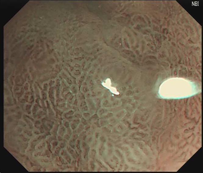
NBI also has utility in identifying gastric preneoplastic lesions. Uedo et al37 reported that the presence of a light blue crest (see figure 1), seen with magnifying NBI, had a sensitivity of 89%, specificity 93% and accuracy of 91% in diagnosing intestinal metaplasia. NBI is generally used for lesion characterisation and not enhanced detection of gastric lesions. However, Ang et al12 have demonstrated, in a multicentre prospective randomised study involving 579 patients, that NBI detected more focal gastric lesions as compared with high-definition WLE (40.6% vs 29%, p=0.003), as well as a greater proportion of intestinal metaplasia (17.7% vs 7.7%, p<0.001). On a similar note, Tongtawee et al's38 study of 200 patients demonstrated that NBI also reliably identifies gastric mucosal morphological patterns associated with H. pylori infection, which correlated significantly with severity of pathological inflammation grading. A subsequent randomised study39 on 500 subjects noted site-specific biopsy guided by NBI had higher yield of diagnosing Helicobacter, as compared with standard WLE biopsy. Findings by Ang and Tongtawee suggest that NBI would be useful to identify sites for high-yield targeted biopsies.
Figure 1.

Light blue crest sign indicating gastric intestinal metaplasia on magnifying NBI. NBI, narrow band imaging.
Blue light imaging (BLI)
Of worthy mention, BLI is a new computer chromoendoscopy method developed by Fujinon, Fujifilm Medical Company, which uses two lasers; one at wavelength 450 nm, to irradiate white colour illumination, and the other at 410 nm, to enhance surface microvasculature patterns.40 Although there are few prospective studies evaluating the utility of BLI in gastric cancer screening, BLI has been shown to provide excellent endoscopic images to better characterise gastric lesions. The BLI-bright mode is the main highlight of this new technology, which allows for vascular contrast detection from a far view, thereby enabling its application in screening endoscopy; Kaneko et al41 demonstrated that BLI-bright mode compared with NBI had appreciable vascular-mucosal contrast from a significantly further distance (26.8 mm vs 16.4 mm, p=0.03), among 14 patients with gastric adenoma. Of note, six of 14 patients with gastric adenoma were identified with BLI at observed distance of 40 mm compared with only one of 14 patients with NBI. Dohi et al42 further validated the utility of magnified BLI in a prospective study of 132 patients; the 127 gastric lesions (of which 34 were histologically proven gastric adenoma) were first identified by WLE, and further characterised using magnified BLI, whereby magnified BLI, compared with WLE, had greater accuracy (92.1% vs 71.7%) in identifying gastric adenoma with sensitivity 93.8%, specificity 91.6%, positive predictive value 78.9% and negative predictive value 97.7%.
Confocal laser endomicroscopy (CLE)
Confocal laser endomicroscopy (CLE) produces high-magnification histological-like images through measuring the gastric tissue reflectance as a low-power laser is used during endoscopy for illumination. CLE may be applied during real-time endoscopy through either using additional probes or dedicated endoscopes with integrated CLE systems.
A recent meta-analysis43 performed on CLE and gastric diseases, focused on three domains: (1) detection and diagnosis of polyps and neoplastic lesions (three studies, sensitivity 85% (95% CI 78% to 91%, I2=52.3%), specificity 99% (95% CI 98% to 99%, I2=92.92%), AUC=0.929), (2) gastritis and gastric metaplasia (six studies, sensitivity 94% (95% CI 92% to 96%, I2=54.8%), specificity 95% (CI 92% to 97%, I2=55.6%), AUC=0.983) and (3) H.pylori-related gastritis (two studies, sensitivity 86% (95% CI 76% to 93%, I2=0), specificity 93% (95% CI 87% to 97%, I2=2.6%)). The pooled analyses demonstrated CLE yields remarkable sensitivity and specificity, in per patient analysis in all three domains. However, the data were based on a limited number of publications.
CLE also allows for targeted biopsies of specific pathological mucosal area, whereby Li et al44 demonstrated that CLE with targeted biopsies is superior to WLE for the detection and surveillance of gastric intestinal metaplasia. The number of biopsies needed to confirm gastric intestinal metaplasia is a third of that needed with WLE. Furthermore, CLE-guided biopsy boasts a higher diagnostic yield for gastric intestinal metaplasia (65.7%), compared with WLE (15.73%; p<0.001).
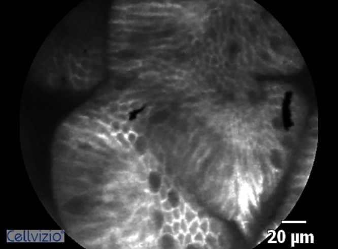
In our local experience,45 we compared the diagnostic performance of WLE, AFI, magnified NBI and CLE for the diagnosis of gastric intestinal metaplasia in 20 patients, examining a total of 125 sites. We found that real-time CLE (88% 95% CI 89.8% to 98.2%) had better accuracy compared with conventional WLE (64.8%, 95% CI 55.7% to 73.1%), AFI (68.8%, 95% CI 59.9% to 76.8%) or magnified NBI (84.8%, 95% CI 81% to93.1%). Off-site review of CLE images further significantly increased the accuracy (95.2%, 95% CI 89.8% to 98.2%, p=0.012) of gastric intestinal metaplasia diagnosis (figure 2).
Figure 2.

Gastric intestinal metaplasia as seen on confocal laser endomicroscopy.
Experience and training greatly affect CLE interpretation.46 We reported that interpretation of CLE images of gastric intestinal metaplasia by experienced endoscopists was associated with higher sensitivity (95.2% vs 61.9%, p=0.039) and higher specificity (93.3% vs 62.2%, p<0.001) than interpretation by inexperienced endoscopists. The agreement between interpretation by experienced endoscopists and histology for gastric intestinal metaplasia was also higher than that for inexperienced endoscopists (K=0.864 vs 0.217). Pittayanon et al47 demonstrated that trainees with no experience in intestinal metaplasia interpretation could achieve high reading accuracy and substantial interobserver agreement after training with 10 sets of CLE video clips. Trainees on achieving high reading accuracy were shown to maintain their high quality of reading skill. Similar findings were noted by Kobayashi et al48 for CLE diagnosis of gastric cancer, whereby correct WLE interpretation and adequate training in WLE was identified to be the most important factor in achieving highly accurate diagnoses of gastric cancer using CLE.
Emerging technologies
Endocytoscopy
Endocytoscopy (ECS) is an ultrahigh magnification technology to enable histopathological diagnosis of the gastrointestinal tract, whereby the magnification lens of a conventional endoscope is gradually increased to visualise the gastric mucosal after an application of methylene blue and crystal violet staining. ECS has been shown to have high diagnostic accuracy (sensitivity 86% specificity 100%) for gastric cancer. However, in this study, 10 of the 82 lesions investigated could not be clearly observed by ECS due to poor staining due to viscous mucus or plaque.49 Sato et al50 further demonstrated that ECS could differentiate between normal antrum mucosa and preneoplastic changes, whereby they were able to identify four different mucosal patterns in the antrum in a small single-centre pilot study consisting of 64 patients, where the positive predictive values for associating type 1 pattern for normal mucosa and type 4 pattern for gastric intestinal metaplasia were both 100%.
Raman spectroscopy
Raman spectroscopy measures inelastic light scattering processes within the gastric mucosa, whereby Teh et al51 was able to differentiate between normal mucosa (sensitivity 80%, specificity 92.7%) and H. pylori infection (sensitivity 91.7%, specificity 80%), as well as between normal mucosa and intestinal metaplasia (sensitivity 80%, specificity 100%). Bergholt et al52 subsequently demonstrated further promise in Raman spectrometry to document gastric carcinogenesis in vivo through accurately identifying areas of gastric dysplasia (sensitivity 83.33%, specificity 95.8%) and adenocarcinoma (sensitivity 84.91%, specificity 95.57%). Raman spectrometry could also identify different subtypes of gastric adenocarcinoma, as first reported by Teh et al53 and reproduced by Kawabata et al.54 Another clinical utility of Raman endoscopy is the diagnosis of benign and malignant gastric ulcers.55
Polarimetry
Polarimetry measures polarisation properties of light beams within the gastric mucosa, whereby it has been proposed to differentiate between normal, dysplasia and cancer. Wang et al56 first demonstrated differences in polarisation parameters, at low magnification, between normal and cancer gastric samples obtained from 20 patients; whereby a combination of polarisation parameters (linear depolarisation and linear retardance) had up to 95% specificity and 95% sensitivity.
Subsequent Wang et al57 demonstrated the potential of polarimetry imaging for gastric tissue diagnosis at the microscopic level, whereby polarimetry was used to diagnose between normal gastric tissue, intestinal metaplasia, dysplasia and cancer, in a total of 84 samples for 59 patients. The accuracy of classification in digital staining using polarimetry imaging on the microscopic level varied from 56–75% for the discrimination of cancer, dysplasia and intestinal metaplasia or normal tissue. With further advances in polarimetry imaging, polarimetry is a promising technique with potential application for endoscopic imaging, such to allow for real-time diagnosis during endoscopy.
Conclusions
Early detection of gastric preneoplastic lesions allows early treatment and leads to better prognosis. Table 1 provides a summary of the enhanced imaging modalities discussed in this review. Building on Muto et al's MESDA-G algorithm, we recommend the use of WLE, in particular HD-WLE, for the detection of suspicious gastric lesions. The demarcation line and subsequent microvascular and microsurface pattern should then be characterised using magnifying NBI or chromoendoscopy. These enhanced imaged modalities will also direct targeted biopsies. The technological advancements in field of endoscopic imaging for gastric lesions are fast growing. Recognition of the various endoscopic appearances of gastric preneoplastic and neoplastic lesions remains the fundamental first step which allows for subsequent early detection and more accurate characterisation, underlining the importance of training.
Table 1.
Summary table of advanced endoscopic imaging modalities
| Methodology | Manufacturers | Clinical performance | Comments | |
|---|---|---|---|---|
| Autofluorescence imaging (AFI) | Based on detection of natural tissue fluorescence emitted by endogenous molecules, to differentiate between normal non-dysplastic tissue (green) and dysplastic tissue (purple) | Olympus Medical Systems; Evis Exera II/III processors | Few studies to demonstrate increase diagnostic yield of gastric cancer and gastric intestinal metaplasia | Inadequate clinical evidence to support routine use of AFI in clinical practice |
| Fuji intelligent chromo endoscopy (FICE) | Video processor synthesises three single-wavelength images to display a composite colour-enhanced image in real-time. 10 preset settings available | Fujinon; Series 500 gastroscopes; VP-4450HD and VP-4400HD processors | No standardised definitions of gastric preneoplastic and neoplastic lesions. Single meta-analysis of seven studies showing more colour contrast compared with WLE alone for gastric cancer and gastric intestinal metaplasia | More clinical evidence required before routine application in clinical practice for gastric lesions |
| i-Scan | Post imaging video processor to provide enhanced images of mucosal surface and blood vessels. three presets mode using a combination of (1) surface enhancement, (2) contrast enhancement and (3) tone enhancement modes | PENTAX Endoscopy; Series I and 90K gastroscopies, EPK-i5010 processor | No standardised definitions of gastric preneoplastic and neoplastic lesions. Few studies to demonstrate better image quality and recognition of gastric cancer and gastric intestinal metaplasia | More clinical evidence required before routine application in clinical practice for gastric lesions |
| Narrow band imaging (NBI) | Placement of NBI filter produces two bands of lights to differentiate between capillaries in the superficial mucosa (brown, 415 nm) and deeper veins in the mucosa/submucosa (cyan, 540 nm) | Olympus Medical Systems; Evis Exera II/III processors | Established NBI classification for gastric lesions and neoplasia. Abundant studies and meta-analysis data available to demonstrate accurate characterisation and diagnostic yield of gastric intestinal metaplasia and gastric cancer | Recommended by ESGE to better improve diagnostic yield of gastric cancer and gastric preneoplastic lesions |
| Blue light imaging (BLI) | Two lasers and a while light phosphor for illumination, with one laser at 450 nm for white light illumination, and another laser at 410 nm to mucosal blood vessels | Fujinon; VP-4450HD processor | Established NBI classification for gastric lesions and neoplasia may be applied. Has two modes (1) contrast mode for magnifying images, (2) bright mode for far view surveillance. Excellent endoscopic images, but few prospective studies | More clinical evidence required before routine application in clinical practice for gastric lesions |
| Confocal laser endomicroscopy (CLE) | Tissue illumination with a low-power laser with subsequent detection of fluorescence of light reflectance |
|
Promising results in improving diagnostic yield of gastric cancer, intestinal metaplasia and Helicobacterpylori-related gastritis. Potentiate the need for fewer biopsies to confirm presence of gastric intestinal metaplasia | Adequate training required for accurate CLE diagnosis |
| Endocytoscopy (ECS) | Fixed-focus, high-power objective lens that project highly magnified images | Olympus Medical Systems;
|
Data from limited number of studies available, but promising results in diagnosing cancer and intestinal metaplasia accurately | More clinical evidence required before routine application in clinical practice for gastric lesions |
Footnotes
Contributors: JWJL was charged with acquisition, analysis and interpretation of the literature review, thereby drafting the work. LGL and KGY revised it critically, and have the final approval for publication.
Competing interests: None declared.
Patient consent: Obtained.
Provenance and peer review: Not commissioned; externally peer reviewed.
Data sharing statement: No additional data are available.
References
- 1.Fock KM. Review article: the epidemiology and prevention of gastric cancer. Aliment Pharmacol Ther 2014;40:250–60. doi:10.1111/apt.12814 [DOI] [PubMed] [Google Scholar]
- 2.de Martel C, Ferlay J, Franceschi S, et al. Global burden of cancers attributable to infections in 2008: a review and synthetic analysis. Lancet Oncol 2012;13:607–15. doi:10.1016/S1470-2045(12)70137-7 [DOI] [PubMed] [Google Scholar]
- 3.Dinis-Ribeiro M, Areia M, de Vries AC, et al. Management of precancerous conditions and lesions in the stomach (MAPS): guideline from the European Society of Gastrointestinal Endoscopy (ESGE), European Helicobacter Study Group (EHSG), European Society of Pathology (ESP), and the Sociedade Portuguesa de Endoscopia Digestiva (SPED). Endoscopy 2012;44:74–94. doi:10.1055/s-0031-1291491 [DOI] [PMC free article] [PubMed] [Google Scholar]
- 4.Atkins L, Benedict EB. Correlation of gross gastroscopic findings with gastroscopic biopsy in gastritis. N Engl J Med 1956;254:641–4. doi:10.1056/NEJM195604052541403 [DOI] [PubMed] [Google Scholar]
- 5.Kato T, Yagi N, Kamada T, et al. Diagnosis of Helicobacter pylori infection in gastric mucosa by endoscopic features: a multicenter prospective study. Dig Endosc 2013;25:508–18. doi:10.1111/den.12031 [DOI] [PubMed] [Google Scholar]
- 6.Cho JH, Chang YW, Jang JY, et al. Close observation of gastric mucosal pattern by standard endoscopy can predict Helicobacter pylori infection status. J Gastroenterol Hepatol 2013;28:279–84. doi:10.1111/jgh.12046 [DOI] [PubMed] [Google Scholar]
- 7.Stathopoulos G, Goldberg RD, Blackstone MO. Endoscopic diagnosis of intestinal metaplasia. Gastrointest Endosc 1990;36:544–5. doi:10.1016/S0016-5107(90)71151-0 [DOI] [PubMed] [Google Scholar]
- 8.Panteris V, Nikolopoulou S, Lountou A, et al. Diagnostic capabilities of high-definition white light endoscopy for the diagnosis of gastric intestinal metaplasia and correlation with histologic and clinical data. Eur J Gastroenterol Hepatol 2014;26:594–601. doi:10.1097/MEG.0000000000000097 [DOI] [PubMed] [Google Scholar]
- 9.Xirouchakis E, Laoudi F, Tsartsali L, et al. Screening for gastric premalignant lesions with narrow band imaging, white light and updated Sydney protocol or both? Dig Dis Sci 2013;58:1084–90. doi:10.1007/s10620-012-2431-x [DOI] [PubMed] [Google Scholar]
- 10.Zhang Q, Wang F, Chen ZY, et al. Comparison of the diagnostic efficacy of white light endoscopy and magnifying endoscopy with narrow band imaging for early gastric cancer: a meta-analysis. Gastric Cancer 2016;19:543–52. doi:10.1007/s10120-015-0500-5 [DOI] [PubMed] [Google Scholar]
- 11.Ezoe Y, Muto M, Uedo N, et al. Magnifying narrowband imaging is more accurate than conventional white-light imaging in diagnosis of gastric mucosal cancer. Gastroenterology 2011;141:2017–25.e3. doi:10.1053/j.gastro.2011.08.007 [DOI] [PubMed] [Google Scholar]
- 12.Ang TL, Pittayanon R, Lau JY, et al. A multicenter randomized comparison between high-definition white light endoscopy and narrow band imaging for detection of gastric lesions. Eur J Gastroenterol Hepatol 2015;27:1473–8. doi:10.1097/MEG.0000000000000478 [DOI] [PubMed] [Google Scholar]
- 13.Dinis-Ribeiro M, da Costa-Pereira A, Lopes C, et al. Magnification chromoendoscopy for the diagnosis of gastric intestinal metaplasia and dysplasia. Gastrointest Endosc 2003;57:498–504. doi:10.1067/mge.2003.145 [DOI] [PubMed] [Google Scholar]
- 14.Areia M, Amaro P, Dinis-Ribeiro M, et al. Estimation of the extent of gastric intestinal metaplasia by methylene blue chromoendoscopy. Eur J Gastroenterol Hepatol 2008;20:939–40. doi:10.1097/MEG.0b013e3282f376ac [DOI] [PubMed] [Google Scholar]
- 15.Tanaka K, Toyoda H, Kadowaki S, et al. Surface pattern classification by enhanced-magnification endoscopy for identifying early gastric cancers. Gastrointest Endosc 2008;67:430–7. doi:10.1016/j.gie.2007.10.042 [DOI] [PubMed] [Google Scholar]
- 16.Kono Y, Takenaka R, Kawahara Y, et al. Chromoendoscopy of gastric adenoma using an acetic acid indigocarmine mixture. World J Gastroenterol 2014;20:5092–7. doi:10.3748/wjg.v20.i17.5092 [DOI] [PMC free article] [PubMed] [Google Scholar]
- 17.Mouzyka S, Fedoseeva A. Chromoendoscopy with hematoxylin in the classification of gastric lesions. Gastric Cancer 2008;11:15–21; discussion 21–2 doi:10.1007/s10120-007-0445-4 [DOI] [PubMed] [Google Scholar]
- 18.Zhao Z, Yin Z, Wang S, et al. Meta-analysis: the diagnostic efficacy of chromoendoscopy for early gastric cancer and premalignant gastric lesions. J Gastroenterol Hepatol 2016;31:1539–45. doi:10.1111/jgh.13313 [DOI] [PubMed] [Google Scholar]
- 19.Olliver JR, Wild CP, Sahay P, et al. Chromoendoscopy with methylene blue and associated DNA damage in Barrett's oesophagus. Lancet 2003;362:373–4. doi:10.1016/S0140-6736(03)14026-3 [DOI] [PubMed] [Google Scholar]
- 20.Davies J, Burke D, Olliver JR, et al. Methylene blue but not indigo carmine causes DNA damage to colonocytes in vitro and in vivo at concentrations used in clinical chromoendoscopy. Gut 2007;56:155–6. doi:10.1136/gut.2006.107300 [DOI] [PMC free article] [PubMed] [Google Scholar]
- 21.Kato M, Kaise M, Yonezawa J, et al. Autofluorescence endoscopy versus conventional White light endoscopy for the detection of superficial gastric neoplasia: a prospective comparative study. Endoscopy 2007;39:937–41. doi:10.1055/s-2007-966857 [DOI] [PubMed] [Google Scholar]
- 22.Kato M, Uedo N, Ishihara R, et al. Analysis of the color patterns of early gastric cancer using an autofluorescence imaging video endoscopy system. Gastric Cancer 2009;12:219–24. doi:10.1007/s10120-009-0529-4 [DOI] [PubMed] [Google Scholar]
- 23.So J, Rajnakova A, Chan YH, et al. Endoscopic tri-modal imaging improves detection of gastric intestinal metaplasia among a high-risk patient population in Singapore. Dig Dis Sci 2013;58:3566–75. doi:10.1007/s10620-013-2843-2 [DOI] [PubMed] [Google Scholar]
- 24.Imaeda H, Hosoe N, Kashiwagi K, et al. Surveillance using trimodal imaging endoscopy after endoscopic submucosal dissection for superficial gastric neoplasia. World J Gastroenterol 2014;20:16311–17. doi:10.3748/wjg.v20.i43.16311 [DOI] [PMC free article] [PubMed] [Google Scholar]
- 25.Kikuste I, Stirna D, Liepniece-Karele I, et al. The accuracy of flexible spectral imaging colour enhancement for the diagnosis of gastric intestinal metaplasia: do we still need histology to select individuals at risk for adenocarcinoma? Eur J Gastroenterol Hepatol 2014;26:704–9. doi:10.1097/MEG.0000000000000108 [DOI] [PubMed] [Google Scholar]
- 26.Pittayanon R, Rerknimitr R, Wisedopas N, et al. Flexible spectral imaging color enhancement plus probe-based confocal laser endomicroscopy for gastric intestinal metaplasia detection. J Gastroenterol Hepatol 2013;28:1004–9. doi:10.1111/jgh.12185 [DOI] [PubMed] [Google Scholar]
- 27.Kikuste I, Marques-Pereira R, Monteiro-Soares M, et al. Systematic review of the diagnosis of gastric premalignant conditions and neoplasia with high-resolution endoscopic technologies. Scand J Gastroenterol 2013;48:1108–17. doi:10.3109/00365521.2013.825315 [DOI] [PubMed] [Google Scholar]
- 28.Osawa H, Yamamoto H, Miura Y, et al. Diagnosis of extent of early gastric cancer using flexible spectral imaging color enhancement. World J Gastrointest Endosc 2012;4:356–61. doi:10.4253/wjge.v4.i8.356 [DOI] [PMC free article] [PubMed] [Google Scholar]
- 29.Qi QQ, Zuo XL, Li CQ, et al. High-definition magnifying endoscopy with i-scan in the diagnosis of Helicobacter pylori infection: a pilot study. J Dig Dis 2013;14:579–86. doi:10.1111/1751-2980.12086 [DOI] [PubMed] [Google Scholar]
- 30.Li CQ, Li Y, Zuo XL, et al. Magnified and enhanced computed virtual chromoendoscopy in gastric neoplasia: a feasibility study. World J Gastroenterol 2013;19:4221–7. doi:10.3748/wjg.v19.i26.4221 [DOI] [PMC free article] [PubMed] [Google Scholar]
- 31.Nishimura J, Nishikawa J, Nakamura M, et al. Efficacy of i-Scan imaging for the detection and diagnosis of early gastric carcinomas. Gastroenterol Res Pract 2014;2014:819395 doi:10.1155/2014/819395 [DOI] [PMC free article] [PubMed] [Google Scholar]
- 32.Jiang H, Tu HM, Qiao Q, et al. Effect of route of preoperative biopsy on endoscopic submucosal dissection for patients with early gastric cancer. Asian Pac J Cancer Prev 2014;15:8917–21. doi:10.7314/APJCP.2014.15.20.8917 [DOI] [PubMed] [Google Scholar]
- 33.Muto M, Yao K, Kaise M, et al. Magnifying endoscopy simple diagnostic algorithm for early gastric cancer (MESDA-G). Dig Endosc 2016;28:379–93. doi:10.1111/den.12638 [DOI] [PubMed] [Google Scholar]
- 34.Yao K, Doyama H, Gotoda T, et al. Diagnostic performance and limitations of magnifying narrow-band imaging in screening endoscopy of early gastric cancer: a prospective multicenter feasibility study. Gastric Cancer 2014;17:669–79. doi:10.1007/s10120-013-0332-0 [DOI] [PubMed] [Google Scholar]
- 35.Yoshida N, Doyama H, Nakanishi H, et al. White globe appearance is a novel specific endoscopic marker for gastric cancer: a prospective study. Dig Endosc 2016;28:59–66. doi:10.1111/den.12519 [DOI] [PubMed] [Google Scholar]
- 36.Hu YY, Lian QW, Lin ZH, et al. Diagnostic performance of magnifying narrow-band imaging for early gastric cancer: a meta-analysis. World J Gastroenterol 2015;21:7884–94. doi:10.3748/wjg.v21.i25.7884 [DOI] [PMC free article] [PubMed] [Google Scholar]
- 37.Uedo N, Ishihara R, Iishi H, et al. A new method of diagnosing gastric intestinal metaplasia: narrow-band imaging with magnifying endoscopy. Endoscopy 2006;38:819–24. doi:10.1055/s-2006-944632 [DOI] [PubMed] [Google Scholar]
- 38.Tongtawee T, Kaewpitoon S, Kaewpitoon N, et al. Correlation between Gastric Mucosal Morphologic Patterns and Histopathological Severity of Helicobacter pylori Associated Gastritis Using Conventional Narrow Band Imaging Gastroscopy. Biomed Res Int 2015;2015:808505 doi:10.1155/2015/808505 [DOI] [PMC free article] [PubMed] [Google Scholar]
- 39.Tongtawee T, Dechsukhum C, Leeanansaksiri W, et al. Improved detection of helicobacter pylori infection and premalignant gastric mucosa using “Site Specific Biopsy”: a randomized control clinical trial. Asian Pac J Cancer Prev 2015;16:8487–90. doi:10.7314/APJCP.2015.16.18.8487 [DOI] [PubMed] [Google Scholar]
- 40.Osawa H, Yamamoto H. Present and future status of flexible spectral imaging color enhancement and blue laser imaging technology. Dig Endosc 2014;26(Suppl 1):105–15. doi:10.1111/den.12205 [DOI] [PubMed] [Google Scholar]
- 41.Kaneko K, Oono Y, Yano T, et al. Effect of novel bright image enhanced endoscopy using blue laser imaging (BLI). Endosc Int Open 2014;2:E212–19. doi:10.1055/s-0034-1390707 [DOI] [PMC free article] [PubMed] [Google Scholar]
- 42.Dohi O, Yagi N, Majima A, et al. Diagnostic ability of magnifying endoscopy with blue laser imaging for early gastric cancer: a prospective study. Gastric Cancer 2016. doi:10.1007/s10120-016-0620-6 [DOI] [PubMed] [Google Scholar]
- 43.Fugazza A, Gaiani F, Carra MC, et al. Confocal laser endomicroscopy in gastrointestinal and pancreatobiliary diseases: a systematic review and meta-analysis. Biomed Res Int 2016;2016:4638683 doi:10.1155/2016/4638683 [DOI] [PMC free article] [PubMed] [Google Scholar]
- 44.Li Z, Zuo XL, Yu T, et al. Confocal laser endomicroscopy for in vivo detection of gastric intestinal metaplasia: a randomized controlled trial. Endoscopy 2014;46:282–90. doi:10.1055/s-0033-1359215 [DOI] [PubMed] [Google Scholar]
- 45.Lim LG, Yeoh KG, Srivastava S, et al. Comparison of probe-based confocal endomicroscopy with virtual chromoendoscopy and White-light endoscopy for diagnosis of gastric intestinal metaplasia. Surg Endosc 2013;27:4649–55. doi:10.1007/s00464-013-3098-x [DOI] [PubMed] [Google Scholar]
- 46.Lim LG, Yeoh KG, Salto-Tellez M, et al. Experienced versus inexperienced confocal endoscopists in the diagnosis of gastric adenocarcinoma and intestinal metaplasia on confocal images. Gastrointest Endosc 2011;73:1141–7. doi:10.1016/j.gie.2011.01.068 [DOI] [PubMed] [Google Scholar]
- 47.Pittayanon R, Rerknimitr R, Wisedopas N, et al. The learning curve of gastric intestinal metaplasia interpretation on the images obtained by probe-based confocal laser endomicroscopy. Diagn Ther Endosc 2012;2012:278045 doi:10.1155/2012/278045 [DOI] [PMC free article] [PubMed] [Google Scholar]
- 48.Kobayashi M, Neumann H, Hino S, et al. Influence of reviewers’ clinical backgrounds on interpretation of confocal laser endomicroscopy findings. Endoscopy 2016;48:521–9. doi:10.1055/s-0042-101408 [DOI] [PubMed] [Google Scholar]
- 49.Kaise M, Ohkura Y, Iizuka T, et al. Endocytoscopy is a promising modality with high diagnostic accuracy for gastric cancer. Endoscopy 2015;47:19–25. doi:10.1055/s-0034-1377965 [DOI] [PubMed] [Google Scholar]
- 50.Sato H, Inoue H, Hayee B, et al. In vivo histopathology using endocytoscopy for non-neoplastic changes in the gastric mucosa: a prospective pilot study (with video). Gastrointest Endosc 2015;81:875–81. doi:10.1016/j.gie.2014.08.019 [DOI] [PubMed] [Google Scholar]
- 51.Teh SK, Zheng W, Ho KY, et al. Near-infrared Raman spectroscopy for optical diagnosis in the stomach: identification of Helicobacter-pylori infection and intestinal metaplasia. Int J Cancer 2010;126:1920–7. doi:10.1002/ijc.24935 [DOI] [PubMed] [Google Scholar]
- 52.Bergholt MS, Zheng W, Ho KY, et al. Fiber-optic Raman spectroscopy probes gastric carcinogenesis in vivo at endoscopy. J Biophotonics 2013;6:49–59. doi:10.1002/jbio.201200138 [DOI] [PubMed] [Google Scholar]
- 53.Teh SK, Zheng W, Ho KY, et al. Near-infrared Raman spectroscopy for early diagnosis and typing of adenocarcinoma in the stomach. Br J Surg 2010;97:550–7. doi:10.1002/bjs.6913 [DOI] [PubMed] [Google Scholar]
- 54.Kawabata T, Kikuchi H, Okazaki S, et al. Near-infrared multichannel Raman spectroscopy with a 1064 nm excitation wavelength for ex vivo diagnosis of gastric cancer. J Surg Res 2011;169:e137–43. doi:10.1016/j.jss.2011.04.032 [DOI] [PubMed] [Google Scholar]
- 55.Bergholt MS, Zheng W, Lin K, et al. Raman endoscopy for in vivo differentiation between benign and malignant ulcers in the stomach. Analyst 2010;135:3162–8. doi:10.1039/c0an00336k [DOI] [PubMed] [Google Scholar]
- 56.Wang W, Lim LG, Srivastava S, et al. Roles of linear and circular polarization properties and effect of wavelength choice on differentiation between ex vivo normal and cancerous gastric samples. J Biomed Opt 2014;19:046020 doi:10.1117/1.JBO.19.4.046020 [DOI] [PubMed] [Google Scholar]
- 57.Wang W, Lim LG, Srivastava S, et al. Investigation on the potential of Mueller matrix imaging for digital staining. J Biophotonics 2016;9:364–75. doi:10.1002/jbio.201500006 [DOI] [PubMed] [Google Scholar]


