Abstract
Metarhizium robertsii is a common soil fungus that occupies a specialized ecological niche as an endophyte and an insect pathogen. Previously, we showed that the endophytic capability and insect pathogenicity of Metarhizium are coupled to provide an active method of insect-derived nitrogen transfer to a host plant via fungal mycelia. We speculated that in exchange for this insect-derived nitrogen, the plant would provide photosynthate to the fungus. By using 13CO2, we show the incorporation of 13C into photosynthate and the subsequent translocation of 13C into fungal-specific carbohydrates (trehalose and chitin) in the root/endophyte complex. We determined the amount of 13C present in root-associated fungal biomass over a 21-day period by extracting fungal carbohydrates and analysing their composition using nuclear magnetic resonance (NMR) spectroscopy. These findings are evidence that the host plant is providing photosynthate to the fungus, likely in exchange for insect-derived nitrogen in a tripartite, and symbiotic, interaction.
The endophytic fungus Metarhizium robertsii is also an insect pathogen and can facilitate transfer of insect-derived nitrogen to host plants. Here, the authors show that carbon is transferred from plant to fungus suggesting a tripartite interaction where nitrogen is exchanged for photosynthate.
Soil fungi form intimate symbioses with over 90% of all vascular plant species1. These interactions have evolved over 400 million years, and are of tremendous ecological importance as they enhance plant nutrient acquisition2. Nitrogen is considered a critical limiting nutrient for plant growth, and generally must be fixed by nitrogen fixing bacteria for plant consumption3. On the other hand, there is a scarcity of easily utilizable carbon in the soil for many microbes, and subsequently, there is tremendous competition amongst them for this resource4. Plants often produce excess photosynthate, particularly when provided with adequate nitrogen, some of which is released into the rhizosphere5. These nutritional circumstances present the opportunity for a potential symbiotic exchange of nutrients between plants and endophytic, insect-pathogenic fungi; plants require nitrogen, and these fungi require accessible carbon.
There are several documented examples of nitrogen-carbon exchange in fungal-plant symbioses. Many fungal-plant symbionts, including mycorrhizal fungi, are obligate biotrophs and are therefore unable to survive outside of the plant. In these associations, the fungus acquires carbon from the plant, as it is unable to efficiently scavenge carbon from soil6. In exchange for plant-derived sugars, the fungus provides the host plant with limiting soil nutrients such as phosphate and/or nitrogen. This reciprocal exchange has been shown using gene expression or transporter-staining experiments7,8,9,10,11. While the movement of carbon has been shown with isotopic CO2, these experiments have often involved obligate biotrophs, or followed the flow of photosynthate into the rhizosphere12,13. The specific movement of atmospheric CO2 from a plant into a root-colonized fungus has previously not been reported.
Metarhizium robertsii and other Metarhizium species occupy a specialized ecological niche as endophytes as well as insect pathogens. Unlike other root colonizers (that is, mycorrhizal fungi), Metarhizium spp. are not obligate biotrophs, and are capable of living freely in soil, surviving as saprotrophs or on insect hosts. However, in their role as endophytes several Metarhizium spp. transfer insect-derived nitrogen to their plant hosts14,15. That is, Metarhizium is able to infect a soil-borne insect and transfer insect-derived nitrogen to plants, via fungal hyphae, through an endophytic association14,15. This transaction is likely widespread in nature as Metarhizium is a globally distributed soil fungus with a broad insect host range, infecting over 200 species of insects16,17. Metarhizium is also capable of forming endophytic associations, under laboratory and field conditions, with many plant species14,18. We hypothesized that in exchange for insect-derived nitrogen, transferred to host plants, M. robertsii (strain ARSEF 2,575) receives plant-derived photosynthate and thus plays a larger role in ecosystem nutrient cycling than previously thought.
Traditional mycological research has placed many soil fungi into a single class, as insect pathogens, endophytes or saprotrophs, for example. However, it is increasingly evident that soil-borne fungi may play multiple roles crucial to ecosystem health19,20,21. The ability of Metarhizium to transfer insect-derived nitrogen to its plant host, and receive photosynthetically fixed carbon, highlights the complex, and potentially unknown, roles of ubiquitous soil fungi.
Here we used 13CO2 to track the incorporation of atmospheric carbon into plant carbohydrates (haricot bean; Phaseolus vulgaris), and the subsequent translocation of 13C into fungal-specific carbohydrates (trehalose and N-acetylglucosamine) in the root/fungal complex. Through NMR analysis, we were able to identify these fungal compounds and found that Metarhizium receives photosynthetically fixed carbon from its plant host. We also report that the amount of carbon received by endophytic Metarhizium is increased when an insect is present in the system, reinforcing the reciprocal relationship between symbionts.
Results
13C incorporation into fungal produced carbohydrates
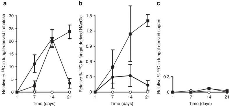
13CO2 was added to airtight plant growth chambers that contained plants with Metarhizium and insects (waxmoth larvae) in soil microcosms (Supplementary Fig. 1). The plants were separated from the insects and fungus by a 30-μm mesh. 13CO2 was provided to plants, in growth chambers, and the transfer of 13C into root-associated fungal biomass was assessed over a 21-day period. At 7, 14, and 21 days, in the presence of 13CO2, we observed 13C incorporation into Metarhizium (fungal-specific) carbohydrates (Fig. 1a,b). All experimental spectra demonstrated 13C resonances present at intensities over what was observed in natural abundance samples, indicating carbon movement from the plant to its fungal partner (Supplementary Fig. 2). Neither 13C labelled-trehalose nor -GlcNAc were observed in samples obtained from plants grown in the absence of Metarhizium (Fig. 1a,b). However, 13C resonances are present in the spectra representative of soluble plant sugars including fructose, glucose and sucrose.
Figure 1. Relative per cent 13C in Metarhizium and plant-derived sugars.
Resonances present in the 13C NMR spectra were integrated relative to an internal standard (100 μl MeOH, added to all samples) and then calculated as a percentage of integration value for the internal standard. (a) Closed circles represent 13C in trehalose extracted from root-colonized Metarhizium without an insect present. Closed squares represent 13C in trehalose extracted from root-colonized Metarhizium with insect present. Open circles represent 13C in trehalose extracted from roots without Metarhizium. (b) Closed circles represent 13C in GlcNAc extracted from root-colonized Metarhizium without an insect present. Closed squares represent 13C in GlcNAc extracted from root colonized Metarhizium with an insect present. Open circles represent 13C in GlcNAc extracted from roots without Metarhizium. (c) Closed circles represent 13C GlcNAc extracted from soil with Metarhizium, but without a plant. Closed squares represent 13C trehalose extracted from soil with Metarhizium, but without a plant. Open circles represent 13C in all sugars extracted from soil without Metarhizium and plant (n=20), bars represent s.e.
At 21 days, in the presence of a host insect, there was a significant increase in the amount of 13C translocated into Metarhizium carbohydrates when compared with microcosms without an insect (analysis of variance, P<0.05) (Fig. 1a). Control plants without 13CO2 did not demonstrate 13C NMR resonances for carbohydrate species above natural abundance levels (Supplementary Fig. 3).
13C fixation by Metarhizium in soil
To ensure there was no CO2 fixation by Metarhizium in the soil, microcosms were set up with Metarhizium without the addition of haricot bean seedlings, and placed in assimilation chambers with 13CO2. The soil was then sampled at 1, 7, 14 and 21 days, and sequentially extracted for trehalose and GlcNAc. 13C-carbohydrates were not observed over what was found at natural abundance levels in soil samples (Fig. 1c) indicating that all 13C present in fungal biomass was obtained through the transfer of photosynthetically fixed carbon into root-colonized Metarhizium.
Plant root weight
When Metarhizium and an insect are present, the root systems were significantly larger at both 14 and 21 days when compared to plants with Metarhizium only (2.9±0.8 g with versus 2.2±0.5 g without insect at 14 days (n=20, ±indicates s.e.), and 7.6±1.4 g with versus 5.8±0.4 g without insect at 21 days (n=20, ±indicates s.e.) analysis of variance, P<0.05). This suggests that Metarhizium is able to provide insect-derived nitrogen for plant-produced carbon, and thus, the root system was overall more robust.
Root section staining
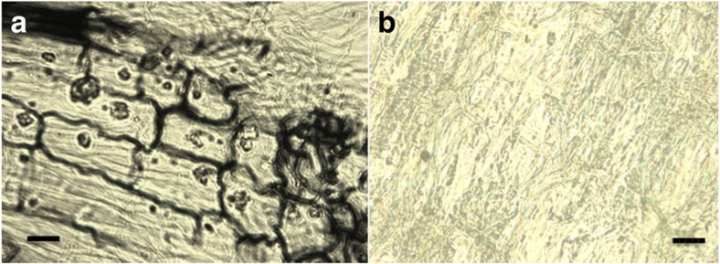
Bean plant roots grown in microcosms with Metarhizium, under experimental conditions, were collected, sectioned, and stained with fungal-specific dye (Fig. 2). The images showed that, under current experimental conditions, Metarhizium was able to internally colonize plant roots.
Figure 2. Internal cross section of M. robertsii colonized haricot bean roots harvested after 10 days and stained with Chlorazol black.
(a) Metarhizium colonized plant roots, sectioned with a microtome and subsequently stained with Chlorazol black. (b) Plant roots grown without Metarhizium stained with Chlorazol black. Images were taken at × 400 magnification. scale bar, 50 μm.
Discussion
Previously, we have shown that a host plant can derive a significant portion of its nitrogen from soil insects through an endophytic association with insect-pathogenic Metarhizium spp.14,15. Here we show that plants reciprocate with an exchange of photosynthate to their endophytic partners. Plants photosynthetically fix carbon, and furnish root-colonized Metarhizium with this carbon, while Metarhizium can access a source of nitrogen (that is, insects) via their specialization as insect pathogens (Fig. 3).
Figure 3. Exchange of insect-derived nitrogen for plant derived photosynthate.
Representation of the exchange of insect-derived nitrogen for plant derived photosynthate between endophytic Metarhizium and its plant host. Metarhizium is able to colonize a plant root, and provide useable, insect-nitrogen in exchange for plant produced carbon.
Plants provided carbon to endophytic Metarhizium even in the absence of a host insect in the microcosm (Fig. 1). However, in the presence of a host insect there was significantly greater 13C transfer to Metarhizium. A similar relationship was observed in Medicago truncatula roots colonized with arbuscular mycorrhizal fungi. In this symbiosis a reciprocal exchange of nutrients stabilized the root/fungal complex and the plant host increased carbon flow to Glomus spp. upon receiving increased nutrients from the fungal partner22. Therefore, the reciprocity seen in the Metarhizium/plant relationship may increase the overall stability of the partnership, ensuring that each symbiont is provided with adequate nutrients.
The ability of Metarhizium to receive plant-derived carbon, suggests the involvement of transporters that allow for fungal assimilation of photosynthate. In Metarhizium, an oligosaccharide transporter, Metarhizium raffinose transporter (mrt), plays a role in the uptake of raffinose from the rhizosphere23. However, in obligate plant/fungal relationships, there is evidence that plant synthesized monosaccharides are shuttled into the fungus directly from the root24. In mycorrhizal fungi, for example, there is a specific increase in the monosaccharide transporter MST2 during plant root colonization25. Metarhizium has three identified monosaccharide transporters (accession numbers: MAA_02403, MAA_03088, MAA_07773), and while these transporters have not been characterized in terms of specific function or location, BLAST analyses show sequence identities with sugar transporters in other root colonizing fungi such as Beauveria bassiana, and Glomerella graminicola, suggesting their role in root colonization and nutrient exchange.
There were no overall differences in root colonization (as measured by CFU per gram root) when Metarhizium and insect were present in the system, compared with only Metarhizium present (Supplementary Fig. 4). Bean plants have previously shown increased root biomass when Metarhizium was present with an insect as compared with when Metarhizium, or an insect, are present singly15. An analogous situation has been observed in arbuscular mycorrhizal fungi (AMF) where AMF-colonized roots typically lead to healthier plant systems, and an overall increase in primary production22. Plants that receive more insect-derived nitrogen through an endophytic association with Metarhizium are potentially able to allocate more resources to root growth, production and photosynthesis, the resultant photosynthate could then be used to reinforce and stabilize nutrient exchange with Metarhizium.
Metarhizium is an insect pathogenic, root colonizing fungus. These two lifestyles are linked through reciprocal nutrient exchange with a host plant, whereby the fungus receives carbon in exchange for insect-derived nitrogen. Interestingly, Metarhizium is phylogenetically related to other endophytes26 and we suggest that their evolution as insect pathogens, as well as their ability to translocate nitrogen27, allowed them to effectively barter a specialized nitrogen source (that is, insects) with host plants for photosynthate. While there has been previous work that has indicated the role that root colonizing fungi play in plant nutrient acquisition, the present study provides information on multi-role lifestyles of insect-pathogenic fungi in soil environments. These unique endophytes may be widespread in nature, and contribute to the overall global cycling of nutrients in a way more profound than previously known.
Methods
Fungal cultures and plant material
Metarhizium robertsii strain ARSEF 2575 was obtained from the US Department of Agricultural Research Service Collection of Entomopathogenic Fungal Cultures, Ithaca, New York. Stock cultures were grown at 27 °C on potato dextrose agar (PDA; Difco laboratories, Mississauga, Ontario, Canada. Phaseolus vulgaris (Haricot Bean) seeds were obtained from OSC seeds, Waterloo, Ontario, Canada. M. robertsii was transformed for expression of the green fluorescent protein (GFP) gene, egfp, utilizing the vector pFBENGFP in Agrobacterium tumefaciens-mediated transformation28.
Seed sterilization and plating
Seeds were surface sterilized before use in order to prevent any unwanted fungal or bacterial growth. Seeds were immersed in sterile distilled water for thirty minutes in a 50 ml capped plastic tube. They were then washed in a 4% sodium hypochlorite solution three times, ten minutes each wash. The seeds were rinsed with sterile distilled water between each wash. The seeds were then placed in 15% hydrogen peroxide for thirty minutes followed by three washes with sterile distilled water. The seeds were then kept overnight at 4 °C to allow for synchronization of growth, then plated on water agar and kept at 25 °C for a photoperiod of 16 h a day for a minimum of seven days in order to obtain seedlings which were used in the study.
Plant growth
Plants were grown in plastic garden pots (height 20 cm). The pots were sterilized with UV light for 2.5 h before use. Soil was sterilized prior to use by autoclaving 3 × on a biohazard cycle and allowed to cool between each cycle (19 h). Pots were partially filled with sterile soil, and five 1cm × 1cm agar plugs were added. Pots were then filled with soil 3 cm from the top and a seedling planted. Where appropriate, Metarhizium infected insects were added to soil microcosms. Galleria mellonella were infected with conidia by placing the larva on a 10-day-old conidiating culture of M. robertsii. The plate was agitated for two minutes allowing the fungus to sufficiently adhere to the insects. Insects were then placed into Petri dishes that had a circle cut into the lid ∼7 cm in diameter, which was subsequently covered with 30 μm mesh. The mesh was adhered to the Petri dish lid with silicon glue. This allowed for the movement of fungus out of the Petri dish while ensuring the insects could not interact with the plant roots. Plants were watered daily with sterile, distilled water and once a week with 50 ml of 50% MMN solution ((0.05 g CaCl2, 0.025 g NaCl, 0.05 g KH2PO4, 0.5 g (NH4)2PO4, 0.15 g MgSO4·7H20, 1 mg FeCl3·6H20, 5 g glucose monohydrate, 10 ml trace elements solution (3.728 g KCl, 1.546 g H3BO3, 0.845 g MnSO4·H20, 0.05 g ZnS04·7H20, 0.0125, g CuS04, 0.05 g (NH4)6Mo7024·4H20 per 1 l) per 1 l).
Assimilation chamber and 13CO2 addition
Plant assimilation chambers were constructed to provide plants with a 1,500 p.p.m. 13CO2 (99% 13C) environment. All chambers were 60 l by volume (Supplementary Fig. 1) and were airtight. Plants were placed in the assimilation chambers and sealed, once inside the chambers, 13CO2 was added until 1,500 p.p.m. was reached. Plants were allowed to grow in a 13CO2-rich environment for 8 h, during maximum photosynthetic period. Plants were provided with 13CO2 every 48 h.
Extraction of soluble sugars and N-acetylglucosamine
Soluble sugars and N-acetylglucosamine extractions were carried out by methanol extraction29. Twenty plants were collected at appropriate time points (1, 7, 14, 21 days), microcosms were dismantled, and insects analysed to ensure infection by Metarhizium. In these microcosms 100% of insects were observed to be infected and killed by Metarhizium. Dead insects were mummified with a layer of fungal hyphae and conidia. Roots were gently washed free of soil, lightly dried, and detached from the stalk with a scalpel blade and weighed. Plant roots were pooled, 5 plants per pool and lyophilized. Plant root tissue pools were frozen in liquid nitrogen and ground with mortar and pestle to a powder in 4–6 ml of MeOH/H20 (70/30). The mixture was then filtered through Fischer brand P4 filter paper, and methanol was removed by evaporation under vacuum. The aqueous solution was freeze-dried and stored at 4 °C for NMR analysis. The solid plant residue was then used for N-acetylglucosamine extraction. Plant tissue was treated for 20 min in boiling 1 M KOH. KOH was then decanted and solids were washed in deionized water and soaked in potassium phosphate buffer (pH 6.0). One unit of chitinase, 1 unit of chitosanase and 0.1 unit of glucanase were added to the samples (each enzyme was prepared as a stock solution in potassium phosphate buffer). To prevent contamination, 0.01% sodium azide was added to each sample. Samples were then incubated at 37 °C for 7 days under continuous agitation. After enzymatic digestion, samples were centrifuged and supernatants were lyophilized and stored at 4 °C prior to NMR analysis.
Nuclear magnetic resonance spectroscopy
All NMR spectra were recorded in D2O on a Bruker Ultra Shield Plus AV-600 spectrometer (1H at 600 MHz, 13C at 151 MHz) with a 5 mm Bruker BBFO probe with methanol (100 μl) as an internal reference for all spectra at 49.5 p.p.m. Acquisition parameters were the same for all samples. Four spectra, each of 27,000 scans, were acquired for each sample pool. This was considered to give sufficient signal-to-noise ratio to identify incorporation of 13C into major metabolites of interest. NMR spectra were analysed using the Bruker Topspin v2.0 software interface. Detailed acquisition parameters can be found in Supplementary Table 1.
Fungus colonized root staining
Harvested roots were washed in sterile distilled water until all soil removed and then stained30. Roots were fixed in a 1:1:1 formalin:acetic acid:alcohol solution for 24 h. The roots were then dehydrated in increasing concentrations of ethanol (30, 50 and 70%) for 30 min, then in 90% ethanol for 60 min. After dehydration, the roots were cut into 2 cm sections and cleared in methyl salicylate for 12 h. The roots were then embedded in paraffin wax and sectioned using a microtome. The wax was removed by washing the samples with xylene for 15 min. The roots were then placed in 10% (wt/vol) KOH solution and heated at 65 °C for 60 min. After 1 h, the KOH was decanted and replaced with Chorazol E Black staining solution (80% lactic acid, glycerol, and distilled water with 0.1% Chlorazol black E, 1:1:1:1) and heated at 65 °C for 60 min.
CFU determination from soil
One gram of soil was taken from each pot, at a distance of 1.0 cm from the root and 2.0 cm below the soil surface, and suspended in 2.0 ml of 0.01% Triton-X. The samples were vortex vigorously and 0.1 ml of suspension was plated, in duplicate, on selective YPD (2 g yeast extract, 10 g peptone, 20 g dextrose, 15 g agar, 0.01 g crystal violet, 0.2 g chloramphenicol, 0.5 g cycloheximide, and 0.3 g dodine adjusted to 1 l with distilled water). Plates were incubated at 27 °C for 10 days. The CFUs were recorded on day 10. To confirm colonies were that of M. robertsii the colonies were examined, before conidiation, for green fluorescence as the strain used (2575-GFP) is a stable lab strain expressing GFP.
CFU determination from roots
Roots were gently washed free of soil, lightly dried and detached from the stalk with a scalpel blade. The roots were sliced into 5 mm sections and placed in a 50 ml tube and weighed. A solution of 0.01% Triton-X was added to the roots at a volume of 2:1 (that is, 2 ml 0.01% Triton-X to every 1 g of root). The roots were homogenized using a Tissue Tearor (Greiner Scientific) and 0.1 ml of the homogenate was plated, in duplicate, on selective YPD. Plates were incubated at 27 °C and the CFUs were recorded on day 10. Colonies were confirmed to be M. robertsii by visualization of GFP fluorescence.
Data availability
The authors declare that all the data supporting the findings of this study, and any relevant subsequent information, is available within the manuscript and its supplementary files or is available from the authors by request.
Additional information
How to cite this article: Behie, S. W. et al. Carbon translocation from a plant to an insect-pathogenic endophytic fungus. Nat. Commun. 8, 14245 doi: 10.1038/ncomms14245 (2017).
Publisher's note: Springer Nature remains neutral with regard to jurisdictional claims in published maps and institutional affiliations.
Supplementary Material
Supplementary Figures and Supplementary Tables
Acknowledgments
This research was conducted with the assistance of a Natural Sciences and Engineering Research Council of Canada Discovery Grant to M.J.B. This manuscript was published with the assistance of a grant from the Brock library open access publishing fund. We also thank Maia Bidochka for the artwork.
Footnotes
Author contributions S.W.B., P.M.Z. and M.J.B. designed the experiments and discussed all results. S.W.B., C.C.M., I.S. and L.B. carried out all the experiments. The manuscript was written with contributions from all authors. All authors have given approval to the final version of the manuscript.
References
- Malloch D. W., Pirozynski K. A. & Raven P. H. Ecological and evolutionary significance of mycorrhizal symbioses in vascular plants (a review). Proc. Natl Acad. Sci. USA 77, 2113–2118 (1980). [DOI] [PMC free article] [PubMed] [Google Scholar]
- Parniske M. Arbuscular mycorrhiza: the mother of plant root endosymbioses. Nat. Rev. Microbiol. 6, 763–775 (2008). [DOI] [PubMed] [Google Scholar]
- Cocking E. C. Endophytic colonization of plant roots by nitrogen-fixing bacteria. Plant Soil. 252, 169–175 (2003). [Google Scholar]
- Pearson J. N., Abbott L. K. & Jasper D. A. Mediation of competition between two colonizing VA mycorrhizal fungi by the host plant. New Phytol. 123, 93–98 (1993). [Google Scholar]
- Hikosaka K. Leaf canopy as a dynamic system: Ecophysiology and optimality in leaf turnover. Ann. Bot. (Lond) 95, 521–533 (2005). [DOI] [PMC free article] [PubMed] [Google Scholar]
- Smith F. A., Grace E. J. & Smith S. E. More than a carbon economy: Nutrient trade and ecological sustainability in facultative arbuscular mycorrhizal symbioses. New Phytol. 182, 347–358 (2009). [DOI] [PubMed] [Google Scholar]
- Fellbaum C. R. et al. Carbon availability triggers fungal nitrogen uptake and transport in arbuscular mycorrhizal symbiosis. Proc. Natl Acad. Sci. USA 109, 1–5 (2012). [DOI] [PMC free article] [PubMed] [Google Scholar]
- Jones D. L., Nguyen C. & Finlay R. D. Carbon flow in the rhizosphere: carbon trading at the soil–root interface. Plant Soil 321, 5–33 (2009). [Google Scholar]
- Balestrini R. & Lanfranco L. Fungal and plant gene expression in arbuscular mycorrhizal symbiosis. Mycorrhiza 16, 509–524 (2006). [DOI] [PubMed] [Google Scholar]
- Karandashov V. & Bucher M. Symbiotic phosphate transport in arbuscular mycorrhizas. Trends Plant. Sci. 10, 22–29 (2005). [DOI] [PubMed] [Google Scholar]
- Guether M. et al. A mycorrhizal-specific ammonium transporter from Lotus japonicus acquires nitrogen released by arbuscular mycorrhizal fungi. Plant Physiol. 150, 73–83 (2009). [DOI] [PMC free article] [PubMed] [Google Scholar]
- Wang G. M. et al. Carbon partitioning patterns of mycorrhizal versus non-mycorrhizal plants-real-time dynamic measurements using (Co2)-C-11. New Phytol. 112, 489–493 (1989). [DOI] [PubMed] [Google Scholar]
- Ostle N. et al. Isotopic detection of recent photosynthate carbon flow into grassland rhizosphere fauna. Soil Biol. Biochem. 39, 768–777 (2007). [Google Scholar]
- Behie S. W., Zelisko P. M. & Bidochka M. J. Endophytic insect-parasitic fungi translocate nitrogen directly from insects to plants. Science 336, 1576–1577 (2012). [DOI] [PubMed] [Google Scholar]
- Behie S. W. & Bidochka M. J. Ubiquity of insect-derived nitrogen transfer to plants by endophytic insect-pathogenic fungi: an additional branch of the soil nitrogen cycle. Appl. Environ. Microbiol. 80, 1553–1560 (2014). [DOI] [PMC free article] [PubMed] [Google Scholar]
- Hajek A. E. & St Leger R. J. Interactions between fungal pathogens and insect hosts. Annu. Rev. Entomol. 39, 293–322 (1994). [Google Scholar]
- Roberts D. W. & Hajek A. E. in Frontiers in Industrial Mycology ed. Leatham G. F. 144–159Springer (1992). [Google Scholar]
- Wyrebek M., Huber C., Sasan R. K. & Bidochka M. J. Three sympatrically occurring species of Metarhizium show plant rhizosphere specificity. Microbiology 157, 2904–2911 (2011). [DOI] [PubMed] [Google Scholar]
- Zhong X. et al. Detection of Ophiocordyceps sinensis in the roots of plants in alpine meadows by nested-touchdown polymerase chain reaction. Fungal Biol. 118, 359–363 (2014). [DOI] [PubMed] [Google Scholar]
- Akello J. et al. Beauveria bassiana (Balsamo) Vuillemin as an endophyte in tissue culture banana (Musa spp.). J. Invertebr. Pathol. 96, 34–42 (2007). [DOI] [PubMed] [Google Scholar]
- Hirsch P. R., Mauchline T. H., Mendum T. A. & Kerry B. R. Detection of the nematophagous fungus Verticillium chlamydosporium in nematode-infested plant roots using PCR. Mycol. Res. 104, 435–439 (2000). [Google Scholar]
- Kiers E. T. et al. Reciprocal rewards stabilize cooperation in the mycorrhizal symbiosis. Science 333, 880–882 (2011). [DOI] [PubMed] [Google Scholar]
- Fang W. & St Leger R. J. Mrt, a gene unique to fungi, encodes an oligosaccharide transporter and facilitates rhizosphere competency in Metarhizium robertsii. Plant Physiol. 154, 1549–1557 (2010). [DOI] [PMC free article] [PubMed] [Google Scholar]
- Javot H., Penmetsa R. V., Terzaghi N., Cook D. R. & Harrison M. J. A Medicago truncatula phosphate transporter indispensable for the arbuscular mycorrhizal symbiosis. Proc. Natl Acad. Sci. USA. 104, 1720–1725 (2007). [DOI] [PMC free article] [PubMed] [Google Scholar]
- Helber N. et al. A versatile monosaccharide transporter that operates in the arbuscular mycorrhizal fungus glomus sp is crucial for the symbiotic relationship with plants. Plant Cell 23, 3812–3823 (2011). [DOI] [PMC free article] [PubMed] [Google Scholar]
- Behie S. W., Padilla-Guerrero I. E. & Bidochka M. J. Nutrient transfer to plants by phylogenetically diverse fungi suggests convergent evolutionary strategies in rhizospheric symbionts. Commun. Integr. Biol. 6, 6–9 (2013). [DOI] [PMC free article] [PubMed] [Google Scholar]
- Hu X. et al. Trajectory and genomic determinants of fungal-pathogen speciation and host adaptation. Proc. Natl Acad. Sci. USA 111, 1–6 (2014). [DOI] [PMC free article] [PubMed] [Google Scholar]
- Fang W., Pei Y. & Bidochka M. J. Transformation of Metarhizium anisopliae mediated by Agrobacterium tumefaciens. Can. J. Microbiol. 52, 623–626 (2006). [DOI] [PubMed] [Google Scholar]
- Bago B. et al. Carbon export from arbuscular mycorrhizal roots involves the translocation of carbohydrate as well as lipid. Plant Physiol. 131, 1496–1507 (2003). [DOI] [PMC free article] [PubMed] [Google Scholar]
- Brundrett M. C., Piché Y. & Peterson R. L. A new method for observing the morphology of vesicular-arbuscular mycorrhizae. Botany 62, 2128–2134 (1984). [Google Scholar]
Associated Data
This section collects any data citations, data availability statements, or supplementary materials included in this article.
Supplementary Materials
Supplementary Figures and Supplementary Tables
Data Availability Statement
The authors declare that all the data supporting the findings of this study, and any relevant subsequent information, is available within the manuscript and its supplementary files or is available from the authors by request.


