Abstract
Background
Syphilis can have many clinical manifestations, including eye involvement, or ‘ocular syphilis’. In 2015, an increase in reported cases of ocular syphilis and potential case clusters raised concern for an oculotropic strain of Treponema pallidum, the infectious agent of syphilis. Molecular typing was used to examine strains found in cases of ocular syphilis in the United States.
Methods
In 2015, following a clinical advisory issued by the Centers for Disease Control and Prevention (CDC), pre-treatment clinical specimens from U.S. patients with ocular syphilis were sent to a research laboratory for molecular analysis of T. pallidum DNA. Molecular typing was conducted on these specimens, and results were compared to samples collected from Seattle patients diagnosed with syphilis, but without ocular symptoms.
Results
Samples were typed from 18 patients with ocular syphilis and from 45 patients with syphilis, but without ocular symptoms. Clinical data were available for 14 ocular syphilis patients: most were men, HIV-infected, and had early syphilis. At least 5 distinct strain types of Treponema pallidum were identified in these patients, and 9 types were identified in the Seattle non-ocular patients. 14d/g was the most common type in both groups. An unusual strain type was detected in a small cluster of ocular syphilis patients in Seattle.
Conclusions
Ocular syphilis is a serious sequela of syphilis. In this preliminary study, clear evidence of a predominant oculotropic strain causing ocular syphilis was not detected. Identification of cases and prompt treatment is critical in the management of ocular syphilis.
Keywords: syphilis, ocular syphilis, molecular typing
Introduction
Syphilis is a genital ulcer disease caused by the spirochete Treponema pallidum subspecies pallidum (T. pallidum), and can result in a variety of clinical manifestations. Reported primary and secondary syphilis cases in the United States have been increasing since 2001, with the total number of primary and secondary syphilis cases reported to the Centers for Disease Control and Prevention (CDC) increasing 15.1% from 2013 to 2014.1
Syphilis can have a variety of clinical manifestations, including involvement of the eye. Ocular syphilis, an inflammatory eye disease, can involve almost any eye structure.2 Uveitis is the most common manifestation, and there have been reports of significant vision loss, including blindness.3-5 Ocular syphilis can occur at any stage, with acute ocular inflammation during early (primary, secondary and early latent) infection and a more indolent ‘chronic’ inflammation during late or tertiary infection.4,6
Historically, limited studies of the expected frequency of ocular syphilis have been performed; results found ocular syphilis in 07 to 7.9%8 of patients with secondary syphilis and from 3%9 to 51%10 of patients with neurosyphilis. In a recent study, 7.9% of syphilis patients reported new onset of visual or hearing disturbances; of these, 44% had cerebrospinal fluid (CSF) findings consistent with neurosyphilis.11 However, most studies have not taken a systematic approach for identification of ocular involvement, and ocular syphilis has not been a reportable manifestation to CDC, therefore no ‘baseline rates’ are available.
Molecular typing of T. pallidum can potentially provide additional information on the epidemiology of syphilis. In 1998, a method was published to distinguish various molecular types of T. pallidum using two genetic targets [variable number of 60-base pair repeats in the acidic repeat protein (arp) gene and restriction fragment length polymorphism (RFLP) analysis of the T. pallidum repeat (tpr) subfamily II genes]12; a third target involving sequence analysis of the tp0548 gene was added in 2010.13 Since the development of this molecular typing system, substantial diversity has been identified in strains of T. pallidum globally.14 In Seattle, a specific strain type (14d/f) was associated with neurosyphilis, suggesting enhanced neurotropism by certain strains,13 which had been suggested previously in observational studies.15
In early 2015, two clusters of ocular syphilis were reported in California and Washington,16 leading CDC to issue a Clinical Advisory in April of that year.17 Since then, many jurisdictions reported an increase in cases of ocular syphilis, raising concern for an ‘outbreak’.17 Clinical specimens from the ocular syphilis cases in Seattle, Washington were typed, and a strain type (8d/g) not previously identified in Seattle nor widely found in the United States was identified in two of the ocular syphilis cases and implicated in a third case. A broader investigation was then initiated to describe molecular types detected in ocular syphilis cases nationally.
Materials and Methods
Ocular syphilis cases
Prompted by the Clinical Advisory,17 clinicians or laboratories with pre-antibiotic clinical samples (whole blood, serum, swabs of lesion exudate, CSF or ocular fluid) from patients with ocular syphilis contacted CDC, which facilitated shipment of the specimens to a research laboratory at the University of Washington. De-identified demographic information was collected on the patients, as well as information on the patients' syphilis diagnoses and ocular involvement. A case of ocular syphilis was defined as a person with clinical symptoms or signs consistent with ocular disease and syphilis of any stage. Specimens from the convenience-sampling of ocular syphilis cases described here were obtained from cases identified in 2015.
Non-ocular syphilis cases from Seattle
Blood and oral or genital swab samples were collected anonymously from 88 patients with syphilis in the Seattle & King County Public Health STD Clinic in Seattle, Washington. None of these individuals had symptoms of ocular syphilis. All samples were collected in 2015.
Laboratory methods
Frozen samples for determination of strain type included blood in EDTA tubes; CSF; sera; ocular fluids; and oral or genital swabs collected in 1 ml 1X lysis buffer (10 mM Tris-HCl, 0.1 M EDTA, 0.5% SDS) before freezing. T. pallidum DNA was extracted from blood using a QIAamp DNA Blood Midi kit (Qiagen, Valencia, CA) according to the manufacturer's instructions, precipitated overnight at -20°C with 2.5 volumes of 100% ethanol, 0.1 volumes of 3 M sodium acetate, and 1 μl of 20 mg/ml glycogen and resuspended in a final volume of 50 μl molecular grade water. T. pallidum DNA was extracted from samples other than blood using a QIAamp DNA Mini kit (Qiagen, Valencia, CA) according to the manufacturer's instructions.
Strain typing was based on analysis of three DNA targets: 1) the number of 60bp repeats in the acidic repeat protein (arp) gene, 2) RFLP analysis of the T. pallidum repeat (tpr) subfamily II genes (tprE [tp0313], tprG [tp0317], tprJ [tp0621]), and 3) sequence analysis of a short region of the tp0548 gene (bp131–215). The first two targets were analyzed according to published methods13 except that the tprE, G, and J amplicon was reamplified for 20 cycles in the case of a low yield of DNA and the PCR products were gel extracted using a QIAquick Gel Extraction kit (Qiagen, Valencia, CA) according to manufacturer's instructions prior to RFLP analysis. The tp0548 sequence from blood samples was determined as previously described.13 For samples other than blood, an alternate antisense primer was used (5′ TGC CCT GGT GTC CGC CGT TCT 3′) with the following touchdown cycling conditions: 95°C for 5 min, then 10 cycles of 95°C for 1 min, 65°C for 30 sec with a 1°C decrease per cycle, and 72°C for 30 sec; then followed by 35 cycles of 95°C for 1 min, 55°C for 30 sec, and 72°C for 30 sec, followed by a final extension for 10 min at 72°C. PCR products were purified using ExoSap-It (Affymetrix, Santa Clara, CA) prior to sequencing. Assignment of molecular type was accomplished as previously described.13 Because of limiting amounts of DNA in the samples, in some cases, only one or two molecular typing targets could be defined. When multiple samples from the same patient were tested, the most complete type was assigned to that individual.
Review Board at CDC determined this investigation not to be human subjects' research, with a primary intent for public health practice or disease control activity. Similarly, samples from syphilis patients in Seattle were collected as a part of disease control activity.
Results
Ocular Syphilis Cases
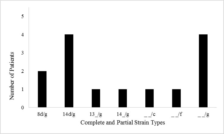
The laboratory received samples from 36 subjects with ocular syphilis and attempted molecular typing on 23 CSF samples, 12 whole blood and 5 serum samples, and 7 ocular fluid samples (either vitreous or aqueous fluid). Complete typing results for all three molecular targets were available for 8 (22%) samples and an additional 10 (27%) samples had a positive result for at least one of the molecular targets. From those 18 patients, clinical information was available for 14 (78%) who had probable ocular syphilis based on examination by an ophthalmologist. Clinical characteristics, laboratory results and molecular typing results for these 14 patients are described in Table 1.
Table 1. Clinical Characteristics, Laboratory Results and Molecular Typing Results for Ocular Syphilis Patients.
| Pt | Location | Gender | Known MSM | HIV Status | Stage of Syphilis | RPR titer | Sample | Strain Type1 | Diagnosis |
|---|---|---|---|---|---|---|---|---|---|
| 1 | Washington | Male | MSM | Positive | Early Latent | 512 | Ocular | 8d/g | Panuveitis |
| 2 | Washington | Male | MSM | Negative | Late Latent | 256 | Ocular | 14d/g | Panuveitis |
| 3 | Washington | Male | MSM | Positive | Early Latent | 1024 | CSF | 8d/g | Panuveitis, retinal detachment |
| 4 | Massachusetts | Male | MSM | Positive | Secondary | 256 | Whole Blood | 14d/g | Chorioretinitis |
| 5 | New York | Male | MSM | Positive | Primary | 256 | Ocular | 14d/g | Uveitis, chorioretinitis, vitritis |
| 6 | Oregon | Male | MSM | Positive | Late Latent | 2048 | CSF | 14d/g | Panuveitis |
| 7 | Massachusetts | Male | MSM | Positive | Secondary | 256 | CSF | 13 __/g | Panuveitis, retinitis |
| 8 | Washington | Male | No | Negative | Secondary | 256 | CSF | 14 __/g | Optic disc edema |
| 9 | Washington | Male | MSM | Positive | Secondary | 64 | CSF & Whole Blood | __ __/g | Papillitis, optic neuritis |
| 10 | Washington | Male | Unknown | Positive | Unknown | 128 | CSF | __ __/g | Panuveitis, chorioretinitis |
| 11 | Illinois | Male | Unknown | Negative | Late Latent | 128 | CSF | __ __/c | Panuveitis, retinitis |
| 12 | Massachusetts | Male | No | Negative | Early Latent | 128 | CSF & Whole Blood | __ __/f | Panuveitis |
| 13 | Washington | Male | No | Negative | Late Latent | 4096 | Whole Blood | __ __/g | Chorioretinitis |
| 14 | Oregon | Female | N/A | Negative | Secondary | 256 | CSF | __ __/g | Posterior Uveitis |
MSM = Men who have sex with men
HIV = Human immunodeficiency virus
RPR = Rapid plasma reagin
CSF = Cerebrospinal fluid
Strain type listed as arp gene (number), tpr RFLP/ tp0548 sequence
Thirteen (93%) of the 14 patients were men, and of those, 8 (62%) reported having sex with men. The median age was 45 years (range 24-63). All patients had been tested for HIV and 8 (57%) were HIV-infected. Ten (71%) had early syphilis (primary, secondary, or early latent); secondary syphilis was the most common stage, seen in 5 (36%). The median rapid plasma reagin (RPR) titer was 256 (range 64-4096). There was no difference in median RPR titers by HIV status. Specific ocular diagnoses were available for all patients. Eight (62%) had panuveitis, and 5 (38%) had retinitis or chorioretinitis.
At least five distinct molecular types of T. pallidum were identified in the ocular syphilis patients (Figure 1). Three types in the arp gene were identified: types 8, 13 and 14. All samples were tpr RFLP type d. Three tp0548 types were identified: types c, f, and g. Type 8d/g was identified in 2 of the first 3 samples tested; however, that type was not subsequently detected. No specific molecular typing result was associated with any specific ocular diagnosis.
Figure 1. Complete and Partial T. pallidum Strain Types in Ocular Syphilis — United States, 2015.
Seattle non-ocular types
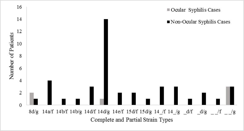
Complete molecular typing results were available for 30 (34%) of 88 patients, including 2 from blood, 5 from oral swabs and 23 from genital swabs; partial results were available for an additional 15 (17%) samples including 3 from blood, 7 from oral swabs and 5 from genital swabs. At least 9 distinct types of T. pallidum were identified among these patients without ocular symptoms. Types from Seattle patients with and without ocular symptoms are displayed in Figure 2.
Figure 2. Complete and Partial T. pallidum Strain Types in Ocular and Non-Ocular Syphilis — Seattle, WA, 2015.
Discussion
We found at least five different T. pallidum types among the ocular syphilis cases from which specimens were collected and able to be typed. This diversity likely represents the breadth of molecular types circulating in local networks, as demonstrated by the typing results from Seattle, and suggests that there is not a single type responsible for an overall increase in reported ocular syphilis. The most common type identified from the Seattle STD clinic was 14d/g. This type was also identified in 4 of the 6 ocular syphilis cases who had clinical information and complete typing results. This is consistent with previous observations that 14d strains are the most commonly identified T. pallidum strains globally.14
While we did not detect a single predominant strain in patients with ocular syphilis, our findings are not inconsistent with the possibility of oculotropic strains. In the initial Seattle cluster,16 two sexual partners were diagnosed with ocular syphilis. Molecular typing results were available for one of these partners, who was infected with type 8d/g. It is reasonable to infer that the ‘untyped partner’ was infected with the same 8d/g strain as his partner. Another ocular syphilis case from Seattle was typed as 8d/g, but no epidemiologic link to the two other cases was found. This finding could represent a focal Seattle cluster of 8d/g ocular syphilis caused by an ‘oculotropic strain’ within a background level of ocular syphilis caused by commonly circulating strains. Such a scenario would be similar to observations of possible T. pallidum strain-specific neurovirulence. Specifically, individuals infected with type 14d/f are more likely to have reactive CSF VDRL than individuals infected with other strains, but reactive CSF VDRLs are not exclusively seen in those infected with type 14d/f.13
Since the Clinical Advisory was issued, CDC has been notified of over 200 cases of ocular syphilis from 20 states over the last 2 years. Similar to the epidemiology of syphilis in the United States, most cases of suspected ocular syphilis reported to CDC (over 90%) were in men, especially in men who have sex with men (MSM). Several of the cases have resulted in significant sequelae, including blindness. In some cases, there was a delay in diagnosis and treatment due to the lack of awareness of ocular syphilis. Given the number of cases reported, CDC has worked with both local and national partners to increase awareness of syphilis as a cause of eye inflammation, which can occur in any stage of infection. Syphilis diagnostic testing with treponemal and non-treponemal tests is encouraged in any patient with visual complaints without another known cause among those at risk of syphilis, especially MSM and those who are HIV-infected. Additionally, all patients diagnosed with syphilis should be assessed for any neurologic complaints, including vision or hearing loss. Patients with syphilis and ocular complains should receive immediate ophthalmologic evaluation.
The 2015 STD Treatment Guidelines state that “Syphilitic uveitis or other ocular manifestations (e.g. neuroretinitis and optic neuritis) can be associated with neurosyphilis. A CSF examination should be performed in all instances of ocular syphilis, even in the absence of clinical neurologic findings. Ocular syphilis should be managed in collaboration with an ophthalmologist and according to the treatment and other recommendations for neurosyphilis, even if a CSF examination is normal.”18 Recommended treatment for neurosyphilis and ocular syphilis is 18–24 million units intravenous (IV) aqueous penicillin G daily, given as continuous infusion or divided into every 4 hour dosing, for 10–14 days. In addition, while CSF examination is recommended, this should not delay initiation of treatment.
There are several limitations of this study that are important to note. The specimens from patients with ocular syphilis represent a convenience sample, without any systematic collection. All syphilis molecular typing must be interpreted in the context of the background of local syphilis molecular epidemiology. While we have provided information on the circulating types in Seattle, Washington in 2015 to provide an example of the diversity of molecular types seen in one city, we do not have current national syphilis molecular epidemiology data for comparison.
Also, as these samples from ocular syphilis patients were not collected with a standardized protocol, some samples were handled in ways that allowed for substantial DNA degradation. This is demonstrated in a lower yield of successful complete molecular typing from the national ocular syphilis samples (22%) compared to the samples collected at the Seattle STD clinic (34%), where samples were frozen immediately after collection. Historically, blood and CSF have lower yield for strain typing compared to samples collected from primary and secondary lesions.14 Additionally, as the molecular methods used to classify T. pallidum strain types are focused on three specific genetic loci, they could potentially miss identifying other genetic signatures that may confer an oculotropic phenotype. Further investigations are warranted, including whole genome sequencing of T. pallidum from patients with uncomplicated and ocular syphilis.
Ocular syphilis is a serious complication of syphilis. The increase in reports of ocular syphilis in 2015 in the United States could be due to increased recognition of ocular manifestations in the setting of increased syphilis rates, or a true increase in the percentage of syphilis cases with ocular disease. Additional epidemiological investigations may shed light on other contributors to risk of ocular syphilis, including demographic or clinical factors, or the possibility that increased awareness of ocular complications of syphilis has led to increased case ascertainment. Further investigation is needed to evaluate molecular epidemiology of the syphilis epidemic in general, and specifically of those cases diagnosed with ocular syphilis. However, based upon this preliminary study, there does not seem to be a single strain responsible for ocular syphilis nationally. A larger sample size is necessary to support these preliminary conclusions. Prompt identification of cases with potential ocular syphilis, ophthalmologic evaluation and appropriate treatment is critical in the prevention and management of visual symptoms and sequelae of ocular syphilis.
Acknowledgments
We would like to thank all patients and clinicians who provided samples for this investigation, as well as state and local health departments who assisted with identification of patients with ocular syphilis. We would also like to thank Julia Dombrowski, MD for assistance in obtaining samples from patients with syphilis in the Seattle & King County Public Health STD Clinic, and Tom Peterman, MD with CDC's Division of STD Prevention for support of this project.
Sources of support: Portions of this work were supported by National Institute of Health, National Institute of Neurological Disorders and Stroke grant number NS082120.
Footnotes
The authors report no conflict of interest
References
- 1.Centers for Disease Control and Prevention. Sexually Transmitted Disease Surveillance 2014. Atlanta: U.S. Department of Health and Human Services; 2015. [Google Scholar]
- 2.Kiss S, Damico FM, Young LH. Ocular manifestations and treatment of syphilis. Semin Ophthalmol. 2005;20(3):161–167. doi: 10.1080/08820530500232092. [DOI] [PubMed] [Google Scholar]
- 3.Mathew RG, Goh BT, Westcott MC. British Ocular Syphilis Study (BOSS): 2-year national surveillance study of intraocular inflammation secondary to ocular syphilis. Invest Ophthalmol Vis Sci. 2014;55:5394–400. doi: 10.1167/iovs.14-14559. [DOI] [PubMed] [Google Scholar]
- 4.Ormerod LD, Puklin JE, Sobel JD. Syphilitic posterior uveitis: correlative findings and significance. Clin Inf Dis. 2001;32:1661–73. doi: 10.1086/320766. [DOI] [PubMed] [Google Scholar]
- 5.Smith JL. Acute blindness in early syphilis. Arch of Ophthal. 1973;90:256–8. doi: 10.1001/archopht.1973.01000050258015. [DOI] [PubMed] [Google Scholar]
- 6.Spoor TC, Wynn P, Hartel WC, et al. Ocular syphilis: Acute and chronic. J Clin Neuroophthalmol. 1983;3:197–204. [PubMed] [Google Scholar]
- 7.Chapel TA. The signs and symptoms of secondary syphilis. Sex Transm Dis. 1980;7:161–4. doi: 10.1097/00007435-198010000-00002. [DOI] [PubMed] [Google Scholar]
- 8.Hira SK, Patel JS, Bhat SG, et al. Clinical manifestations of secondary syphilis. Int J Dermatol. 1987;26:103–7. doi: 10.1111/j.1365-4362.1987.tb00532.x. [DOI] [PubMed] [Google Scholar]
- 9.Bennett JE, Dolin R, Blaser MJ. Principles and practice of Infectious Disease. Elsevier Health Sciences. 2014 [Google Scholar]
- 10.Centers for Disease Control and Prevention (CDC) Symptomatic early neurosyphilis among HIV-positive men who have sex with men — four cities, United States, January 2002–June 2004. MMWR Morb Mortal Wkly Rep. 2007;56:625–8. [PMC free article] [PubMed] [Google Scholar]
- 11.Dombrowski JC, Pederson R, Marra CM, et al. Prevalence estimates of complicated syphilis. Sex Transm Dis. 2015;42(12):702–4. doi: 10.1097/OLQ.0000000000000368. [DOI] [PubMed] [Google Scholar]
- 12.Pillay A, Liu H, Chen C, et al. Molecular subtyping of Treponema pallidum subspecies pallidum. Sex Transm Dis. 1998;25(8):408–414. doi: 10.1097/00007435-199809000-00004. [DOI] [PubMed] [Google Scholar]
- 13.Marra CM, Sahi SK, Tantalo LC, et al. Enhanced molecular typing of Treponema pallidum: geographical distribution of strain types and association with neurosyphilis. J Infect Dis. 2010;202(9):1380–1388. doi: 10.1086/656533. [DOI] [PMC free article] [PubMed] [Google Scholar]
- 14.Peng RR, Wang AL, Li J, et al. Molecular typing of Treponema pallidum: a systematic review and meta-analysis. PLos Neg Trip Dis. 2011;5:e1273. doi: 10.1371/journal.pntd.0001273. [DOI] [PMC free article] [PubMed] [Google Scholar]
- 15.Stokes JH, Beerman H, Ingram NR. Modern Clinical Syphilology. Philadelphia, Pennsylvania, USA: W.B. Saunders; 1944. [Google Scholar]
- 16.Woolston S, Cohen SE, Fanfair RN, et al. A cluster of ocular syphilis cases — Seattle, Washington, and San Francisco, California, 2014-2015. MMWR Morb Mortal Wkly Rep. 2015;64:1150–1. doi: 10.15585/mmwr.mm6440a6. [DOI] [PMC free article] [PubMed] [Google Scholar]
- 17.Centers for Disease Control and Prevention (CDC) [15 February 2016];Clinical advisory: Ocular syphilis in the United States. 2015 http://www.cdc.gov/std/syphilis/clinicaladvisoryos2015.htm.
- 18.Workowski KA, Bolan GA. Centers for Disease Control and Prevention (CDC). Sexually transmitted diseases treatment guidelines, 2015. MMWR Recomm Rep. 2015;64:1–137. [PMC free article] [PubMed] [Google Scholar]


