Abstract
Epirubicin is an anthracycline chemotherapy agent used for treatment of several cancers including oesophageal, breast and gastric. Extravasation is a well-recognised and serious complication of any intravenous therapies but especially chemotherapeutic agents. Signs of the injury can be subtle and without prompt recognition and treatment there can be extensive tissue damage and depending on location of injury this can result in significant functional loss. In this article, a case of delayed management of epirubicin extravasation from a cannula situated at the dorsum of the hand is discussed. Successful surgical reconstruction of the resulting substantial tissue damage using a radial forearm flap 21 days following injury is described.
Background
Extravasation is defined as the process of fluid leaking from a vessel into surrounding tissues.1 Any intravenous agent can potentially cause extravasation injuries. In some cases, in particular with chemotherapeutic agents, damage from extravasation can be severe. The incidence of peripheral extravasation injuries caused by chemotherapy agents is estimated to be between 0.01% and 7.0%.1
Epirubicin is a chemotherapy agent used in the treatment of many neoplastic conditions including breast, gastric and oesophageal cancer. Epirubicin is an anthracycline agent. The mechanism of action of anthracyclines is unclear, and suggestions include DNA damage (through lipid peroxidation and DNA cross-linking), formation of free radicals, binding to proteasomes (complexes involved in protein degradation) and inhibition of topoisomerase II (enzymes involved in DNA breaks after altering the twisting status of the DNA helix).2 Anthrayclines can remain in tissues for months following an extravasation injury.3 Without prompt treatment, epirubicin continues to damage surrounding tissues and manifests as progressive skin ulceration.3 In some cases, damage can be severe enough to cause destruction of joints and nerves.3 The consequences of delayed detection of extravasation may include pain, physical defect, psychological distress, cost of hospitalisation and more extensive tissue damage as a result of delay in the treatment.4
Extravasation injuries can present to medical and surgical specialties, so it is important that all doctors are able to recognise the features and the requirement for prompt treatment of the injury. In many trusts, local guidelines describe measures to ensure early detection of extravasation and immediate management steps.4 Preventative measures include checking the patency of the cannula and regular monitoring of the administration site. Additionally, some hospitals advise the use of central venous access devices for administration of epirubicin.5 Signs indicative of extravasation are sensory changes, pain, swelling, blanching of the skin during administration and erythema. If any of these signs develop, the infusion should be stopped and immediate action taken.
Case presentation
A 77-year-old woman with a diagnosis of lower oesophageal cancer presented to the plastic outpatient department 11 days following an epirubicin extravasation injury. She had received her fifth cycle of epirubicin at a dose of 100 mg (50 mg/m2) through a peripheral cannula on the dorsum of her right hand. Following the infusion, there was some mild swelling around the administration site with no sensory changes except some pain around the cannula site immediately after the infusion. Although there was some concern of an extravasation injury at this point, the skin changes were minimal so it was thought to only be a minor injury and immediate treatment was not started. Instead, she was admitted to hospital for a period of observation. The following day she was reviewed by a member of the plastics team who reported a small blister, mild erythema and persistent mild swelling of the dorsum of the hand. She was reviewed daily and received hydrocortisone cream and dimethylsulfoxide dressings in line with the local hospital extravasation guidelines. After 6 days, she was discharged with a follow-up appointment in plastics outpatient clinic.
Treatment
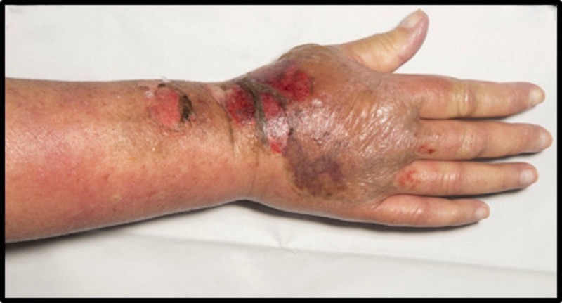
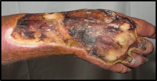
At time of presentation to the plastic surgery outpatient clinic (11 days after the initial injury), there were early signs of permanent tissue damage of the skin and underlying extensor tendons of all fingers. There was marked swelling of the hand and the radial and dorsal sides of the forearm (figure 1). There were no signs of sepsis. She was then reviewed on a twice weekly basis in order to allow the area of necrosis to accurately demarcate itself. On review at day 20, there was a large amount of slough overlying the extravasation site and an offensive smelling discharge was noted (figure 2). At this point, an emergency admission to hospital was made for intravenous antibiotics and urgent surgical debridement. The following day she was taken to the operating theatre for a debridement of the necrotic tissue on the hand and forearm. All non-viable tissue was removed, and the wound left open. The debridement extended down to expose extensor tendons of the hand. Five days later (day 21 after the injury), she underwent further debridement and reconstruction with a radial forearm flap and split thickness skin grafts to the donor site and distal forearm. A split skin graft could not be used alone as primary reconstruction on the dorsum of the hand as the drug had eroded through the paratenons. The postoperative course was uneventful, and the flap was healthy and well perfused. She was discharged with follow-up in plastic surgery outpatient clinic and hand therapy.
Figure 1.

Day 11 postinjury on first reviewed in plastics outpatient clinic.
Figure 2.

Day 20 postinjury—necrotic area demarcated with slough and an offensive discharge.
Outcome and follow-up
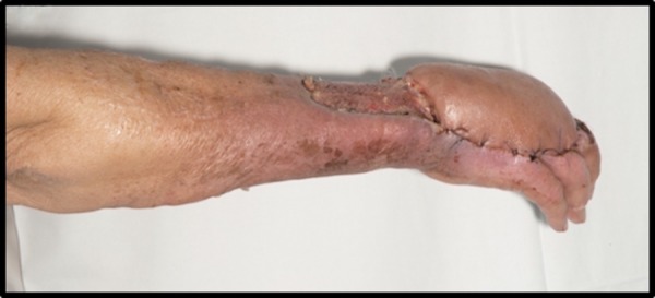
The patient is being actively followed-up in the plastic surgery outpatients’ clinic and is making extremely good progress. The flap had taken well at the first dressing change (figure 3). Subsequently, there were two small areas of minor dehiscence on the second, third and fourth digits which have healed with conservative management alone. Functionally, her range of movement is good. She is pleased with the outcome and has been offered a debulking of the flap in the future.
Figure 3.

Postoperative review following reconstruction with a radial forearm flap and split thickness skin graft to the distal forearm and donor site.
Discussion
This case demonstrates a delayed recognition of an epirubicin extravasation injury with a missed opportunity for early aggressive management. The ideal management would have been immediately thoroughly washing out the area with normal saline via the cannula, making multiple puncture sites at the extravasation site to allow the effluent out and injecting the affected area with hyaluronidase to increase the tissue permeability and speed up the dispersion of the remaining drug.1 4
Most of the cases in the literature describe earlier recognition of the injury, when cases have been amenable to non-surgical treatment. However, there are also several cases of delayed detection of these injuries which result in variable outcomes.
In one case, a patient with an injury over the left wrist was treated with a 2-month course of collagenase clostridiopeptidase A and protease ointment. She had been given an infusion of epirubicin (dose of 90 mg/ square meter body-surface area), fluorouracil and cyclophosphamide through a cannula situated in her anterior forearm proximal to the wrist for treatment of infiltrative ductal carcinoma of the breast. The treatment was started 10 days following the injury, and the wound healed within 6 months.5
In severe injuries, or when tissues have already become non-viable, surgery may be required. A patient, who developed an injury at the anterior aspect of the forearm following infusion of epirubicin for metastatic carcinoma of the breast, was referred to the plastic team within 2 days of receiving the infusion. She underwent surgical debridement on days 2 and 3 following the injury. A split skin graft on day 4 resulted in good wound coverage and no functional loss in the arm.6
In another case, the patient had not been referred to the plastic surgery team until 4 weeks following the injury. She presented with an injury at the dorsum of her left wrist secondary to epirubicin infusion for treatment of metastatic carcinoma of the breast. Surgical debridement was undertaken 4 weeks after the injury the patient had extensive necrotic tissue and slough reaching the tendons. The patient underwent multiple debridements. The wound was closed using a fascial forearm flap covered by split thickness skin graft. She had good wound coverage, but her hospital stay was 9 weeks and she had poor functional recovery.6
In addition to time to diagnosis of extravasation, a number of other factors determine the extent of injury following extravasation. These include patient factors, site of cannulation and properties of the substance administered.
First, patient factors associated with a greater risk of extravasation are as follows: elderly or young patients (owing to their veins being more fragile and susceptible to rupture), patients who have received multiple intravenous therapies (thrombosed veins are more likely to leak), reduced Glasgow Coma Scale (GCS) (unable to communicate sensory changes), obese patients (more difficult to site the cannula or recognition of skin changes) and patients with diseases resulting in sensory neuropathies such as diabetes, Raynaud’s and peripheral vascular disease (the patient is less likely to experience the sensory changes associated with extravasation).7
Second, selecting an appropriate cannulation site can reduce the risk of extravasation and the extent of complications. Areas with limited soft tissue to protect underlying structures (tendons, bones and nerves) and areas with small and fragile veins, such as lower extremities, should be avoided.8 9 European Society for Medical Oncology guidelines advise using large veins in the forearm and against using sites over joints, the inner wrist, lower extremities, antecubital fossa or on the dorsum of the hand.1 In many units, chemotherapy agents are administered using an intravenous pump to ensure a safe flow rate is achieved. A flow rate that is too fast risks damaging fragile vessels. As well as this a reduced flow rate may suggest that the cannula has displaced and that there is a greater risk for extravasation.8
Finally, the extent of injury is also determined by properties of the substance administered. Chemotherapy agents can be classified into five different categories according to their potential for tissue damage. Epirubicin is classified as a vesicant since it can cause necrosis and the formation of blisters. Other chemotherapy agents classified as veiscants include ‘actinomycin D, dactinomycin, daunorubicin, doxorubicin, idarubicin, mitomycin C, vinblastine, vindesine, vincristine and vinorelbine’. The other four categories are exfoliants (may cause tissue shedding without necrosis), irritants (irritation with no blister formation), inflammitants (can cause mild-to-moderate inflammation) and neutrals (no tissue damage).10
In conclusion, the extent of injury following extravasation of epirubicin cannot be predicted at the time of injury. Without prompt action, there can be significant tissue damage. If the 24 hour treatment window is missed, regular assessment of the patient is important in determining whether surgical intervention is necessary and the extent of tissue debridement required. In the cases documented in the literature, where extensive surgical debridement has been undertaken there was significant morbidity for the patient with a protracted hospital stay and functional impairment.6 In this report, we have described a case of delayed management of a significant epirubicin extravasation injury. We have also highlighted the importance of regular review and a reconstructive option of a radial forearm flap with good functional outcome and reduced surgical morbidity.
Patient's perspective.
I wish the problem had been picked up earlier to prevent me needing the operations but I am very pleased with the result that I have.
Learning points.
There should be a low threshold of treatment for patients presenting with any signs of extravasation: sensory changes, pain, swelling, blanching of the skin and erythema.
Superficial appearance of injury is not a good indicator of depth or severity of injury.
Management should be early and aggressive to prevent extensive tissue damage.
Early treatment has several benefits including limited tissue damage, less invasive reconstruction and a better functional outcome.
Steps to prevent extravasation injuries from a peripheral cannula include placement of the cannula into a large vein, checking the patency with a saline flush, patient education of suspicious symptoms and regular monitoring of the administration site for any signs of extravasation.
Footnotes
Contributors: All authors have contributed substantially to the preparation of the manuscript. All authors have reviewed the current manuscript and are happy for submission. The report has been written jointly by OH and PGD (background, treatment and discussion by OH, and case presentation and editing/review by PGD). Planning of the paper and patient consent were performed by PGD. The paper was conceptualised, edited and reviewed by the senior author AL who also managed the patient and collected the images.
Competing interests: None declared.
Patient consent: Obtained.
Provenance and peer review: Not commissioned; externally peer reviewed.
References
- 1.Pérez Fidalgo J, García Fabregat L et al. , ESMO Guidelines Working Group. Management of chemotherapy extravasation: ESMO-EONS Clinical Practice Guidelines. Ann Oncol 2012;23(Suppl 7):vii167–73. 10.1093/annonc/mds294 [DOI] [PubMed] [Google Scholar]
- 2.Minotti G, Menna P, Salvatorelli E et al. Anthracycline molecular advances and pharmacological developments in antitumor activity and cardiotoxicity. Pharmacol Rev 2004;56:185–229. 10.1124/pr.56.2.6 [DOI] [PubMed] [Google Scholar]
- 3.Jordan K, Behlendorf T, Mueller F et al. Anthracycline extravasation injuries: management with dexrazoxane. Ther Clin Risk Manag 2009;5:361 10.2147/TCRM.S3694 [DOI] [PMC free article] [PubMed] [Google Scholar]
- 4.Pan Birmingham Cancer Network. Guidelines for the management of extravasation 2014. (cited 7 April 2016). http://www.uhb.nhs.uk/Downloads/pdf/CancerPbExtravasation.pdf
- 5.Vano-Galvan S, Jaen P. Images in clinical medicine. Extravasation of epirubicin. New Engl J Med 2009;360:2117 10.1056/NEJMicm0800614 [DOI] [PubMed] [Google Scholar]
- 6.Heitmann C, Durmus C, Ingianni G. Surgical management after doxorubicin and epirubicin extravasation. J Hand Surg Br 1998;23:666–8. 10.1016/S0266-7681(98)80024-7 [DOI] [PubMed] [Google Scholar]
- 7.Lake C, Beecroft C. Extravasation injuries and accidental intra-arterial injection. Contin Educ Anaesth Crit Care Pain 2010;10:109–13. 10.1093/bjaceaccp/mkq018 [DOI] [Google Scholar]
- 8.Al-Benna S, O‘Boyle C et al. Extravasation Injuries in Adults. ISRN Dermatol 2013;2013:856541 10.1155/2013/856541 [DOI] [PMC free article] [PubMed] [Google Scholar]
- 9.Boschi R, Rostagno E. Extravasation of antineoplastic agents: prevention and treatments. Pediatr Rep 2012;4:e28 10.4081/pr.2012.e28 [DOI] [PMC free article] [PubMed] [Google Scholar]
- 10.Kreidieh FY, Moukadem HA, El Saghir NS. Overview, prevention and management of chemotherapy extravasation. World J Clin Oncol 2016;7:87–97. 10.5306/wjco.v7.i1.87 [DOI] [PMC free article] [PubMed] [Google Scholar]


