Abstract
Background
The derivative of caffeamide exhibits antioxidant and antityrosinase activity. The activity and mechanism of N-(4-methoxyphenyl) caffeamide (K36E) on melanogenesis was investigated.
Methods
B16F0 cells were treated with various concentrations of K36E; the melanin contents and related signal transduction were studied. Western blotting assay was applied to determine the protein expression, and spectrophotometry was performed to identify the tyrosinase activity and melanin content.
Results
Our results indicated that K36E reduced α-melanocyte-stimulating hormone (α-MSH)-induced melanin content and tyrosinase activity in B16F0 cells. In addition, K36E inhibited the expression of phospho-cyclic adenosine monophosphate (cAMP)-response element-binding protein, microphthalmia-associated transcription factor (MITF), tyrosinase, and tyrosinase-related protein-1 (TRP-1). K36E activated the phosphorylation of protein kinase B (AKT) and glycogen synthase kinase 3 beta (GSK3β), leading to the inhibition of MITF transcription activity. K36E attenuated α-MSH induced cAMP pathways, contributing to hypopigmentation.
Conclusions
K36E regulated melanin synthesis through reducing the expression of downstream proteins including p-CREB, p-AKT, p-GSK3β, tyrosinase, and TRP-1, and activated the transcription factor, MITF. K36E may have the potential to be developed as a skin whitening agent.
Keywords: N-(4-methoxyphenyl) caffeamide, Melanogenesis, Propolis, Microphthalmia-associated transcription factor, cAMP response element-binding protein, Glycogen synthase kinase 3 beta
Background
Melanin plays a pivotal role in preventing photodamage and photocarcinogenesis of the skin; however, abnormal accumulation of melanin induces hyperpigmentation disorders such as age spots and melasma [1, 2]. Melanogenesis is a series of complex process with many participating factors. Genetic background is the most crucial factor for skin pigmentation; more than 150 genes have been found to regulate melanin biosynthesis [3–5]. Moreover, nongenetic factors, such as medication, hormonal changes, inflammation, ageing, and exposure to ultraviolet (UV) irradiation, affect skin pigmentation [4, 5]. Melanogenesis is regulated by various proteins and enzymes including tyrosinase, microphthalmia-associated transcription factor (MITF), tyrosinase-related protein-1 (TRP-1), and tyrosinase-related protein-2 (TRP-2) [4, 6–8]. UV irradiation stimulates the secretion of α-melanocyte-stimulating hormone (α-MSH) in keratinocytes, which binds to the melanocortin 1 receptor (MC1R) and catalyses adenosine triphosphate conversion to cyclic adenosine monophosphate (cAMP) [9]. cAMP stimulates protein kinase A (PKA), and PKA translocates into the nucleus and activates cAMP-response element-binding protein (CREB) [10, 11]. Phospho-cAMP-response element binding protein (p-CREB) increases the expression of MITF to induce the expression of tyrosinase, TRP-1, and TRP-2. Tyrosinase undergoes maturation and activation through multiple mechanisms, including copper binding, glycosylation, and phosphorylation, resulting in melanin synthesis [12].
Substantial research has studied the regulation of melanin biosynthesis for the development of hypopigmenting agents. To inhibit tyrosinase activity and reduce melanin synthesis, several tyrosinase inhibitors that prevent hyperpigmenation have been developed through either synthesis or isolation from natural sources [1, 2, 7, 13]. N-(4-methoxyphenyl) caffeamide (K36E; Fig. 1) is an analogue of caffeic acid phenethyl ester, an active component of propolis. In our previous study, a caffeamide derivative exhibited antioxidant properties, prevented the degradation of skin collagen after UVB exposure, and stimulated collagen synthesis in human skin fibroblasts and in hairless mice [14, 15]. Another caffeamide derivative exhibited antimelanogenic activity by inhibiting tyrosinase activity and expression [16]. Moreover, caffeic acid derivatives such as trans-N-caffeoyltyramine and trans-N-dihydro-p-hydroxycinnamoyltyramine inhibited tyrosinase activity in melanocytes [17]. Thus, we speculated that K36E inhibits melanogenesis. In the current study, we investigated the effect of K36E activity on melanin synthesis in B16F0 cells, which is a well-established model for the investigation of skin whitening agents [18–20]. In addition, we investigated whether the antimelanogenic activity of K36E depends on the regulation of TRP-1, AKT/glycogen synthase kinase 3 beta (GSK3β)/CREB, and MITF.
Fig. 1.
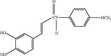
Structure of K36E
Methods
Materials and chemicals
K36E was synthesized and identified by Professor Yueh-Hsiung Kuo with a purity of 99.9% [21]. α-MSH was purchased from Merck (Darmstadt, Germany). Arbutin, 3,4-dihydroxy-l-phenylalanine (L-DOPA), DL-dithiothreitol, H-89 dihydrochloride hydrate, phenylmethanesulfonyl fluoride, and L-tyrosine were purchased from Sigma-Aldrich Chemical Co. (St. Louis, MO, USA). Fetal bovine serum (FBS), Dulbecco’s modified Eagle’s medium (DMEM) and trypsin- ethylenediaminetetraacetic acid (EDTA) were purchased from the GIBCO Invitrogen Corporation (NY, USA). An antibody recognising MITF was obtained from Abcam (Cambridge, MA, USA). Antibodies recognising p-CREB and CREB were purchased from Cell Signalling Technology, Inc. (Danvers, MA, USA). Antibodies recognising phospho-AKT, AKT, and phospho-glycogen synthase kinase 3 beta (p-GSK3β) were obtained from GeneTex, Inc. (CA, USA). Antibodies recognising actin, GSK3β, TRP-1, and tyrosinase were obtained from Santa Cruz Biotechnology, Inc. (Santa Cruz, CA, USA).
Effect of K36E on mushroom tyrosinase inhibition
The activity of mushroom tyrosinase was spectrophotometrically determined with minor modifications to the previously described procedure [7, 21–23]. Arbutin (2 mM) was the positive control. The test sample and L-tyrosine in phosphate buffer saline (PBS) and were added to a 96-well microplate (Nunc, Denmark), and mushroom tyrosinase was added. After incubation, the amount of dopachrome produced in the reaction mixture was determined at the optical density of 492 nm by using a microplate reader (Tecan, Grodig, Austria).
Cell cultures
B16F0 cells were purchased from the Bioresource Collection and Research Centre in Taiwan and cultured in DMEM supplemented with 10% FBS and 100 units/mL of penicillin and streptomycin at 37 °C in 5% CO2.
Cell viability assay
Cell growth experiments were performed using a 3-(4, 5-dimethylthiazol-2-yl)-2, 5-diphenyltetrazolium bromide (MTT) assay with minor modifications to the previously described procedure [7, 8, 24, 25]. Hydrogen peroxide was used as the positive control. The cells were cultured overnight and treated with various concentrations of K36E for 48 h, and an MTT solution was then added to each well. After incubation, a sodium dodecyl sulphate (SDS) solution was added, dissolving the formazan crystals produced in the cells. The optical density was measured at 570 nm by using a microplate reader (Tecan, Grodig, Austria).
Cellular melanin content
The melanin content in B16F0 cells was measured by using a method modified from previous studies [7, 8, 23]. The B16F0 cells were seeded in 6-well plates at a density of 7 × 10⁴ cells per well and incubated overnight. The cells were exposed to a medium containing α-MSH and K36E for 48 h. Arbutin (1 mM) was the positive control. NaOH (2 N) was added to each well to lyse the cells, which were then centrifuged. The melanin content in the supernatant was measured at 405 nm by using an ELISA reader (Tecan, Grodig, Austria).
Cellular tyrosinase activity assay
The tyrosinase activity of B16F0 cells after K36E treatment was measured with slight modification on the method described in previous studies [7, 26, 27]. B16F0 cells were plated in 24-well multidishes and incubated overnight. The cells were treated with various concentrations of K36E and were incubated for another 48 h. They were washed with PBS and lysed with 1% Triton X-100 mixed in 100 mM PBS (pH 6.8); the resultant mixture was frozen during incubation at −80 °C for 15 min and thawed at room temperature. Subsequently, the samples were centrifuged. A freshly prepared substrate (15 mM L-DOPA in a 48 mM pH 7.1 sodium phosphate buffer) was added to the supernatant and incubated. The absorbance was subsequently measured at 405 nm by using a microplate reader (Tecan, Grodig, Austria).
The rate of tyrosinase activity was calculated using the following equation:
Western blot analysis
Western blot analysis was used to demonstrate the effects of K36E on the expression of melanogenesis-related proteins in B16F0 cells as previously described [7, 8, 22, 28, 29]. B16F0 cells were seeded in a 10-cm dish for 24 h and treated with α-MSH alone (control group) or with α-MSH plus various concentrations of K36E for 48 h. The lysates were centrifuged and the protein content was determined using a Bradford reagent (Bio-Rad, Hercules, CA, USA). Twenty micrograms of protein were separated on a sodium dodecyl sulfate polyacrylamide gel electrophoresis (SDS-PAGE) gel and blotted using a polyvinylidene difluoride (PVDF) membrane (Hybond ECL, Amersham Pharmacia Biotech Inc., Piscataway, NJ, USA). The blots were blocked with 5% (w/v) skimmed milk in Tris-buffered saline containing 0.05% Tween 20 and with specific antibodies: actin (1:1000), AKT (1:5000), p-AKT (1:5000), CREB (1:1000), p-CREB (1:1000), GSK3β (1:500), p-GSK3β (1:500), MITF (1:1000), TRP-1 (1:500), and tyrosinase (1:200). The PVDF membranes were incubated with the corresponding conjugated anti-immunoglobulin G horseradish peroxidase (Santa Cruz Biotechnology, Inc.). Immunoreactive proteins were detected using an Enhanced Chemiluminescence Plus kit (Fujifilm, LAS-4000), and signal strengths were quantified using a densitometric program (MultiGauge V2.2). The results of western blot assays represented at least three individual experiments.
Statistical analyses
Values were expressed as the mean ± standard deviation from the results of at least three individual experiments. Differences in the effects of various treatments were compared using the Student’s t-test or ANOVA as well as Scheffe’s test through SPSS software (version 12.0). P values <0.05 indicated significance.
Results
Inhibition of mushroom tyrosinase activity by K36E
K36E at 1000 μM significantly reduced the mushroom tyrosinase activity. The mushroom tyrosinase inhibitory effect at 500, 750 and 1000 μM was 6.8% ± 1.6%, 14.0% ± 7.1% and 36.8% ± 1.1%, respectively. In addition, the inhibition rate of 2 mM arbutin on mushroom tyrosinase activity was 65.3% ± 2.5%.
Effect of K36E on the viability of B16F0 cells
Cell viability after treatment with 1, 1.5, 2, 2.5, 4, 5, 10, 25, and 50 μM K36E was 92.7% ± 2.0%, 91.7% ± 2.1%, 90.9% ± 2.2%, 87.8% ± 4.2%, 72.8% ± 1.0%, 68.5% ± 2.4%, 54.6% ± 1.6%, 38.3% ± 0.8%, and 28.4% ± 2.3%, respectively. Hydrogen peroxide was the positive control, and the cell viability of 0.1 μM H2O2 was 48.9% ± 7.5% after 48 h treatment. The cell viability was acceptable for developing a material for cosmetics. According to International Organization for Standardization (ISO) 10993–5:2009 (Biological Evaluation of Medical Devices), cell viability higher than 80% is considered as noncytotoxicity. The results indicated that treatment with 0.5 to 2.5 μM K36E for 48 h had no cytotoxic effect on the B16F0 cells.
Inhibition of melanin biosynthesis by K36E in B16F0 cells
Figure 2a shows the effects of K36E on melanin biosynthesis after stimulation by 0.5 μM α-MSH in B16F0 cells. The intracellular melanin content increased to 124.6% ± 13.0% after treatment with α-MSH. K36E at doses higher than 1.0 μM significantly reduced the melanin content, which decreased to 97.5% ± 1.9%, 96.6% ± 3.3%, 94.4% ± 2.8%, and 90.8% ± 1.4% (Fig. 2a). The effect of K36E on melanin biosynthesis was similar to that of 1 mM of arbutin.
Fig. 2.
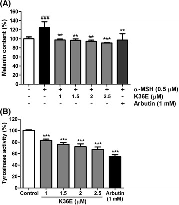
Effects of K36E on melanogenesis of B16F0 cells. a K36E reduced α-MSH-induced melanin content (%) of B16F0 cells. (Significant difference vs. control: ###, P < 0.001; significant difference vs. α-MSH-treated group: **, P < 0.01; ***, P < 0.001) and (b) tyrosinase activity (%) of B16F0 cells treated with K36E. (Significant difference vs. control: ***, P < 0.001)
Inhibition of tyrosinase activity by K36E in B16F0 cells
K36E significantly inhibited tyrosinase activity in B16F0 cells after treatment for 48 h (Fig. 2b). The levels of tyrosinase activity were 83.2% ± 2.1%, 76.3% ± 2.9%, 72.0% ± 5.0%, and 67.2% ± 4.4% after treatment with 1, 1.5, 2, and 2.5 μM K36E, respectively, for 48 h. The results indicated that K36E inhibited the melanin content of B16F0 cells through the inhibition of tyrosinase activity.
Effects of K36E on melanogenesis-related proteins
K36E downregulated tyrosinase and TRP-1 expression
To examine whether the inhibition of melanogenesis by K36E was related to the expression levels of melanogenesis-related proteins, including tyrosinase and TRP-1, B16F0 cells were incubated with α-MSH (0.5 μM) and various concentrations of K36E (1–2.5 μM) for 48 h. Although tyrosinase expression exhibited a 2.7-fold increase compared with that in the control after treatment with α-MSH, K36E significantly suppressed tyrosinase expression in a dose-dependent manner (Fig. 3). In addition, K36E significantly reduced α-MSH-stimulated TRP-1 expression at doses higher than 1.5 μM (Fig. 3).
Fig. 3.
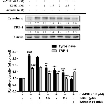
Effect of K36E on α-MSH-induced expression of tyrosinase and TRP-1 in B16F0 cells. (Significant difference vs. control: ###, P < 0.001; significant difference vs. α-MSH-treated group: **, P < 0.01; ***, P < 0.001)
K36E downregulated MITF expression
MITF expression in B16F0 cells exhibited a 1.5-fold increase compared with that in the control after treatment with α-MSH (Fig. 4). K36E treated for 4 h dose-dependently inhibited MITF expression and significantly downregulated MITF expression in the B16F0 cells at a concentration of 1 μM (Fig. 4).
Fig. 4.
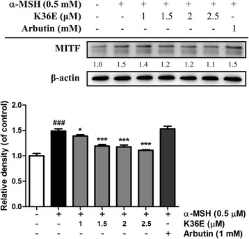
Effect of K36E on α-MSH-induced expression of MITF in B16F0 cells. (Significant difference vs. control: ###, P < 0.001; significant difference vs. α-MSH treated group: *, P < 0.05 ***, P < 0.001)
K36E downregulated p-CREB expression
p-CREB expression in B16F0 cells exhibited a 1.4-fold increase compared with that in the control after α-MSH treatment (Fig. 5). K36E significantly inhibited p-CREB expression at concentrations higher than 1.5 μM and subsequently downregulated MITF expression in the B16F0 cells.
Fig. 5.
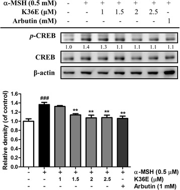
Effect of K36E on α-MSH-induced expression of p-CREB in B16F0 cells. (Significant difference vs. control: ###, P < 0.001; significant difference vs. α-MSH-treated group: **, P < 0.005)
Effects of K36E on the melanogenesis signalling pathway
K36E-inhibited melanogenesis was associated with PKA regulation
To determine whether K36E-inhibited melanogenesis was associated with PKA, B16F0 cells were incubated with 10 μM H-89, a PKA inhibitor [30], and 2.5 μM K36E for 48 h. Treatment with K36E and H-89 separately caused a 1.2- and 1.5-fold decrease in α-MSH-induced tyrosinase expression, respectively, compared with that in the control (Fig. 6). In addition, cotreatment with K36E and H-89 cause a 0.9-fold decrease in tyrosinase expression compared with that in the control. In addition, the tyrosinase expression following cotreatment was significant lower than that of K36E or H-89 treatment separately. The results indicated that the PKA pathway may be involved in the antimelanogenic effect of K36E.
Fig. 6.
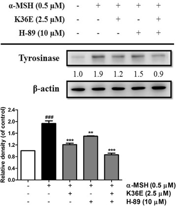
Effect of K36E on α-MSH-induced expression of tyrosinase in B16F0 cells after treatment with H-89. (Significant difference vs. control: ###, P < 0.001; significant difference vs. α-MSH-treated group: **, P < 0.01; ***, P < 0.001)
K36E inhibited melanogenesis by upregulating p-AKT and p-GSK3β expression
As shown in Fig. 7, treatment with 2.5 μM K36E for 1 h markedly increased p-AKT and p-GSK3β expression. The level of p-AKT reached a maximum (1.6-fold increase compared with that in the control) after 1 h and 1.5-fold increase after 2 h; the level of p-GSK3β also displayed a 1.2-fold increase after 1 h compared with that in the control, suggesting that K36E suppressed melanogenesis in B16F0 cells by activating the AKT and GSK3β signalling pathways, resulting in the inhibition of MITF expression and transcription activity and, thus, inhibiting the expression of the tyrosinase gene.
Fig. 7.
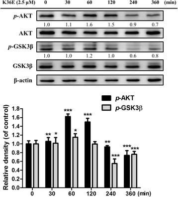
Effect of K36E on expression of p-AKT and p-GSK3β in B16F0 cells. (Significant difference vs. control: *, P < 0.05; **, P < 0.01; ***, P < 0.001)
Discussion
Tyrosinase and its activity play a major role in controlling melanogenesis [31–33]. Agents or products that inhibit tyrosinase activity have been used in skin whitening cosmetics and cosmeceuticals [1, 2, 34]. Quercetin and vanillic acid inhibited α-MSH induced the expression of MITF, tyrosinase, TRP-1, and TRP-2, causing melanogenesis inhibition [35, 36]. Resveratrol derivatives inhibited melanin synthesis through the inhibition of melanogenic enzyme expressions such as tyrosinase and TRP-1 [37]. Our results indicated that K36E inhibited tyrosinase activity and α-MSH-induced protein expression, thereby suppressing melanin biosynthesis. In addition, K36E inhibited melanogenesis-related proteins such as TRP-1. TRP-1 is considered to play a vital role both as a structural protein and catalytic enzyme in the eumelanic pathway of melanosomes [38, 39]. The results mentioned above suggested that the decrease in melanogenesis of K36E could be achieved through its inhibition on the signalling pathway that regulates tyrosinase expression and activity.
MITF is the most critical transcription factor that regulates melanogenesis by inducing the expression of melanogenic genes [5, 40]. MITF activation upregulates the expression of tyrosinase and TRP-1, and it consequently increases melanin synthesis. In this study, K36E suppressed melanogenesis by inhibiting α-MSH induced MITF expression. The cAMP pathway plays a pivotal role in α-MSH-induced melanogenesis. In previous study, cAMP-elevating agents inhibited PI3K/AKT. GSK3β may stimulate the binding of MITF to its target sequence to stimulate the expression of melanogenic enzymes and facilitate melanin production [41]. cAMP inhibits PI3K and AKT phosphorylation and activity, and reduces GSK3β phosphorylation to stimulate its activity. The activation of cAMP signal transduction results in the binding of MITF to the tyrosinase promoter, thereby leading to the stimulation of melanogenesis [41, 42]. Activation of the AKT pathway suppressed melanin synthesis by decreasing melanogenic enzymes [41]. Cordycepin was reported to inhibit α-MSH and IBMX induced melanin biosynthesis by inhibiting melanin synthesis related enzymes, such as tyrosinase, TRP-1, and TRP-2, suppressing CREB and MITF activity, and activating the PI3K/AKT pathway in B16F10 melanoma cells [43]. In our results, K36E inhibited CREB phosphorylation. K36E elevated the expression of p-AKT and p-GSK3β, possibly reducing MITF transcription to suppress tyrosinase gene expression. Previous studies have reported that the activation of AKT inhibited melanogenesis in melanocytes [33, 44]. Thus, K36E inhibited α-MSH induced hyperpigmentation caused by AKT and GSK3β activation, and subsequently downregulated MITF, CREB, tyrosinase, and TRP-1 production (Fig. 8).
Fig. 8.
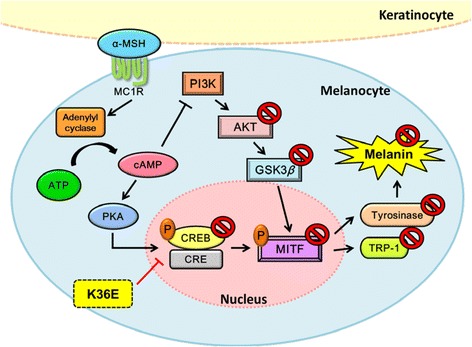
Schematic diagram showing inhibitory effects of K36E in α-MSH-induced melanogenesis
UV exposure stimulates the secretion of α-MSH in keratinocytes. α-MSH binds to MC1R in melanocytes, resulting in cAMP production and PKA activation [10]. The signal transduction related to the cAMP pathway, including the activation of PKA and CREB transcription factors, leads to the upregulation of MITF [45]. PKA subsequently phosphorylates CREB to activate MITF gene expression [46, 47]. Nicotinic acid hydroxamate inhibited melanin synthesis through the activation of the MEK/ERK and AKT/GSK3β signalling pathways in B16F10 melanoma cells [48]. Dried pomegranate concentration powder exerts whitening effects by effectively decreasing tyrosinase activity and melanin production in B16F10 cells through inactivation of the p38 and PKA/CREB signalling pathways in B16F10 cells [49]. cAMP-induced PI3K inhibition decreases AKT phosphorylation and its activation. In the present study, α-MSH-induced MITF expression was inhibited by K36E and H-89, which is a PKA inhibitor. In addition, cotreatment with K36E and H-89 significantly attenuated the K36E-induced reduction of melanin synthesis. Our results suggested that the antimelanogenic activity of K36E is associated with PKA pathway and thus leads to downregulation of MITF (Fig. 8).
Conclusion
K36E reduced MITF expression by inhibiting CREB phosphorylation. Additionally, K36E inhibited MITF expression by upregulating the phosphorylation of AKT and GSK3β, which subsequently inhibited the expression of tyrosinase and TRP-1 and thereby reduced melanin biosynthesis. Normal melanocytes and in vivo studies may be applied for further investigation into the effect of K36E on melanogenesis. In conclusion, K36E may be a candidate for the regulation of melanogenesis and it is likely to have various applications in skin whitening products in the future.
Acknowledgments
This research was supported by grants from the Ministry of Science and Technology (NSC100-2320-B-039-002-MY3; MOST104-2320-B-039-006), CMU under the Aim for Top University Plan of the Ministry of Education, Taiwan, and Taiwan Ministry of Health and Welfare Clinical Trial and Research Center of Excellence (MOHW105-TDU-B-212-133019), and China Medical University (CMU102-ASIA-18).
Availability of data and materials
The datasets supporting the conclusions of this article are included within the article.
Authors’ contributions
YHK, CYL, and HMC were responsible for the design of study and providing the research funding. PYW, CSW, and PJS designed the experiments and provided technical guidance. CCC and HMC performed the experimental operation. YHK, YHK, CYL, and HMC wrote the paper. All authors read and approved the final manuscript.
Competing interests
The authors declare that they have no competing interests.
Consent for publication
Not applicable.
Ethics approval and consent to participate
This study used commercially available cell lines; thus, ethical approval was not required.
Footnotes
Yueh-Hsiung Kuo and Po-Yuan Wu contributed equally, while Chien-Yih Lin and Hsiu Mei Chiang shared equal contribution
Contributor Information
Yueh-Hsiung Kuo, Email: kuoyh@mail.cmu.edu.tw.
Chien-Chia Chen, Email: ben00691206@hotmail.com.
Po-Yuan Wu, Email: wu.poyuan@gmail.com.
Chin-Sheng Wu, Email: cswu@mail.cmu.edu.tw.
Ping-Jyun Sung, Email: pjsung@nmmba.gov.tw.
Chien-Yih Lin, Email: yihlin@asia.edu.tw.
Hsiu-Mei Chiang, Phone: +886-4-22053366-5302, Email: hmchiang@mail.cmu.edu.tw.
References
- 1.Solano F, Briganti S, Picardo M, Ghanem G. Hypopigmenting agents: an updated review on biological, chemical and clinical aspects. Pigment Cell Res. 2006;19(6):550–71. doi: 10.1111/j.1600-0749.2006.00334.x. [DOI] [PubMed] [Google Scholar]
- 2.Chiang HM, Chen HW, Huang YH, Chan SY, Chen CC, Wu WC, Wen KC. Melanogenesis and natural hypopigmentation agents. 2012. [Google Scholar]
- 3.Costin GE, Hearing VJ. Human skin pigmentation: melanocytes modulate skin color in response to stress. FASEB J. 2007;21(4):976–94. doi: 10.1096/fj.06-6649rev. [DOI] [PubMed] [Google Scholar]
- 4.Kondo T, Hearing VJ. Update on the regulation of mammalian melanocyte function and skin pigmentation. Expert Rev Dermatol. 2011;6(1):97–108. doi: 10.1586/edm.10.70. [DOI] [PMC free article] [PubMed] [Google Scholar]
- 5.Yamaguchi Y, Hearing VJ. Physiological factors that regulate skin pigmentation. Biofactors. 2009;35(2):193–9. doi: 10.1002/biof.29. [DOI] [PMC free article] [PubMed] [Google Scholar]
- 6.Hearing VJ. Milestones in melanocytes/melanogenesis. J Invest Dermatol. 2011;131(E1):E1. doi: 10.1038/skinbio.2011.1. [DOI] [PubMed] [Google Scholar]
- 7.Chiang HM, Chien YC, Wu CH, Kuo YH, Wu WC, Pan YY, Su YH, Wen KC. Hydroalcoholic extract of Rhodiola rosea L. (Crassulaceae) and its hydrolysate inhibit melanogenesis in B16F0 cells by regulating the CREB/MITF/tyrosinase pathway. Food Chem Toxicol. 2014;65:129–39. doi: 10.1016/j.fct.2013.12.032. [DOI] [PubMed] [Google Scholar]
- 8.Wen KC, Chang CS, Chien YC, Wang HW, Wu WC, Wu CS, Chiang HM. Tyrosol and its analogues inhibit alpha-melanocyte-stimulating hormone induced melanogenesis. Int J Mol Sci. 2013;14(12):23420–40. doi: 10.3390/ijms141223420. [DOI] [PMC free article] [PubMed] [Google Scholar]
- 9.Cui R, Widlund HR, Feige E, Lin JY, Wilensky DL, Igras VE, D’Orazio J, Fung CY, Schanbacher CF, Granter SR, et al. Central role of p53 in the suntan response and pathologic hyperpigmentation. Cell. 2007;128(5):853–64. doi: 10.1016/j.cell.2006.12.045. [DOI] [PubMed] [Google Scholar]
- 10.Hocker TL, Singh MK, Tsao H. Melanoma genetics and therapeutic approaches in the 21st century: moving from the benchside to the bedside. J Invest Dermatol. 2008;128(11):2575–95. doi: 10.1038/jid.2008.226. [DOI] [PubMed] [Google Scholar]
- 11.Drira R, Sakamoto K. Isosakuranetin, a 4′-O-methylated flavonoid, stimulates melanogenesis in B16BL6 murine melanoma cells. Life Sci. 2015;143:43–9. doi: 10.1016/j.lfs.2015.10.009. [DOI] [PubMed] [Google Scholar]
- 12.Chou TH, Ding HY, Lin RJ, Liang JY, Liang CH. Inhibition of melanogenesis and oxidation by protocatechuic acid from Origanum vulgare (oregano) J Nat Prod. 2010;73(11):1767–74. doi: 10.1021/np100281g. [DOI] [PubMed] [Google Scholar]
- 13.Yeom GG, Min S, Kim SY. 2,3,5,6-Tetramethylpyrazine of Ephedra sinica regulates melanogenesis and inflammation in a UVA-induced melanoma/keratinocytes co-culture system. Int Immunopharmacol. 2014;18(2):262–9. doi: 10.1016/j.intimp.2013.11.028. [DOI] [PubMed] [Google Scholar]
- 14.Chiang HM, Chen CW, Lin TY, Kuo YH. N-phenethyl caffeamide and photodamage: Protecting skin by inhibiting type I procollagen degradation and stimulating collagen synthesis. Food Chem Toxicol. 2014;72C:154–61. doi: 10.1016/j.fct.2014.07.007. [DOI] [PubMed] [Google Scholar]
- 15.Kuo YH, Chen CW, Chu Y, Lin P, Chiang HM. In Vitro and In Vivo Studies on Protective Action of N-Phenethyl Caffeamide against Photodamage of Skin. PLoS One. 2015;10(9):e0136777. doi: 10.1371/journal.pone.0136777. [DOI] [PMC free article] [PubMed] [Google Scholar]
- 16.Shimoda H, Shan SJ, Tanaka J, Maoka T. beta-Cryptoxanthin suppresses UVB-induced melanogenesis in mouse: involvement of the inhibition of prostaglandin E2 and melanocyte-stimulating hormone pathways. J Pharm Pharmacol. 2012;64(8):1165–76. doi: 10.1111/j.2042-7158.2012.01495.x. [DOI] [PubMed] [Google Scholar]
- 17.Okombi S, Rival D, Bonnet S, Mariotte AM, Perrier E, Boumendjel A. Analogues of N-hydroxycinnamoylphenalkylamides as inhibitors of human melanocyte-tyrosinase. Bioorg Med Chem Lett. 2006;16(8):2252–5. doi: 10.1016/j.bmcl.2006.01.022. [DOI] [PubMed] [Google Scholar]
- 18.Bellei B, Pitisci A, Izzo E, Picardo M. Inhibition of melanogenesis by the pyridinyl imidazole class of compounds: possible involvement of the Wnt/beta-catenin signaling pathway. PLoS One. 2012;7(3):e33021. doi: 10.1371/journal.pone.0033021. [DOI] [PMC free article] [PubMed] [Google Scholar]
- 19.Wang L, Lu AP, Yu ZL, Wong RN, Bian ZX, Kwok HH, Yue PY, Zhou LM, Chen H, Xu M, et al. The melanogenesis-inhibitory effect and the percutaneous formulation of ginsenoside Rb1. AAPS PharmSciTech. 2014;15(5):1252–62. doi: 10.1208/s12249-014-0138-3. [DOI] [PMC free article] [PubMed] [Google Scholar]
- 20.Suwannalert P, Kariya R, Suzu I, Okada S. The effects of Salacia reticulata on anti-cellular oxidants and melanogenesis inhibition in alpha-MSH-stimulated and UV irradiated B16 melanoma cells. Nat Prod Commun. 2014;9(4):551–4. [PubMed] [Google Scholar]
- 21.Chou YC, Sheu JR, Chung CL, Chen CY, Lin FL, Hsu MJ, Kuo YH, Hsiao G. Nuclear-targeted inhibition of NF-kappaB on MMP-9 production by N-2-(4-bromophenyl) ethyl caffeamide in human monocytic cells. Chem Biol Interact. 2010;184(3):403–12. doi: 10.1016/j.cbi.2010.01.010. [DOI] [PubMed] [Google Scholar]
- 22.Chiang HM, Lin JW, Hsiao PL, Tsai SY, Wen KC. Hydrolysates of citrus plants stimulate melanogenesis protecting against UV-induced dermal damage. Phytother Res. 2011;25(4):569–76. doi: 10.1002/ptr.3302. [DOI] [PubMed] [Google Scholar]
- 23.Chiang H-M, Ko Y, Shih I, Wen K. Development of Wine Cake as a Skin-Whitening Agent and Humectant. J Food Drug Anal. 2011;19(2):223–9. [Google Scholar]
- 24.Chiang HM, Chen HC, Lin TJ, Shih IC, Wen KC. Michelia alba extract attenuates UVB-induced expression of matrix metalloproteinases via MAP kinase pathway in human dermal fibroblasts. Food Chem Toxicol. 2012;50(12):4260–9. doi: 10.1016/j.fct.2012.08.018. [DOI] [PubMed] [Google Scholar]
- 25.Salucci S, Burattini S, Battistelli M, Buontempo F, Canonico B, Martelli AM, Papa S, Falcieri E. Tyrosol prevents apoptosis in irradiated keratinocytes. J Dermatol Sci. 2015;80(1):61–8. doi: 10.1016/j.jdermsci.2015.07.002. [DOI] [PubMed] [Google Scholar]
- 26.Zhang Y, Helke KL, Coelho SG, Valencia JC, Hearing VJ, Sun S, Liu B, Li Z. Essential role of the molecular chaperone gp96 in regulating melanogenesis. Pigment Cell Melanoma Res. 2014;27(1):82–9. doi: 10.1111/pcmr.12165. [DOI] [PMC free article] [PubMed] [Google Scholar]
- 27.Park H, Song KH, Jung PM, Kim JE, Ro H, Kim MY, Ma JY. Inhibitory Effect of Arctigenin from Fructus Arctii Extract on Melanin Synthesis via Repression of Tyrosinase Expression. Evid Based Complement Alternat Med : eCAM. 2013;2013:965312. doi: 10.1155/2013/965312. [DOI] [PMC free article] [PubMed] [Google Scholar]
- 28.Chiang HM, Lin TJ, Chiu CY, Chang CW, Hsu KC, Fan PC, Wen KC. Coffea arabica extract and its constituents prevent photoaging by suppressing MMPs expression and MAP kinase pathway. Food Chem Toxicol. 2011;49(1):309–18. doi: 10.1016/j.fct.2010.10.034. [DOI] [PubMed] [Google Scholar]
- 29.Chiang H-M, Chiu H-H, Liao S-T, Chen Y-T, Chang H-C, Wen K-C. Isoflavonoid-Rich Flemingia macrophylla Extract Attenuates UVB-Induced Skin Damage by Scavenging Reactive Oxygen Species and Inhibiting MAP Kinase and MMP Expression. Evid Based Complement Alternat Med. 2013;2013:12. doi: 10.1155/2013/696879. [DOI] [PMC free article] [PubMed] [Google Scholar]
- 30.Bertolotto C, Bille K, Ortonne JP, Ballotti R. Regulation of tyrosinase gene expression by cAMP in B16 melanoma cells involves two CATGTG motifs surrounding the TATA box: implication of the microphthalmia gene product. J Cell Biol. 1996;134(3):747–55. doi: 10.1083/jcb.134.3.747. [DOI] [PMC free article] [PubMed] [Google Scholar]
- 31.Korner A, Pawelek J. Mammalian tyrosinase catalyzes three reactions in the biosynthesis of melanin. Science (New York, NY) 1982;217(4565):1163–5. doi: 10.1126/science.6810464. [DOI] [PubMed] [Google Scholar]
- 32.Tripathi RK, Hearing VJ, Urabe K, Aroca P, Spritz RA. Mutational mapping of the catalytic activities of human tyrosinase. J Biol Chem. 1992;267(33):23707–12. [PubMed] [Google Scholar]
- 33.Slominski A, Tobin DJ, Shibahara S, Wortsman J. Melanin pigmentation in mammalian skin and its hormonal regulation. Physiol Rev. 2004;84(4):1155–228. doi: 10.1152/physrev.00044.2003. [DOI] [PubMed] [Google Scholar]
- 34.Fu YT, Lee CW, Ko HH, Yen FL. Extracts of Artocarpus communis decrease alpha-melanocyte stimulating hormone-induced melanogenesis through activation of ERK and JNK signaling pathways. ScientificWorldJournal. 2014;2014:724314. doi: 10.1155/2014/724314. [DOI] [PMC free article] [PubMed] [Google Scholar]
- 35.Chou TH, Ding HY, Hung WJ, Liang CH. Antioxidative characteristics and inhibition of alpha-melanocyte-stimulating hormone-stimulated melanogenesis of vanillin and vanillic acid from Origanum vulgare. Exp Dermatol. 2010;19(8):742–50. doi: 10.1111/j.1600-0625.2010.01091.x. [DOI] [PubMed] [Google Scholar]
- 36.Kumar KJ, Yang JC, Chu FH, Chang ST, Wang SY. Lucidone, a novel melanin inhibitor from the fruit of Lindera erythrocarpa Makino. Phytother Res. 2010;24(8):1158–65. doi: 10.1002/ptr.3018. [DOI] [PubMed] [Google Scholar]
- 37.Liu Q, Kim C, Jo YH, Kim SB, Hwang BY, Lee MK. Synthesis and Biological Evaluation of Resveratrol Derivatives as Melanogenesis Inhibitors. Molecules (Basel, Switzerland) 2015;20(9):16933–45. doi: 10.3390/molecules200916933. [DOI] [PMC free article] [PubMed] [Google Scholar]
- 38.Jimenez-Cervantes C, Solano F, Kobayashi T, Urabe K, Hearing VJ, Lozano JA, Garcia-Borron JC. A new enzymatic function in the melanogenic pathway. The 5,6-dihydroxyindole-2-carboxylic acid oxidase activity of tyrosinase-related protein-1 (TRP1) J Biol Chem. 1994;269(27):17993–8000. [PubMed] [Google Scholar]
- 39.Kobayashi T, Urabe K, Winder A, Jimenez-Cervantes C, Imokawa G, Brewington T, Solano F, Garcia-Borron JC, Hearing VJ. Tyrosinase related protein 1 (TRP1) functions as a DHICA oxidase in melanin biosynthesis. EMBO J. 1994;13(24):5818–25. doi: 10.1002/j.1460-2075.1994.tb06925.x. [DOI] [PMC free article] [PubMed] [Google Scholar]
- 40.Gaggioli C, Busca R, Abbe P, Ortonne JP, Ballotti R. Microphthalmia-associated transcription factor (MITF) is required but is not sufficient to induce the expression of melanogenic genes. Pigment Cell Res. 2003;16(4):374–82. doi: 10.1034/j.1600-0749.2003.00057.x. [DOI] [PubMed] [Google Scholar]
- 41.Khaled M, Larribere L, Bille K, Aberdam E, Ortonne JP, Ballotti R, Bertolotto C. Glycogen synthase kinase 3beta is activated by cAMP and plays an active role in the regulation of melanogenesis. J Biol Chem. 2002;277(37):33690–7. doi: 10.1074/jbc.M202939200. [DOI] [PubMed] [Google Scholar]
- 42.Khaled M, Larribere L, Bille K, Ortonne JP, Ballotti R, Bertolotto C. Microphthalmia associated transcription factor is a target of the phosphatidylinositol-3-kinase pathway. J Invest Dermatol. 2003;121(4):831–6. doi: 10.1046/j.1523-1747.2003.12420.x. [DOI] [PubMed] [Google Scholar]
- 43.Shao YY, Chen CC, Wang HY, Chiu HL, Hseu TH, Kuo YH. Chemical constituents of Antrodia camphorata submerged whole broth. Nat Prod Res. 2008;22(13):1151–7. doi: 10.1080/14786410601132410. [DOI] [PubMed] [Google Scholar]
- 44.Oka M, Nagai H, Ando H, Fukunaga M, Matsumura M, Araki K, Ogawa W, Miki T, Sakaue M, Tsukamoto K, et al. Regulation of melanogenesis through phosphatidylinositol 3-kinase-Akt pathway in human G361 melanoma cells. J Invest Dermatol. 2000;115(4):699–703. doi: 10.1046/j.1523-1747.2000.00095.x. [DOI] [PubMed] [Google Scholar]
- 45.Busca R, Ballotti R. Cyclic AMP a key messenger in the regulation of skin pigmentation. Pigment Cell Res. 2000;13(2):60–9. doi: 10.1034/j.1600-0749.2000.130203.x. [DOI] [PubMed] [Google Scholar]
- 46.Shibahara S, Takeda K, Yasumoto K, Udono T, Watanabe K, Saito H, Takahashi K. Microphthalmia-associated transcription factor (MITF): multiplicity in structure, function, and regulation. J Invest Dermatol Symp Proc/Soc Invest Dermatol, Inc [and] Eur Soc Dermatol Res. 2001;6(1):99–104. doi: 10.1046/j.0022-202x.2001.00010.x. [DOI] [PubMed] [Google Scholar]
- 47.Lee JY, Choi HJ, Chung TW, Kim CH, Jeong HS, Ha KT. Caffeic Acid Phenethyl Ester Inhibits Alpha-Melanocyte Stimulating Hormone-Induced Melanin Synthesis through Suppressing Transactivation Activity of Microphthalmia-Associated Transcription Factor. J Nat Prod. 2013;76(8):1399–405. doi: 10.1021/np400129z. [DOI] [PubMed] [Google Scholar]
- 48.Huang GJ, Huang SS, Lin SS, Shao YY, Chen CC, Hou WC, Kuo YH. Analgesic effects and the mechanisms of anti-inflammation of ergostatrien-3beta-ol from Antrodia camphorata submerged whole broth in mice. J Agric Food Chem. 2010;58(12):7445–52. doi: 10.1021/jf1013764. [DOI] [PubMed] [Google Scholar]
- 49.Tsai TC, Tung YT, Kuo YH, Liao JW, Tsai HC, Chong KY, Chen HL, Chen CM. Anti-inflammatory effects of Antrodia camphorata, a herbal medicine, in a mouse skin ischemia model. J Ethnopharmacol. 2015;159:113–21. doi: 10.1016/j.jep.2014.11.015. [DOI] [PubMed] [Google Scholar]
Associated Data
This section collects any data citations, data availability statements, or supplementary materials included in this article.
Data Availability Statement
The datasets supporting the conclusions of this article are included within the article.


