Abstract
The aim of this study was to analyse transfer of mechanical energy between body segments during the glide shot put. A group of eight elite throwers from the Polish National Team was analysed in the study. Motion analysis of each throw was recorded using an optoelectronic Vicon system composed of nine infrared camcorders and Kistler force plates. The power and energy were computed for the phase of final acceleration of the glide shot put. The data were normalized with respect to time using the algorithm of the fifth order spline and their values were interpolated with respect to the percentage of total time, assuming that the time of the final weight acceleration movement was different for each putter. Statistically significant transfer was found in the study group between the following segments: Right Knee – Right Hip (p = 0.0035), Left Hip - Torso (p = 0.0201), Torso – Right Shoulder (p = 0.0122) and Right Elbow – Right Wrist (p = 0.0001). Furthermore, the results of cluster analysis showed that the kinetic chain used during the final shot acceleration movement had two different models. Differences between the groups were revealed mainly in the energy generated by the hips and trunk.
Key words: track and field, inverse dynamics, mechanical power, kinematic chain
Introduction
One example of balistic movements in sport is the shot put, which is a complex movement that involves segments’ translational and rotational motions (Lanka, 2000). The goal of the shot put is to release the shot at maximum forward velocity at an angle of approximately forty degrees (Hubbard et al., 2001; Linthorne, 2001). Nowadays, two putting styles are in general use by shot put competitors: the glide and the spin. With all putting styles, the objective is to reach a high rotational body speed and to transfer the energy to the shot (Linthorne, 2001). Transfer of mechanical energy plays an essential role in analysis of the movement. Many have examined this parameter in order to get insight into a normal human gait (Siegel et al., 2004), a gait with abnormalities (McGibbon et al., 2001) and to analyse sports performance (Zatsiorsky, 2000).
The parameter which well describes the flow of the energy through the body is mechanical power (Winter and Robertson, 1978). It has been shown that the energy transfer between two segments occurs when both segments rotate in the same direction and when there is a net moment of force acting across the joint (Winter and Robertson, 1978). The flow of mechanical energy also occurs when there is a translational movement of the joint. In movements requiring high speed generation such as the shot put, the rates of energy transfer are much higher than muscles can generate. Therefore, joint translational power is critical. In a discrete system, power is transferred through joints by joint torques and through reaction forces. The reaction forces perform work, with its power quantified as a scalar product of joint reaction force (F) and joint velocity (v). This term represents the rate of passive transfer of mechanical energy into or out of the segment from an adjacent segment, but it contains no information about which force or torque was responsible for the energy transfer. Therefore, a limitation of the segmental power technique is that the effect of a joint torque on the energy level of anatomically remote segments cannot be determined directly. Despite this limitation, some researchers have used segmental power analysis to make inferences about the mechanical energy flow (Hof et al., 1992; Meinders et al., 1998). Their techniques are based on the assumption that the increase in segmental energy can occur only if joint power is positive. Therefore, the purpose of the present study was to use a similar segmental power analysis technique to evaluate transfer of mechanical energy through all leg segments to the trunk and upper limb during the glide shot put.
Material and Methods
Participants
The study evaluated 8 male athletes with mean age of 22.3 ± 4.2 years, body mass of 108.2 ± 25.2 kg and height of 194 ± 5.1 cm. The subjects had previously participated in the national team competitions and practiced the shot put for at least five years.
The study was conducted according to the ethical guidelines and principles of the Declaration of Helsinki. The subjects provided written consent to participate in the experimental procedures, which were approved by the Senate Committee for Ethics in Scientific Research of the Józef Piłsudski Academy of Physical Education in Warsaw.
Measures
Before the experiment, anthropometric measurements were taken for each person. Next, thirty-four spherical markers were placed at anatomical landmarks according to the biomechanical model PlugInGait standards available within the motion capture system (Vicon Motion Systems Ltd, Oxford, UK). Three force plates (Kistler Holding AG, Winterthur, Switzerland) embedded into the floor were used to measure ground reaction force (GRF) data at a sampling rate of 1000 Hz. The motion capture system consisting of nine infra-red cameras was employed to collect kinematic data at a sampling rate of 100 Hz. The force plates were synchronized with the motion capture system. Before the trials were conducted, both systems were calibrated according to the manufacturers’ recommendations.
Procedures
The experiment was carried out in an indoor hall adapted to conduct biomechanical tests of the shot put. Each subject performed three trials of the shot put using the glide technique. The athletes threw a special 7.26 kg ball made of durable flexible polyvinyl chloride (PVC) and filled with pellets. A net was suspended at a distance of 6 m from the platforms to catch a shot put ball after the spin was executed. The analysis was performed based on the trials without any random mistakes, with the individual performing the task naturally and the trial evaluated by the coach as good. Further analysis was based on power and energy profiles obtained during the shot put performance.
Data analysis
Kinetic data of the shot put glide were obtained from the Vicon system. The signals were filtered conventionally with a fourth-order zerophase Butterworth filter to eliminate noise from the raw data. Using MatLab (MathWorks, USA) software, the data were normalized with respect to time using the algorithm of the fifth order spline and their values were interpolated with respect to the percentage of total time, assuming that the time used during the final weight acceleration movement was different for each putter. The first foot contact with the Kistler plate immediately after the glide was adopted as the beginning of the movement. The end of the movement was the moment when the ball left the athlete’s hand. Joint power (P) calculated from Vicon was the dot product of the moment vector (M) and the angular velocity vector (ω), i.e.
P = Mxωx + Myωy + Mzωz.
A detailed motion analysis was focused on energy transfer. Evaluation of mechanical energy transfer between body segments required an analysis of actual sources of mechanical power. The Riemann integral was used to compute mechanical energy expenditure:
where: P(ti) – joint power at the point ti.
The interval [t1, t2], where t1 denotes the initial point and t2 is the end of the movement was divided into smaller intervals with the length of 0.01 s consistent with the frequency of recording. Each term in the sum was the product of the value of the function at a given point and the length of an interval. Consequently, each term represented the area of a rectangle with height P(ti) and width xi+1 – xi = 0.01. The following assumptions were made for the analysis:
The Riemann sum is the area of all the rectangles.
Maximum energy value in the individual joints in the whole movement is represented by P(ti)(xi+1 – xi).
Statistical Analysis
The first step was to calculate Pearson’s correlation in order to find the dependence between personal best and body mass. Furthermore, the t-test for the Riemann sum was used to calculate statistical significance of energy transfer from segment to segment. In order to perform a more detailed analysis of the energy flow, the highest values of energy P(ti)(xi+1 – xi) for lower limbs and right upper limb joints were exported to Statistica 12.0 software (StatSoft, PL). Based on these data, the k-means clustering method was used to find differences in the shot put technique among the analyzed athletes. The procedure aimed to classify a given data set through a certain number of clusters. The number of clusters was chosen automatically by the software. The main idea was to define k number of centroids (one for each cluster) in a way that the centroids were placed as far from each other as possible. The next step was to take each point belonging to a given data set and associate it to the nearest centroid. The program moved objects between those clusters with the goal to minimize variability within clusters and maximize variability between clusters. The clustering method used Euclidean distances between objects when forming the clusters.
Results
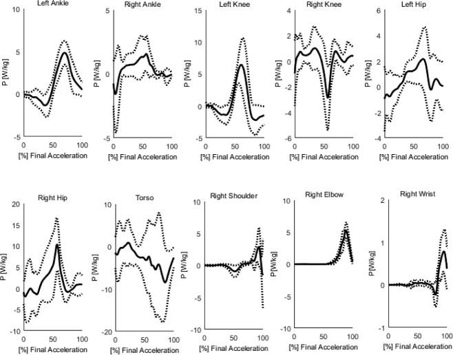
The shot put kinetic variables, consisting of ankle, knee, hip, torso, shoulder, elbow and wrist joint power are shown for each subject in Figure 1. These data were expressed relative to body mass. Furthermore, power in time domain was positive when the body generated energy through concentric muscle activity and negative when the body absorbed the energy through eccentric muscle activity or extension of the soft tissue. The between-subject variability, reflected by the magnitude of the standard deviation envelopes, was greater for the torso and hip variables than for the knee or ankle in the lower limbs, whereas the envelopes for the upper limbs were relatively small. The highest average maximum value of normalized power was observed in the hip joint whereas the lowest was found for the wrist.
Figure 1.
Mean profiles and standard deviations of power for the final acceleration for lower and upper limbs during the shot put.
Table 1.
Characteristics of individuals with personal best in the shot put, from: * http://www.domtel-sport.pl/statystykaLA/.
| Athlete | Age (years) | Body mass (kg) | Height (cm) | Personal best (m)* |
|---|---|---|---|---|
| Subject 1 | 19 | 71 | 192 | 9.01 |
| Subject 2 | 21 | 82 | 193 | 13.74 |
| Subject 3 | 31 | 141 | 204 | 21.95 |
| Subject 4 | 24 | 92 | 197 | 14.41 |
| Subject 5 | 22 | 132 | 199 | 18.71 |
| Subject 6 | 17 | 119 | 191 | 16.47 |
| Subject 7 | 23 | 88 | 192 | 14.55 |
| Subject 8 | 25 | 120 | 191 | 19.67 |
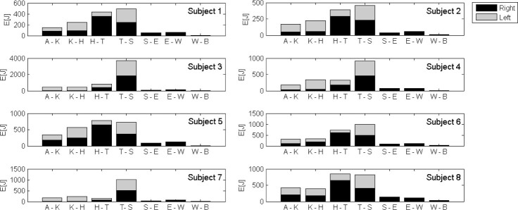
The energy of motion was defined as the work that would be performed by the body possessing the energy when it was brought to rest or the velocity was changed. A strong correlation was found in the group between personal best and body mass (r = 0.9414). Therefore, an alternative approach was to express shot put energy transfer in a dimensionless form as presented in Figure 2. In the study group, energy transfer was statistically significant between the following body segments: Right Knee – Right Hip (p = 0.0035), Left Hip – Torso (p = 0.0201), Torso – Right Shoulder (p = 0.0122) and Right Elbow – Wrist (p = 0.0001). Average values of energy transfer between Right Knee and Right Hip rose from 107.82 to 401.11 J, and between Left Hip and Torso from 159.20 to 1137.06 J. For the other two segments, transfer decreased significantly: Torso – Right Shoulder (1137 06 – 68.73 J) and Right Elbow – Wrist (92.75 – 15.67 J).
Figure 2.
Transfer of energy for each subject from the distal to the proximal segments according to the pattern: A – K (Ankle – Knee), K – H (Knee – Hip), H – T (Hip – Torso), T – S (Torso – Shoulder), S – E (Shoulder – Elbow), E – W (Elbow – Wrist), W – B (Wrist – Ball).
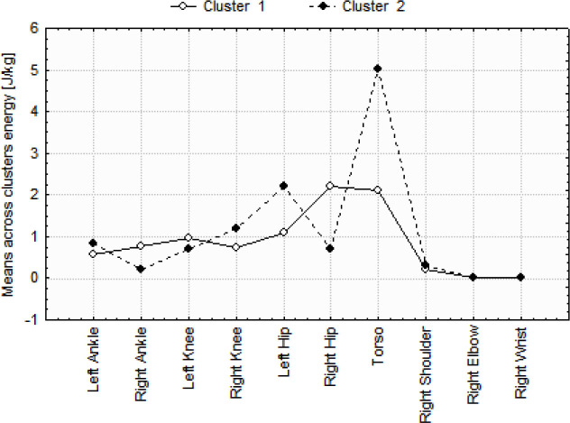
The subjects did not generate the same mechanical energy in the individual joints during the shot put. Therefore, the cluster method was used to examine whether there were subgroups of athletes who preferred different techniques of the shot put (Figure 3).
Figure 3.
Division of the shot put athletes according to the criterion of the maximal value of energy in the joints of lower and upper limbs.
The analysis revealed that the athletes formed two clusters, with cluster 2 represented by subjects 3 and 6 and other athletes forming cluster 1. Differences between the groups were found mainly for the values of energy generated by the hips and trunk.
Discussion
The study of biomechanics is critical for understanding the way in which the human body moves when engaged with a multitude of different activities (Watkins, 2014). The shot put seems to be a relatively easy task. However, there are numerous biomechanical factors that determine whether the performance will be successful. These factors include: the kinetic chain, optimum angle/height of release, dynamics and speed, as well as throwing technique i.e. gliding or rotational (Blazevic, 2010). The shot put is placed into the open kinetic chain movement category because the individual is able to move the hand freely when pushing the shot; however, the shoulder movement is constricted as it is attached to the body. A considerable disadvantage is that the individual is not able to produce a shot at a greater speed due to the muscle movement within the body (Blazevic, 2010). To execute a successful movement in the shot put, the individual rotates the torso and the rear knee is bent. There is an upward motion that relies on the power to be generated through extending the knee that is bent. As argued by Zatsiorsky (2000), a key component of the shot put performance is the position of specific segments of the lower limb for right-handed athletes. Angles in the ankle and knee should be such that the resultant vector of gravity of the whole body is in the area of the forefoot. One of the mistakes in the shot put technique is the withdrawal of the pelvis backwards. This reduces the likelihood of generating the maximum muscle force by the hip extensors and results in unwanted lowering of the position of the center of mass. Consequently, the athlete loses the distance of the throw by up to 0.5 meters. The athlete applies the force to the ground with the bent knee and the ground reacts with an opposite force which is transferred through the body to the throwing arm as the shot is released. The front leg remains straight and the shot is pushed by the tips of the fingers at an optimal angle.
Byun et al. (2008) presented a biomechanical review of the performance of world top shot putters. The authors analysed the above mentioned release parameters, shot trajectory and velocity, linear and angular momentum of body segments and duration of the movement. All of the parameters differed from athlete to athlete, but the authors pointed out that proper acceleration of the athlete-shot system was the key factor in ensuring the energy source necessary for the delivery. Therefore, together with body mass, the body speed at the end of the approach has a strong influence on the kinetic energy obtained by the thrower. The kinetic energy is energy of motion and is defined as the work that will be performed by the body possessing the energy when it is brought to rest or the velocity is changed. Energy transfer between segments plays an important role in performance of a wide variety of human motions. For example, it is important for the economy of human walking (Umberge et al., 2013) and essential in any sport performance involving high speed movements like kicks, pitches and strokes. Commonly used methods for investigating the mechanical energy flow during the gait are based on kinematics of the center of mass (Bennett et al., 2005), segment kinematics (Olney et al., 1987) or joint power (Robertson and Winter, 1980).
Zelik and Kuo (2010) showed the values of joint power generated during the gait. Extreme values of power obtained in our study in the group of throwers were twice as great in the ankle joint than those recorded for walking, six times greater in the knee joint and 10 times in the hip joint. Although joint power does represent the net effect of a muscle group on the mechanical energy of the entire body, it does not adequately reveal the role of a muscle group in changing the energy level of individual body segments. The local effects of energy transfer can be several times greater than the magnitude of the net joint power and even opposite in sign (Blazevic, 2010). In our experiment, average values of energy transfer between Right Knee and Right Hip increased significantly from 107.82 to 401.11 J whereas for the transfer between Left Hip and Torso, they increased from 159.20 to 1137.06 J. For other two pairs of segments, the values decreased substantially, from 1137.06 to 68.73 J for Torso – Right Shoulder and from 92.75 to 15.67 J for Right Elbow – Wrist. Hence, incorrect timing of the shot put can lead to an injury of any of the rotator cuff muscles. Various tears of the long head tendon of biceps brachii and the wrist and finger flexors and extensors originating from the humeral epicondyles are associated with several shot-put technique faults. These include: poor coordination of arm and trunk muscles, the putting elbow being too low or ahead of the shot and ‘dropping’ the shoulder on the non-throwing side. Incorrect positioning of the thumb can lead to the injury of the extensor policis longus muscle (Bartlett and Bussey, 2011). Therefore, performance of a throwing technique is impossible without power and conversely, high power cannot be generated without a good technique. This has to be the overriding principle in both training and performance diagnosis.
In conclusion, the flow of mechanical energy throughout the kinematic chain is one of the most important criteria for evaluation of the shot put skills. The direction and value of the mechanical energy indicate the segments where energy is dissipated. The results in the shot put are substantially affected by the movements of the right leg and trunk for right-handed athletes. The limb acts as a stabilizer and has to transfer to the entire energy generated by the main muscle groups of the lower limb and trunk to the ball. Analysis of the mechanical energy flow showed a large variation in the inter-individual technique of gliding shot put technique between the athletes who volunteered to participate in our experiment.
Acknowledgements
Funding for this project was provided by the Ministry of Science and Higher Education under Grant RSA2 011 52.
Authors submitted their contribution to the article to the editorial board.
References
- Bartlett R, Bussey M. Sports Biomechanics: Reducing Injury Risk and Improving Sports Performance. Routledge; Chapman Hall: 2011. [Google Scholar]
- Bennett B, Abel M, Wolovick A, Franklin T, Allaire P, Kerrigan D.. Center of mass movement and energy transfer during walking in children with cerebral palsy. Arch Phys Med Rehabil. 2005;86:2189–94. doi: 10.1016/j.apmr.2005.05.012. [DOI] [PubMed] [Google Scholar]
- Blazevic A. Sports Biomechanics. The Basics: Optimising Human Performance. London: A&C Black Publishers Ltd; 2010. [Google Scholar]
- Byun K, Fujii H, Murakami M, Endo K, Takesako H, Gomi K, Tauchi K.. A biomechanical analysis of the men’s shot put at the 2007 World Championships in Athletics. New Studies in Athletics. 2008;2:53–62. [Google Scholar]
- Hof A, Nauta J, van der Knaap E, Schallig M, Struwe D.. Calf muscle work and segment energy changes in human treadmill walking. J Electromyogr Kinesiol. 1992;2(4):203–16. doi: 10.1016/1050-6411(92)90024-D. [DOI] [PubMed] [Google Scholar]
- Hubbard M, De Mestre N, Scott J.. Dependance of release variables in the shot put. Journal of Biomechanics. 2001;34:449–456. doi: 10.1016/s0021-9290(00)00228-1. [DOI] [PubMed] [Google Scholar]
- Lanka J. Zatsiorsky VM. Shot putting. Oxford: Blackwell Science; 2000. Biomechanics in Sport, Performance Enhancement and Injury Prevention; pp. 435–457. [Google Scholar]
- Linthorne N.. Optimum release angle in the shot put. Journal of Sports Sciences. 2001;19:359–72. doi: 10.1080/02640410152006135. [DOI] [PubMed] [Google Scholar]
- McGibbon C, Puniello M, Krebs D.. Mechanical energy transfer during gait in relation to strength impairment and pathology in elderly women. Clinical Biomechanics. 2001;16(4):324–33. doi: 10.1016/s0268-0033(01)00004-3. [DOI] [PubMed] [Google Scholar]
- Meinders M, Gitter A, Czerniecki J.. The role of ankle plantar flexor muscle work during walking. Scandinavian Journal of Rehabilitation Medicine. 1998;30(1):39–46. doi: 10.1080/003655098444309. [DOI] [PubMed] [Google Scholar]
- Olney S, Costigan P, Hedden D.. Mechanical energy patterns in gait of cerebral palsied children with hemiplegia. Phys Ther. 1987;67:1348–54. doi: 10.1093/ptj/67.9.1348. [DOI] [PubMed] [Google Scholar]
- Robertson D, Winter D.. Mechanical energy generation, absorption and transfer amongst segments during walking. Journal of Biomechanics. 1980;13(1):845–854. doi: 10.1016/0021-9290(80)90172-4. [DOI] [PubMed] [Google Scholar]
- Siegel K, Kepple T, Stanhope S.. Joint moment control of mechanical energy flow during normal gait. Gait and Posture. 2004;19(1):69–75. doi: 10.1016/s0966-6362(03)00010-9. [DOI] [PubMed] [Google Scholar]
- Umberger B, Augsburger S, Resig J, Oeffinger D, Shapiro R, Tylkowski C.. Generation, absorption, and transfer of mechanical energy during walking in children. Medical Engineering Physics. 2013;35(1):644–651. doi: 10.1016/j.medengphy.2012.07.010. [DOI] [PubMed] [Google Scholar]
- Watkins J. Fundamental Biomechanics of Sport and Exercise. New York: Hoboken Taylor and Francis; 2014. [Google Scholar]
- Winter DA, Robertson DG.. Joint torque and energy patterns in normal gait. Biological Cybernetics. 1978;29(3):137–142. doi: 10.1007/BF00337349. [DOI] [PubMed] [Google Scholar]
- Zatsiorsky V. Biomechanics in sport. Performance enhancement and injury prevention. Oxford: Blackwell Science; 2000. [Google Scholar]
- Zelik K, Kuo A.. Human walking isn’t all hard work: evidence of soft tissue contributions to energy dissipation and return. The Journal of Experimental Biology. 2000;213:4257–64. doi: 10.1242/jeb.044297. [DOI] [PMC free article] [PubMed] [Google Scholar]


