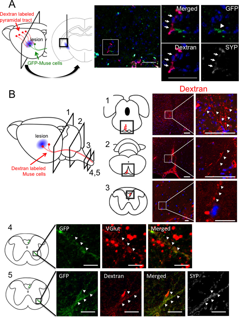
Figure 4. Involvement of Muse cells into reconstruction of pyramidal tract.

(A) Eight weeks after engraftment of FACS-isolated GFP-Muse cells. Motor cortical neurons were anterogradely labeled with dextran. Interruption of nerve axons in pyramidal tract was confirmed in Fig. 1B. FACS-isolated GFP-Muse cells (arrowhead) transplanted nearby the lesion was observed to connect to dextran-labeled motor neuron axons (red, arrows) which merged with synaptophysin (SYP, arrows), suggesting that Muse cells connected with motor cortex neurons and participated in the restoration of pyramidal tract. (B) Eight weeks after engraftment of MACS-sorted Muse-rich cells (data from level 1–3) and FACS-isolated GFP-Muse cells (data from level 4, 5). Dextran labeled axons (red, arrowheads) were detected at level 1: midbrain and level 2: medulla in the ipsilateral side, and in cervical spinal cord at level 3–5 in the contralateral side. Level 4 and 5 show the area of anterior horn in the spinal cord where pyramidal tract axons formed synapses with motor neurons. GFP+ neurite were positive for VGlut, suggesting differentiation of Muse cells into glutamatergic neuron (Level 4). Dextran (red)-labeled GFP (green) positive Muse cells were shown to be positive for synaptophysin (white) in upper cervical spinal cord (Level 5). Scale bars, A = 100 µm; B-Level 1 and 2 = 50 µm; Level 3–5 = 10 µm.
