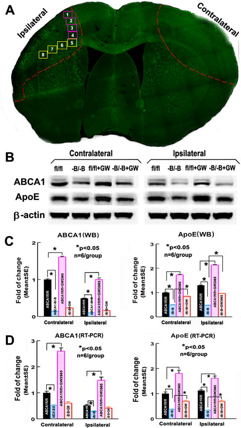
Figure 1.

ABCA1−B/−B decreases brain ABCA1/ApoE level; GW3965-treatment increases ABCA1/ApoE level in the ABCA1fl/fl-brain, but not in ABCA1−B/−B-brain 14 days after dMCAo. A: Confocal-micrograph photo schematically shows the areas where the images were taken for Syn (square 1–4) or LFB/BS/SMI31/PDGFRα (square 5–8), and ipsilateral-ischemic tissue and contralateral brain tissue (outlined areas); B: Western blot (WB) image; C: quantitative data of WB; D: RT-PCR assay. *p<0.05, n=6/group.
