Abstract
It has been described that the frequency ranges at which theta, mu and alpha rhythms oscillate is increasing with age. The present report, by analyzing the spontaneous EEG, tries to demonstrate whether there is an increase with age in the frequency at which the cortical structures oscillate. A topographical approach was followed. The spontaneous EEG of one hundredand seventy subjects was recorded. The spectral power (from 0.5 to 45.5 Hz) was obtained by means of the Fast Fourier Transform. Correlations of spatial topographies among the different age groups showed that older groups presented the same topographical maps as younger groups, but oscillating at higher frequencies. The results suggest that the same brain areas oscillate at lower frequencies in children than in older groups, for a broad frequency range. This shift to a higher frequency with age would be a trend in spontaneous brain rhythm development.
Electronic supplementary material
The online version of this article (doi:10.1007/s11571-016-9402-4) contains supplementary material, which is available to authorized users.
Keywords: Spectral power, Development, Individual alpha peak frequency, Frequency shifting, Topography, Children
Introduction
The spontaneous EEG is a continuous and rhythmic activity recorded at scalp electrodes, related to the neurofunctional brain states. It can provide clues about the brain state in normal and pathological subjects and about developmental changes across the lifespan. Two main descriptors of developmental changes from childhood to adulthood are the spectral power (SP) and the frequency at which the alpha peak is reached (iAPF: individual alpha peak frequency).
The decrease in absolute spectral power in all frequency bands has been widely found to be one of the landmarks during EEG child development, and it is fulfilled at all the frequencies investigated for the absolute SP (Matousek and Petersén 1973; Gasser et al. 1988; Cragg et al. 2011; Lüchinger et al. 2011; Rodríguez-Martínez et al. 2012). The most common hypothesis proposed to explain the decrease in the absolute SP during development refers to the reduction in gray matter associated with maturation (Whitford et al. 2007; Feinberg and Campbell 2010; Shaw et al. 2006, 2008). In fact, a parallel developmental trajectory between the reduction in cortical thickness and the decrease in spectral power with age has been observed (Whitford et al. 2007). This relationship, although not necessarily causal, suggests a direct link between the reduction in gray matter with age and the decrease in brain rhythm amplitude. The most likely neuronal mechanism would be the process of synaptic pruning during maturation (Keshavan et al. 2002; Giedd 2004), where a large number of synapses that do not receive enough trophic factors are eliminated.
Quite often, and in order to eliminate the high individual variability in the EEG, the EEG SP is normalized as the so-called relative SP. The most frequent result consists of an increase in relative SP for high frequencies (Alpha, Beta and Gamma) and a decrease in relative SP for low frequencies (Delta and Theta) as maturation progresses. The results can be interpreted as an increase in the preponderance of high frequencies in the action mode of cortical circuits as maturation progresses (Segalowitz et al. 2010). This pattern of maturation of the normalized amplitude of EEG rhythms during development has also been replicated during sleep, although an increase in theta rhythm with age was also reported in this case (Chu et al. 2014).
Another important parameter is the iAPF, which is more visible in posterior scalp areas. The iAPF value increases during development, and the value of 10 Hz is reached at primary school age. This value corresponds to the most common frequency of the alpha adult’s EEG. Studies have proposed that adult values for iAPF are achieved at the age of 11 (Lindsley 1939; Niedermeyer and Lopes da Silva 1999). In children from 7 to 11 years old, there is an increase of 1 Hz in the iAPF (Miskovic et al. 2015), but it was recently found that the increase in alpha frequency continues at least until the age of 15 (Marcuse et al. 2008). Cragg et al. (2011) found that during adolescence there is a decrease in the amplitude of slow waves, an increase in fast rhythms, and an increase in the iAPF. They did not find any iAPF differences based on gender.
The iAPF parameter is not only important as a physiological marker, but it has also been shown to be directly related to behavioral measures such as reaction times and short-term memory performance in adults (Klimesch 1999). In children, it has also been demonstrated to be related with locomotor activities (Mierau et al. 2016). The iAPF has been considered one of the most robust markers of neurological maturation (Valdés et al. 1990). It has been suggested that the increase from low to high frequencies in the brain rhythms and the increase in iAPF from childhood to adulthood would be due to the increase in the speed of neural communication, basically produced by the increase in speed of action potentials, due to myelination and/or an increase in the axon diameter (Segalowitz et al. 2010; Barriga-Paulino et al. 2011; Thorpe et al. 2016).
The central mu rhythm, related to motor inactivity, has also shown a consistent frequency oscillation increase with maturation. During the first years of life, mu oscillates at around 6.4–8.4 Hz, and in pre-school children it increases to 8.4–10.4 Hz (Orekhova et al. 2006; Marshall et al. 2002; Thorpe et al. 2016). Berchicci et al. (2011) also found an increase in mu frequency from infants to preschool children and adults, where mu frequency stabilizes around 10 Hz. Orekhova et al. (2006) used a functional method to characterize the frequency of theta rhythm in infants and pre-school children, by provoking theta reactivity with speech and toy inspection. They found an increase in the upper frequency bound of theta rhythm from infants to pre-school children. The increase ranged from 3.6 to 5.6 Hz in infants to 4–8 Hz in pre-school children.
Alpha, consistently studied across age, and mu and theta, studied in more limited age groups, have demonstrated increases in frequency oscillations related to child development. These rhythms have been characterized as independent entities from the background EEG, thanks to the presence of defined spectral peaks and topographies and their reactivity to external illumination (alpha), visualized or produced movements (mu), and cognitive and emotionally demanding stimulation (theta). However, increases in brain rhythm frequency might not be exclusively restricted to these rhythms, and other rhythms, such as delta, beta and gamma, could also increase their frequency with age. This possibility is also suggested by the Principal Component Analysis (PCA) of SP in a limited sample of preadolescents and young adults, where the loading components representing the variance related to rhythms from theta to beta showed a slight shift to higher frequencies from preadolescents to young adults (Rodríguez-Martínez et al. 2012).
The present report tries to demonstrate whether there is a systematic increase with age in the frequency at which the cortical structures spontaneously oscillate. The hypothesis is that in a broad frequency range, from 0.5–45.5 Hz, there are similar topographies at different ages, but with children oscillating at lower frequencies than older subjects. This hypothesis would be explored through spatial correlations of absolute SP maps. The confirmation of the hypothesis would imply that one of the natural trends of brain maturation is to increase the frequency of cortical oscillations across the scalp in a broad frequency range.
Results on amplitude of power spectral density of this same group of data have been presented elsewhere for other scientific proposals (Rodríguez-Martínez et al. 2015).
Methodology
Experimental human subjects
The study included a sample of 172 human subjects between 6 and 26 years old (4 of each gender for each year of age). Five subjects were excluded from the analysis due to excessive EEG artifacts in the recordings. The final sample comprised 154 right-handed subjects and 13 left-handed. The left-handed subjects were maintained in the analyzed population in order to increase the generalizability of the results. The male group consisted of 82 subjects (mean ± standard deviation of age: 15.94 ± 6.06), 75 right-handers and 7 left-handers. The female group consisted of 85 subjects (mean ± standard deviation of age: 15.73 ± 6.10), 79 right-handers and 6 left-handers.
Subjects did not report any neurological disease or psychological impairments, and they were extracted from a middle socio-economic background. The children had normal academic records, and the young adults were college students. Experiments were conducted with the informed and written consent of each participant (parents/tutors in the case of children), following the Helsinki Protocol. The study was approved by the Ethical Committee of the University of Seville.
EEG recording
The EEG was recorded during 3 min of spontaneous activity in an eyes-open condition. This period of time is considered sufficient for recording, given that test–retest reliability for EEG in periods of 60 s is 92 % (Salinsky et al. 1991).
Subjects were asked to stay calm and look at the screen, blink as little as possible, and keep their eyes focused on a white cross at the center of the black screen. They were recorded at different times of the day between 12 and 8 pm. No information about previous sleep was required. Recordings were obtained from an average reference of 32 scalp sites from the international system (Fp1, Fpz, Fp2, F7, F3, Fz, F4, F8, FC5, FC1, FC2, FC6, M1, T7, C3, Cz, C4, T8, M2, CP5, CP1, CP2, CP6, P7, P3, Pz, P4, P8, POz, O1, Oz, O2), using tin electrodes mounted on an electrode cap designed for this purpose (ElectroCap). Eye movements were recorded by two electrodes at the outer canthus of each eye for horizontal movements and by electrodes placed above and below the left eye for vertical movements.
All the scalp electrodes were re-referenced offline to the mastoid average (M1 + M2)/2. Impedance was maintained below 10 KiloOhm (KΩ) throughout the recording time. Data were recorded in direct current mode at 512 Hz, with a 20,000 amplification gain using a commercial analog digital acquisition and analysis board (ANT). Data were not filtered during recording.
Data analysis
EEG recordings were analyzed with the EEGLAB (Delorme and Makeig 2004) and Matlab 2010a software packages. To eliminate alternating current power line interference and blink artifacts on the EEG, an independent components analysis was performed. These components were discarded, and the EEG signal was reconstructed. The segmented epochs lasted 2000 ms. All the epochs where the EEG exceeded ±100 Microvolts (μV) in any channel were automatically discarded.
The analyzed time in the power spectral analysis was less than what was initially recorded (90 epochs of 2 s), due to the elimination of epochs containing artifacts. The average time analyzed in the sample’s EEG recordings was 2′32″ (mean ± standard deviation: 2′32″ ± 35.89; minimum time 46″), while the average time recorded in the different age groups was: (a) Children: 6–9 years old, 2′9″ (mean ± standard deviation: 2′9″ ± 44.2, minimum time 48″); (b) Pre-adolescents: 10–13 years old, 2′34″ (mean ± standard deviation: 2′34″ ± 32.45, minimum time 48″); (c) Adolescents: 14–17 years old, 2′32″ (mean ± standard deviation: 2′32″ ± 38, minimum time 46″); (d) Youngsters: 18–21 years old, 2′47″ (mean ± standard deviation: 2′47″ ± 23.51, minimum time 72″); and (e) Young adults: 22–26 years old, 2′39″ (mean ± standard deviation: 2′39″ ± 28.33, minimum time 66″).
The SP of individual epochs was computed by means of the EEGLAB function spectopo. This function uses the pwelch function of matlab which applies a hamming window and computes PSD with the frequency resolution of 1 Hz. In order to increase the frequency resolution, one single value was linearly interpolated between each two recorded points. This procedure permitted to increase frequency resolution to 0.5 Hz. The spectopo function was also applied to the EEG recordings without data interpolation, and the same results of frequency shifting with age, described in the results section, were obtained. However, the lack of interpolation produced lower frequency resolution (1 Hz). The SP was computed in windows of 2 s and expressed in logarithms. The SP obtained from individual trials was averaged in each individual subject.
The EEG frequencies in the range from 0.5 to 45.5 Hz were selected for analysis. EEG frequencies above 45.5 Hz were excluded from the analysis in order to reduce the impact of possible electromyographical signals and the 50 Hz power line contamination of EEG recordings.
Analysis of the SP: differences in SP in different age groups
Using the Statistical Package for the Social Sciences (SPSS) 20.0, a mixed-model Analysis of Variance (ANOVA) was applied to the logarithm of the SP to compare the SP of the five age groups. The age groups were considered the between-subjects factor, and electrodes were considered the within-subject factor. The electrodes FPz, Fz, Cz, Pz, POz, Oz were the levels of the electrodes factor. Eight independent ANOVAs were computed for the eight different brain rhythms considered: low-delta (frequency range: 0–1 Hz); delta (frequency range: 2–3 Hz); theta (frequency range: 4–7 Hz); low-alpha (8–10 Hz); high alpha (11–14 Hz); low-beta (15–20 Hz); high-beta (21–35 Hz) and gamma (36–45.5 Hz). For each frequency band, the Bonferroni corrected means comparison t tests were computed for the different groups of subjects when the group factor was significant.
Finally, and for representation purposes, we computed the average of the logarithm of the SP in each age group for the eight different brain rhythms considered, and the associated standard error (2* SE).
The results relative to the amplitude of SP are presented as supplemental table and figure, given that they are marginal for the increased frequency of oscillations with age hypothesis of present report. The report of changes of SP with age intend to demonstrate that the different signal analysis used in present report did not change the results previously obtained of a reduction of SP power with age in most frequency bands (Rodríguez-Martínez et al. 2015).
Alpha peaks: differences in alpha frequency for different age groups
The specific frequency at which each of the analyzed subjects reached the peak amplitude in the alpha range was calculated using an automated algorithm. This was done in the three most occipital electrodes, where the alpha rhythm appears with greater intensity (O1, Oz and O2).
The iAPF was considered to be the frequency with the highest amplitude in the frequency range between 7–13 Hz. Then, two ANOVAs were computed, with age group as between-subjects factor (five levels) and the occipital electrodes (O1, Oz and O2) as within-subjects factor. In the first ANOVA, the iAPF was analyzed, and in the second ANOVA, the alpha amplitudes at the iAPF were analyzed. For the post hoc tests, the Bonferroni correction was applied to make comparisons between different age groups. Finally, the logarithmic regression of the alpha peak frequency with the age of the subjects was computed.
Absolute SP topography of the different frequencies
The topographies of absolute SP from 0.5 to 27.5 Hz were represented to appreciate whether different age groups showed similar topographies at different frequencies. These topographies were computed to assess the hypothesis of the present report: the same brain areas oscillate at different frequencies at different ages.
Using EEGLAB, the topography of the average of the SP logarithm obtained in each of the analyzed electrodes was represented from 0.5 to 27.5 Hz in each age group. The 27.5 Hz frequency was chosen as the frequency limit for the analysis because only very small topographical changes appeared after 27.5 Hz.
Inter-group/inter-frequency correlations
In order to quantify whether similar SP topographies appeared at different oscillatory frequencies in different age groups, correlation matrices of the SP topographies between different age groups were computed. The SP topography of each computed frequency (0.5–45.5 Hz) for each age group was correlated with the topographies of the other age groups. This approach would make it possible to observe whether similar topographies were reached at different frequencies in different age groups.
Correlations were computed with Spearman’s rank correlation coefficient. The two-tailed statistical significance of the correlations between different frequencies was estimated. The correlation values were thresholded using the Bonferroni correction for multiple comparisons p < (0.05/(91 × 91) = 0.000006). This p value was applied as a threshold value for the rejection of the null hypothesis: there is no significant correlation between analyzed frequencies and groups.
Correlations between specific frequencies
In order to obtain the magnitude of the frequency differences in the SP topographies of different age groups and obtain a similar topography, the SP topographies in certain predefined frequencies (1, 5, 10, 15, 20, 30 Hz) of the children’s group were correlated with the SP topographies of all the other groups. These frequencies were selected in order to cover delta, theta, alpha, low beta and high beta frequency ranges. In addition, the group of youngsters was also correlated with the young adult group.
Results
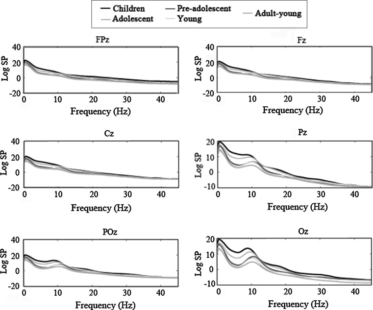
Figure 1 shows the logarithm of the absolute SP for each age group in six midline electrodes. This figure shows a decrease in SP whit age. The inter-group differences in SP are higher at low frequencies: delta, theta, alpha, and in the more posterior electrodes: Pz, POz and Oz. In addition, in the alpha band there is an increase of peak frequency with age, resulting in a peak that occurs at slightly higher frequencies in older subjects than in younger subjects.
Fig. 1.
Grand average of the Logarithm of the spectral power (Log SP) in each age group (children: 6–9 years, preadolescents: 10–13 years, adolescents: 14–17 years, youngsters: 18–21 years, young adults: 21–26 years); from 0.5 to 45.5 Hz, in six midline electrodes (FPz, Fz, Cz, Pz, POz, Oz). The spectral power is represented across all the analyzed frequency ranges (0.5–45.5 Hz)
In Supplemental Fig. 1, the average of the SP logarithm for each age group in different frequency bands and the associated standard error are represented. The mean SP value decreases with the increase in age for the eight frequency bands considered.
In Supplemental Table 1 shows the inter-group results for the ANOVA in each frequency band and Bonferroni comparisons of SP among all age groups. The data presented in this table indicate that the group factor was statistically significant (p = 0.001) in all the frequency ranges considered, with the exception of gamma (p = 0.242).
The Bonferroni comparisons showed differences between groups in all the ANOVA significant frequency ranges. Interestingly, there are significant differences for low delta, high delta, and theta between the groups of adolescents and young adults, but there is no difference between youngsters and young adults, suggesting that SP maturation ends in the transition from adolescence to young for the low frequency range. For higher frequency ranges, the stabilization of mean SP occurs at earlier ages (see Suppl. Table 1), suggesting a slower maturation rate for low frequency bands than for high frequency bands.
In Fig. 2, the logarithmic regression of the iAPF with the age of each subject is presented for three occipital electrodes (O1, Oz and O2). When the age of subjects increases, the iAPF also increases. The logarithmic regression of peak frequency with age was statistically significant for the three electrodes (see values in Fig. 2).
Fig. 2.
Logarithmic regression between the frequencies at which the alpha peak (iAPF) occurs and age, in the posterior electrodes (O1, Oz and O2). Age is expressed in days. The p values and the Pearson coefficient (R2), indicating the proportion of explained variance, are also displayed
Table 1 shows the alpha peak frequency and the alpha peak amplitude ANOVA results. In both cases, the group factor was statistically significant. Table 1 reveals the mean comparisons between the different age groups. The value of iAPF stabilizes around preadolescence, and the alpha amplitude stabilizes in the young group.
Table 1.
Statistical comparisons of the different age groups for the alpha peak frequency (iAPF) and alpha peak amplitude
| Alpha peak frequency | Alpha peak amplitude | |
|---|---|---|
| F | 12.602 | 21.92 |
| DF | 4 | 4 |
| P | 0.001* | 0.001* |
| 1–2 | 0.21 | 0.301 |
| 1–3 | 0.001* | 0.001* |
| 1–4 | 0.001* | 0.001* |
| 1–5 | 0.001* | 0.001* |
| 2–3 | 0.24 | 0.061 |
| 2–4 | 0.481 | 0.001* |
| 2–5 | 0.134 | 0.001* |
| 3–4 | 1 | 1 |
| 3–5 | 1 | 0.015* |
| 4–5 | 1 | 1 |
The asterisk indicates statistically significant difference
The ANOVA F values, degrees of freedom (DF) and p for the age group factor are presented. Additionally, the p-values for the Bonferroni corrected mean comparisons between the different age groups are also displayed (1-children, 2-pre-adolescents, 3-adolescents 4-youngsters, 5-young adults). The frequency range to measure the alpha peak frequency and amplitude was between 7–13 Hz
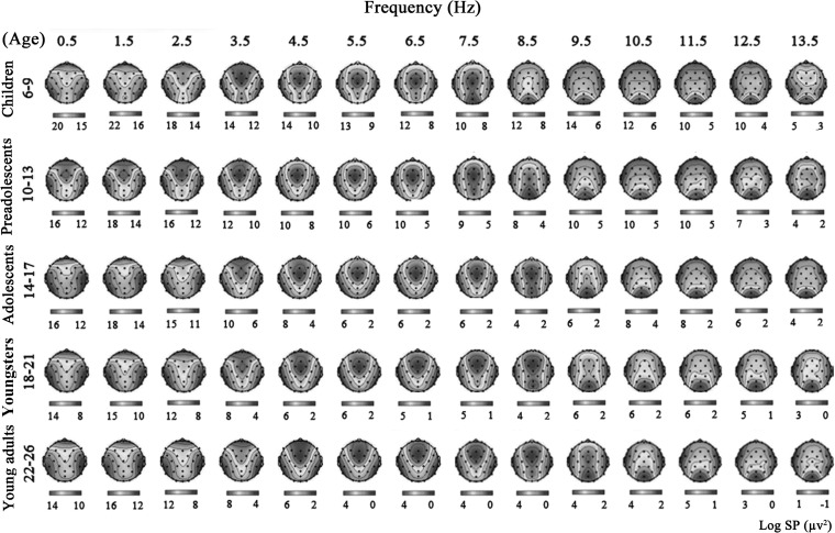
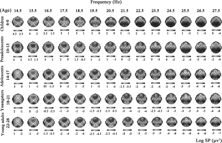
Figures 3 and 4 show the logarithmic SP topographies obtained as an average of individual topographies for the different age groups. The represented frequencies vary from 0.5 to 27.5 Hz. Low delta and delta ranges presented an anterior and posterior topography. Theta displayed a central distribution, alpha was posterior, low beta was anterior and posterior, and high beta presented an anterior distribution. No topographical changes were appreciated in the gamma range compared to the high beta range. These figures show that the scalp topography of the SP varies with age. The topographies of certain frequencies in children appear at higher frequencies in adults; for example, in the children age group (from 6 to 9 years old), the 8.5 Hz topography is similar to the topography observed in the adults group (from 22 to 26 years old) in the 10.5 Hz frequency. Another example that can be easily observed is in the 20.5 and 25.5 Hz frequencies for the children and adult groups, respectively. The same frequency shifting with age can be observed across the different frequencies. Frequency shifting can also be appreciated when other age groups are compared, although to a lesser extent.
Fig. 3.
Topographies of the logarithm for the spectral power for each age group (children: 6–9 years, preadolescents: 10–13 years, adolescents: 14–17 years, youngsters: 18–21 years, young adults: 22–26 years) expressed in years, for frequencies between 0.5 and 13.5 Hz. Scores on the color bar indicate the logarithm for the spectral power represented in each topography. (Color figure online)
Fig. 4.
Identical to Fig. 3 for frequencies 14.5–27.5 Hz
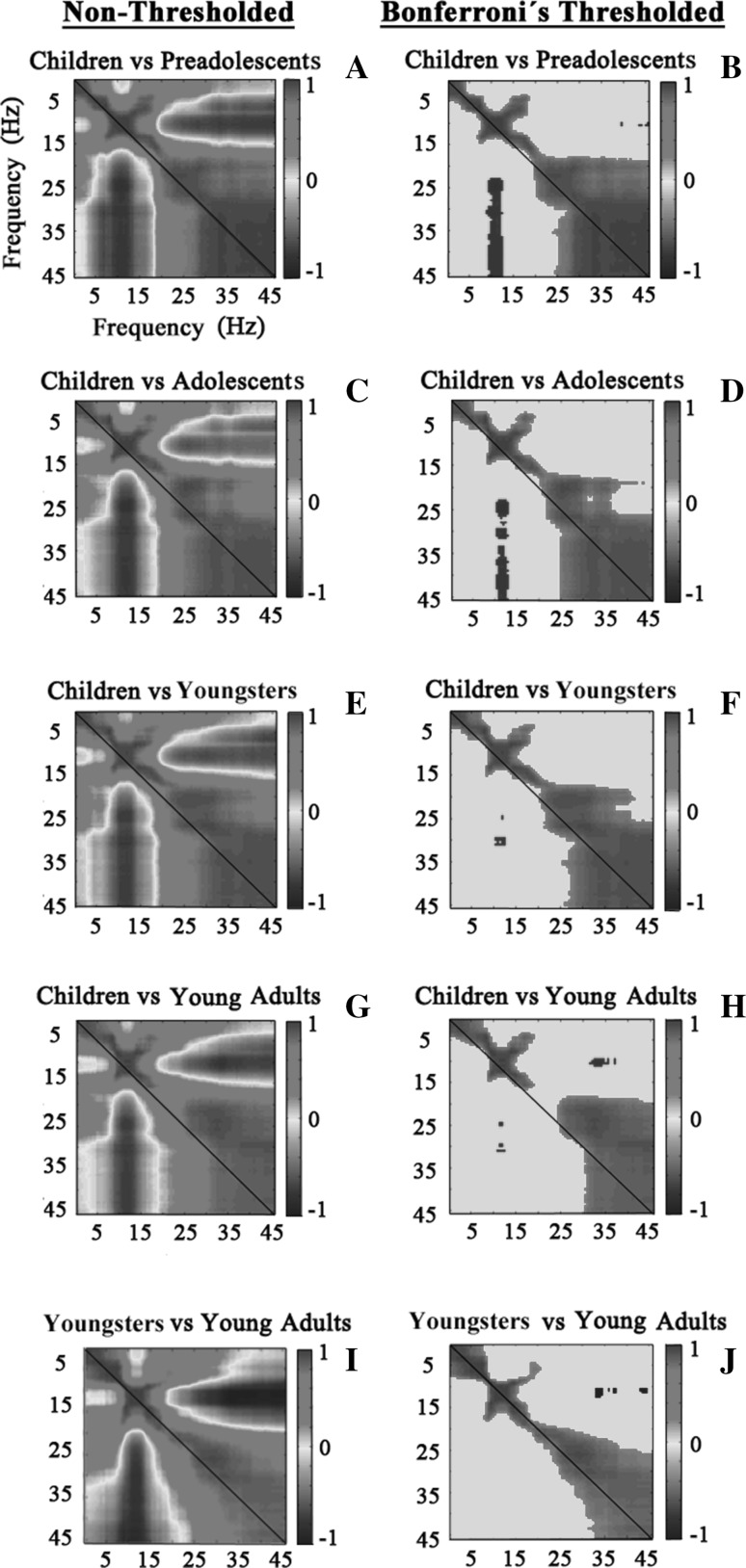
In order to quantify the displacement of topographies to higher frequencies with age, all the SP topographies from 0.5 to 45.5 Hz in the children’s group was correlated with the topographies for each of the other groups. The correlation between the young group and the young adult group is also displayed for the sake of comparison. The correlations are represented in Fig. 5 without applying the Bonferroni correction (first column: a, c, e, g, i) and after applying it (second column: b, d, f, h, j). A diagonal is also represented in each graph, indicating the point at which the value of correlation 1 would be obtained if the topographies of the correlated groups were equal at the same frequencies. In this regard, a high degree of symmetry around the diagonal in the correlation matrices in 5a (and 5b) and 5i (and 5j) can be observed, corresponding to the correlations between the SP topographies of children with preadolescents and of youngsters with young adults, respectively. These results can be interpreted as a great similarity in the SP topographies of the same frequencies in these age adjacent groups. However, the SP topography correlation matrices of children versus adolescents, youngsters and young adults (Fig. 5c, e, g), show a clear displacement in the positive correlations above the diagonal, indicating the older groups present similar topographies to children, but at higher frequencies. The latter results are also clear when the Bonferroni correction for multiple comparisons is applied (Fig. 5d, f, h). In addition, the negative correlations obtained in alpha topographies compared to higher frequencies are due to an inverse topography between these frequency ranges (high power for alpha in posterior sites, and high power for high frequencies in anterior sites).
Fig. 5.
Inter-frequency/inter-group spectral power topography correlations between the groups of children and all the other age groups (children: 6–9 years, preadolescents: 10–13 years, adolescents: 14–17 years, youngsters: 18–21 years, young adults: 21–26 years) (a, b, c, d, e, f, g, h), and correlations between young and young adults groups (i, j) for the analyzed frequencies (0.5–45.5 Hz). On the X and Y axes, the frequencies for each comparison group are represented. On the Y axis, the first comparison group is represented, and on the X axis, the second comparison group is represented. The diagonal marks the points at which the maximum correlations should be expected if both correlated age groups have the same SP topography. a, c, e, g, i represent the correlation matrices for non-threshold correlations, and b, d, f, h, j, represent the correlation matrices, applying a threshold based on Bonferroni correction of p values
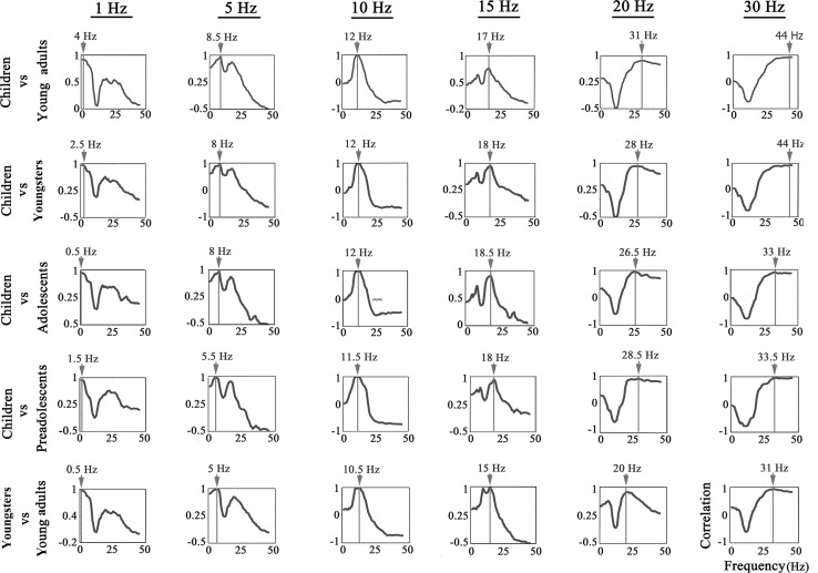
Figure 6 shows the correlations between the SP topographies of children at some predetermined frequencies, which acted as seed for inter-group inter-frequency correlations (1, 5, 10, 15, 20, 30 Hz), and the SP topographies of the other age groups in all frequencies, in orderto check the value of the frequency displacement of similar SP topographies with age. In addition, the youngsters group and the young adult group were compared to check that no differences in frequency appear in age adjacent groups. Topography correlations between age groups were computed throughout the analyzed frequency range (0.5–45.5 Hz). Figure 6 shows the shift to higher frequencies that occurs in older age groups. The frequency of the highest correlation between the SP topography of children and the SP topography of the other groups is represented. The frequency displacement is always from the younger to the older group, except in 1 Hz between children and adolescents. However, the frequency shifting in topographies detected by the correlational analysis is not found when comparing the youngsters group with the young adult group, where the frequency of the maximum correlation is practically equal to the compared frequency, which indicates that these age groups have already stabilized the displacement of topographies with age.
Fig. 6.
Inter-frequency/inter-group correlations between SP topographies of the children’s group and SP topographies of all the other age groups (preadolescents: 10–13 years, adolescents: 14–17 years, youngsters: 18–21 years, young adults: 21–26 years), and of the young group with young adults for the whole analyzed frequency range (0.5–45.5 Hz). The Y axis represents the degree of correlation between the compared groups, and the X axis represents the analyzed frequencies. The analysis between groups is performed in six specific frequencies in the children group: 1, 5, 10, 15, 20, 25 and 30 Hz, which acts as a seed for correlations with the SP maps of all the other frequencies. The red line and the top score indicate the frequency that produces the maximum correlation between the two compared groups. (Color figure online)
Discussion
The study focuses on the maturation of frequency oscillations in spontaneous brain activity from childhood to young adulthood. Accompanying the classically described reduction in brain rhythm amplitude with age, an increase in frequency oscillation around the whole scalp is observed from intergroup/interfrequency correlations. The results suggest that a shifting to higher frequencies occurs with maturation. Using a spatial criterion, these results complement previous results indicating an increase in the higher frequency contribution to the spectral content as maturation progresses.
The different age groups showed significant differences in the absolute SP in all the analyzed EEG rhythms, with the exception of the analyzed fraction of gamma (36–46 Hz). This finding supports the well-established result that there is a decrease in the absolute SP with age (Saby and Marshall 2012; Segalowitz et al. 2010). The reason the maturation of gamma is not observed as clearly as in other frequency bands is probably due to the fact that the gamma rhythm is especially sensitive to external stimuli, such as complex or salient visual stimuli (Lachaux et al. 2005) or intense sounds (Edwards et al. 2005), and cognitive processes such as attention and working memory (Boreom et al. 2007). As we recorded spontaneous EEG, it is possible that this rhythm did not have enough power to allow us to observe differences in amplitude between age groups. However, more sensitive techniques, such as magnetoencephalography, have been able to record an increase with age in absolute spectral power in the beta and low gamma signals from MEG sources obtained from beamformer (Schäfer et al. 2014). In addition, in EEG spontaneous recordings, an increase in the beta rhythm with age has been described (Lüchinger et al. 2011). Therefore, the possibility of high frequency increases in absolute SP during spontaneous activity remains an open question. With regard to the age-dependent brain rhythm amplitude (from low-delta to high beta) obtained in the present report, the general trends corresponded to great changes (decreases) until adolescence, and then a stabilization in amplitude, but with more prolonged changes (slower maturation) in low frequency rhythms than in high frequency rhythms, which has also been repeatedly observed in EEG recordings (John 1977; Rodríguez-Martínez et al. 2012). The results obtained, with the aforementioned exception of high frequency signals, confirm most of the previously obtained results showing that absolute SP decreases with age.
The increase in frequency of the spontaneous EEG, the main interest of the present report, has been partially demonstrated. The most replicated result was the increase in iAPF with age (Lindsley 1939; Niedermeyer and Lopes da Silva 1999). For instance, Bickford (1973) proposed that alpha peak frequency was already present at the age of 6. The present results extend these results about an increase in iAPF with age, using ANOVA and developmental trajectories, and they support previous results that indicated an increase in the iAPF during childhood. However, the present ANOVA results indicated that the only group that presented a lower iAPF than the other groups was the children’s group (6–9 years old). This result agrees with classic results indicating that maturation of the iAPF occurs early in life. However, more modern results using fine-grained frequencies and longitudinal studies that have reduced the contribution of individual variability to iAPF variability have found an increase in iAPF that continues during adolescence (Marcuse et al. 2008; Cragg et al. 2011). The latter two factors would explain the reduced period of time obtained for iAPF in the present report, which in any case supports the increase in the frequency oscillation of the alpha rhythm with age. As indicated in the introduction section, other brain rhythms like theta and mu have also shown an increase in frequency oscillations with age (Orekhova et al. 2006; Marshall et al. 2002; Thorpe et al. 2016; Berchicci et al. (2011). The classic result of an increased contribution of higher frequencies to the spectral content of normalized spectra points in the same direction (Saby and Marshall 2012; Segalowitz et al. 2010). Moreover, when brain rhythms are extracted through Principal Component Analysis, a systematic shift from lower to higher frequencies occurs in the loading components representing brain rhythms from 0 to 20 Hz in the transition from childhood to young adulthood (Rodríguez-Martínez et al. 2012). However, how these results can be generalized to the rest of the spectrum, and whether the shift to higher frequencies is due to a generalized process in which the cortical patches oscillate at higher frequencies as maturation progresses, are issues that have not been previously addressed.
The present results provide new insight into this issue. The correlation matrices between groups and frequencies (intergroup/interfrequency) showed that similar topographies were obtained by the different age groups, but they were obtained at different frequencies depending on age: Younger ages obtained the same SP topography, but at lower frequencies than older groups. This result is not only observed in the correlation matrices, but a careful inspection of the maps also shows this age-related frequency shifting of the SP topography. The fact that the same topographies occur at higher frequencies in the older groups supports that brain maturation causes an increase in the oscillation frequency of the EEG. Thus, for example, the predominance of occipital activity in alpha, which occurs between 8 and 12 Hz in children, occurs between 10 and 13 Hz in young adults. In addition, the frontal activity in beta, which is produced around 20 Hz in children, appears at around 25 Hz in young adults. When correlations between the SP topographies of the young adults and youngsters were computed for each frequency, most of the significant correlations occurred symmetrically around the diagonal: the expected result if both groups had the same SP topography for each oscillatory frequency. When the comparison is made between the most distant age groups, children versus young adults, the greater concentration of significant correlations is displaced to above the diagonal: the expected result if the same brain areas in older subjects are oscillating at higher frequencies than in younger subjects. The spatial correlations (inter group/interfrequency correlations) were high, indicating that SP topographies in the different age groups were very similar after applying the frequency shifting. The low to high frequency shifting with age described in the previous paragraph extends to frequencies from low-delta to high beta. The high correlation value between SP topographies at different ages, once frequency shifting is applied, suggests that the basic process underlying frequency shifting with age is the increase in frequency oscillations of the cortical tissue underlying the generation of brain rhythms (Niedermeyer and Lopes da Silva 1999; Gómez et al. 2006). As the results are based on topographies and are not differences in amplitude between the different age groups, the spatial element included in the correlations of spatial maps complements previous results showing an increase in the contribution of higher frequencies to the spectrum as maturation progresses (Segalowitz et al. 2010). In addition, the negative correlations obtained between the alpha band versus the beta and gamma band topographies would reflect that alpha is present in posterior areas and beta and gamma in anterior regions.
In a recent paper (Lea-Carnall et al. 2016), the authors proposed that the resonant frequency of the EEG (maximal response to a driving frequency) is modified by network size (inversely), proportion of excitatory and inhibitory synapses, and transmission delays (inversely). Although resonant frequencies are not the same as the spectral power content of the EEG, they are produced by similar networks. Given that brain size does not change very much in children, but myelination does, it can be proposed that the increase in the frequency of oscillation of cortical patches underlying scalp recordings, as demonstrated in the present report, would be due to the increase in myelination with age, which would reduce transmission delays and, consequently, increase the frequency of oscillations. This argument suggesting the role of myelination in the increase in the frequency of the oscillations with age has been presented previously (Segalowitz et al. 2010), although the role of the number of connections and the proportion and type of circuitry of excitatory and inhibitory connections cannot be ruled out, given the complex dynamics underlying spontaneous EEG generation.
Electronic supplementary material
Below is the link to the electronic supplementary material.
Supplemental Figure 1: Mean of the Logarithm of the spectral power in different age groups (1-children, 2-preadolescents, 3-adolscents, 4-youngsters, 5-young adults) for each frequency band: low delta (0-1 Hz); high delta (2-3 Hz); theta (4-7 Hz); low alpha (8-10 Hz); high alpha (11-14 Hz); low beta (15-20 Hz); high beta (21-35 Hz) and gamma (36-46 Hz). Each single point represents the mean SP value of the electrodes represented in Figure 1. The bars represent 2* Standard Error. (TIFF 4147 kb)
Acknowledgments
This work was supported by the Spanish Ministry of Science and Innovation, grant number PSI2013-47506-R funded by the FEDER program of the UE, and Junta de Andalucía, grant number CTS-153.
References
- Barriga-Paulino CI, Flores AB, Gómez CM. Developmental changes in the EEG rhythms of children and young adults: analyzed by means of correlational, brain topography and principal components analysis. J Psychophysiol. 2011;25(3):143–158. doi: 10.1027/0269-8803/a000052. [DOI] [Google Scholar]
- Berchicci M, Zhang T, Romero L, Peters A, Annett R, Teuscher U, et al. Development of mu rhythm in infants and preschool children. Dev Neurosci. 2011;33:130–143. doi: 10.1159/000329095. [DOI] [PMC free article] [PubMed] [Google Scholar]
- Bickford RG. Clinical Electroencephalography. New York: Medcom; 1973. [Google Scholar]
- Boreom L, Kwang SP, Do-Hyung K, Kyung WK, Young YK, Jun SK. Generators of the gamma-band activities in response to rare and novel stimuli during the auditory oddball paradigm. Neurosci Lett. 2007;413:210–215. doi: 10.1016/j.neulet.2006.11.066. [DOI] [PubMed] [Google Scholar]
- Chu CJ, Leahy J, Pathmanathan J, Kramer MA, Cash SS. The maturation of cortical sleep rhythms and networks over early development. Clin Neurophysiol. 2014;125:1360–1370. doi: 10.1016/j.clinph.2013.11.028. [DOI] [PMC free article] [PubMed] [Google Scholar]
- Cragg L, Kovacevic N, McIntosh AR, Poulsen C, Martinu K, Leonard G, Paus T. Maturation of EEG power spectra in early adolescence: a longitudinal study. Dev Sci. 2011;14(5):935–943. doi: 10.1111/j.1467-7687.2010.01031.x. [DOI] [PubMed] [Google Scholar]
- Delorme A, Makeig S. EEGLAB: an open source toolbox for analysis of single-trial EEG dynamics including independent component analysis. J Neurosci Methods. 2004;134(1):9–21. doi: 10.1016/j.jneumeth.2003.10.009. [DOI] [PubMed] [Google Scholar]
- Edwards E, Soltani M, Deouell LY, Berger MS, Knight RT. High gamma activity in response to deviant auditory stimuli recorded directly from human cortex. J Neurophysiol. 2005;94(6):4269–4280. doi: 10.1152/jn.00324.2005. [DOI] [PubMed] [Google Scholar]
- Feinberg I, Campbell IG. Sleep EEG during adolescence: an index of fundamental brain reorganization. Brain Cogn. 2010;72:56–65. doi: 10.1016/j.bandc.2009.09.008. [DOI] [PubMed] [Google Scholar]
- Gasser T, Verleger R, Bächer P, Sroka L. Development of the EEG of school-age children and adolescents. I. Analysis of band power. Electroencephalogr Clin Neurophysiol. 1988;69(2):91–99. doi: 10.1016/0013-4694(88)90204-0. [DOI] [PubMed] [Google Scholar]
- Giedd JN. Structural magnetic resonance imaging of the adolescent brain. Ann N Y Acad Sci. 2004;1021:77–85. doi: 10.1196/annals.1308.009. [DOI] [PubMed] [Google Scholar]
- Gómez CM, Marco-Pallarés J, Grau C. Location of brain rhythms and their modulation by preparatory attention estimated by current density. Brain Res. 2006;1107(1):151–160. doi: 10.1016/j.brainres.2006.06.019. [DOI] [PubMed] [Google Scholar]
- John ER. Neurometrics functional neuroscience. Hillsdale: Lawrence Erlbaum Associates; 1977. [Google Scholar]
- Keshavan MS, Diwadkar VA, DeBellis M, Dick E, Kotwal R, Rosenberg DR, Pettegrew JW. Development of the corpus callosum in childhood, adolescence and early adulthood. Life Sci. 2002;70:1909–1922. doi: 10.1016/S0024-3205(02)01492-3. [DOI] [PubMed] [Google Scholar]
- Klimesch W. EEG alpha and theta oscillations reflect cognitive and memory performance: a review and analysis. Brain Res Rev. 1999;29:169–195. doi: 10.1016/S0165-0173(98)00056-3. [DOI] [PubMed] [Google Scholar]
- Lachaux JP, George N, Tallon-Baudry C, Martinerie J, Hugueville L, Minotti L. The many faces of the gamma band response to complex visual stimuli. Neuroimage. 2005;25(2):491–501. doi: 10.1016/j.neuroimage.2004.11.052. [DOI] [PubMed] [Google Scholar]
- Lea-Carnall CA, Montemurro MA, Trujillo-Barreto NJ, Parkes LM, El-Deredy W. Cortical resonance frequencies emerge from network size and connectivity. PLoS Comput Biol. 2016;12(2):e1004740. doi: 10.1371/journal.pcbi.1004740. [DOI] [PMC free article] [PubMed] [Google Scholar]
- Lindsley DB. A longitudinal study of the occipital alpha rhythm in normal children: frequency and amplitude standards. J Genet Psychol. 1939;55:197–213. [Google Scholar]
- Lüchinger R, Michels L, Martin E, Brandeis D. EEG–BOLD correlations during post-adolescent brain maturation. Neuroimage. 2011;56:1493–1505. doi: 10.1016/j.neuroimage.2011.02.050. [DOI] [PubMed] [Google Scholar]
- Marcuse LV, Schneider M, Mortati KA, Donnelly KM, Arnedo V, Grant AC. Quantitative analysis of the EEG posterior-dominant rhythm in healthy adolescents. Clin Neurophysiol. 2008;119:1778–1781. doi: 10.1016/j.clinph.2008.02.023. [DOI] [PubMed] [Google Scholar]
- Marshall PJ, Bar-Haim Y, Fox NA. Development of the EEG from 5 months to 4 years of age. Clin Neurophysiol. 2002;113:1199–1208. doi: 10.1016/S1388-2457(02)00163-3. [DOI] [PubMed] [Google Scholar]
- Matousek M, Petersén I (1973) Frequency analysis of the EEG in normal children and adolescents. In: Kellaway P, Petersén I (eds) Automation of clinical electroencephalography. Rave, New York, pp 75–102
- Mierau A, Felsch M, Hülsdünker T, Mierau J, Bullermann P, Weiß B, Strüder K. The interrelation between sensorimotor abilities, cognitive performance and individual EEG alpha peak frequency in young children. Clin Neurophysiol. 2016;127:270–276. doi: 10.1016/j.clinph.2015.03.008. [DOI] [PubMed] [Google Scholar]
- Miskovic V, Ma X, Chou CA, Fan M, Owens M, Sayama H, Gibb B. Developmental changes in spontaneous electrocortical activity and network organization from early to late childhood. NeuroImage. 2015;118:237–247. doi: 10.1016/j.neuroimage.2015.06.013. [DOI] [PMC free article] [PubMed] [Google Scholar]
- Niedermeyer E, Lopes da Silva F. Electroencephalography: basic principles, clinical applications, and related fields. 4. Baltimore: Williams and Wilkins; 1999. [Google Scholar]
- Orekhova EV, Stroganova TA, Posikera IN, Elam M. EEG theta rhythm in infants and preschool children. Clin Neurophysiol. 2006;117:1047–1062. doi: 10.1016/j.clinph.2005.12.027. [DOI] [PubMed] [Google Scholar]
- Rodríguez-Martínez EI, Barriga-Paulino CI, Zapata MI, Chinchilla C, López- Jiménez AM, Gómez CM. Narrow band quantitative and multivariate electroencephalogram analysis of peri-adolescent period. BMC Neurosci. 2012;13:104. doi: 10.1186/1471-2202-13-104. [DOI] [PMC free article] [PubMed] [Google Scholar]
- Rodríguez-Martínez EI, Barriga-Paulino CI, Rojas-Benjumea MA, Gómez CM. Co-maturation of theta and low-beta rhythms during child development. Brain Topogr. 2015;28:205–260. doi: 10.1007/s10548-014-0369-3. [DOI] [PubMed] [Google Scholar]
- Saby JN, Marshall PJ. The utility of EEG band power analysis in the study of infancy and early childhood. Dev Neuropsychol. 2012;37:253–273. doi: 10.1080/87565641.2011.614663. [DOI] [PMC free article] [PubMed] [Google Scholar]
- Salinsky MC, Oken BS, Morehead L. Test–retest reliability in EEG frequency analysis. Electroencephalogr Clin Neurophysiol. 1991;79:383–392. doi: 10.1016/0013-4694(91)90203-G. [DOI] [PubMed] [Google Scholar]
- Schäfer C, Morgan B, Ye A, Taylor MJ, Doesburg SM. oscillations, networks, and their development: mEG connectivity changes with age. Hum Brain Mapp. 2014;35:5249–5261. doi: 10.1002/hbm.22547. [DOI] [PMC free article] [PubMed] [Google Scholar]
- Segalowitz SJ, Santesso DL, Jetha MK. Electrophysiological changes during adolescence: a review. Brain Cogn. 2010;72(1):86–100. doi: 10.1016/j.bandc.2009.10.003. [DOI] [PubMed] [Google Scholar]
- Shaw P, Greenstein D, Lerch J, Clasen L, Lenroot R, Gogtay N, Giedd J. Intellectual ability and cortical development in children and adolescents. Nature. 2006;440:676–679. doi: 10.1038/nature04513. [DOI] [PubMed] [Google Scholar]
- Shaw P, Kabani NJ, Lerch JP, Eckstrand K, Lenroot R, Gogtay N, Wise SP. Neuro developmental trajectories of the human cerebral cortex. J Neurosci. 2008;28:3586–3594. doi: 10.1523/JNEUROSCI.5309-07.2008. [DOI] [PMC free article] [PubMed] [Google Scholar]
- Thorpe SG, Cannon EN, Fox NA. Spectral and source structural development of mu and alpha rhythms from infancy through adulthood. Clin Neurophysiol. 2016;127:254–269. doi: 10.1016/j.clinph.2015.03.004. [DOI] [PMC free article] [PubMed] [Google Scholar]
- Valdés P, Virués T, Szava S, Galán L, Biscay R. High resolution spectral EEG norms topography. Brain Topogr. 1990;3:281–282. [Google Scholar]
- Whitford TJ, Rennie CJ, Grieve SM, Clark CR, Gordon E, Williams LM. brain maturation in adolescence: concurrent changes in neuroanatomy and neurophysiology. Hum Brain Mapp. 2007;28:228–237. doi: 10.1002/hbm.20273. [DOI] [PMC free article] [PubMed] [Google Scholar]
Associated Data
This section collects any data citations, data availability statements, or supplementary materials included in this article.
Supplementary Materials
Supplemental Figure 1: Mean of the Logarithm of the spectral power in different age groups (1-children, 2-preadolescents, 3-adolscents, 4-youngsters, 5-young adults) for each frequency band: low delta (0-1 Hz); high delta (2-3 Hz); theta (4-7 Hz); low alpha (8-10 Hz); high alpha (11-14 Hz); low beta (15-20 Hz); high beta (21-35 Hz) and gamma (36-46 Hz). Each single point represents the mean SP value of the electrodes represented in Figure 1. The bars represent 2* Standard Error. (TIFF 4147 kb)


