Abstract
The mandibular foramen is a landmark for procedures like inferior alveolar nerve block, mandibular implant treatment, and mandibular osteotomies. The present study was aimed to identify the precise location of the mandibular foramen and the incidence of accessory mandibular foramen in dry adult mandibles of South Indian population. The distance of mandibular foramen from the anterior border of the ramus, posterior border of the ramus, mandibular notch, base of the mandible, third molar, and apex of retromolar trigone was measured with a vernier caliper in 204 mandibles. The mean distance of mandibular foramen from the anterior border of ramus of mandible was 17.11±2.74 mm on the right side and 17.41±3.05 mm on the left side, from posterior border was 10.47±2.11 mm on the right side and 9.68±2.03 mm on the left side, from mandibular notch was 21.74±2.74 mm on the right side and 21.92±3.33 mm on the left side, from the base of the ramus was 22.33±3.32 mm on the right side and 25.35±4.5 mm on the left side, from the third molar tooth was 22.84±3.94 mm on the right side and 23.23±4.21 mm on the left side, from the apex of retromolar trigone was 12.27±12.13 mm on the right side and 12.13±2.35 mm on the left side. Accessory mandibular foramen was present in 32.36% of mandibles. Knowledge of location mandibular foramen is useful to the maxillofacial surgeons, oncologists and radiologists.
Keywords: Mandible, Mandibular foramen, Mandibular notch, Accessory Mandibular foramen, Inferior alveolar nerve block
Introduction
The mandibular foramen (MF) is an irregular foramen located a little above the centre of the medial surface of the mandibular ramus. The inferior alveolar nerve and vessels pass through the MF and traverse the mandibular canal and divides into mental and incisive branches to supply the mandibular teeth and participates in the formation of the anterior loop [1,2]. Inferior alveolar nerve block is a common local anaesthetic technique used in dental practice. But the failure rate of this technique is reported to be as high as 20%–25% [3]. The commonest cause for inferior alveolar nerve block failure is inaccurate localization of MF [4]. The main complications during this technique are haemorrhage, injury to the neurovascular bundle, fractures, and necrosis of mandibular ramus [5]. Hence, thorough knowledge of the mandibular ramus is very essential.
Clarke and Holmes [6], have reported that the position of the MF is 1cm above the occlusal plane of the lower molars, and is also at the same height of the gingival papillae of the upper teeth when the individual is with his mouth closed. But, Nicholson [7], has said that, there is variability of the two mandibular rami in the same person, and it is not possible to standardize the foramen identification. Studies have conclusively proved that there are significant morphological differences in the mandibular anatomy among the three major racial groups—Caucasoid, Mongoloid, and Negroid [8,9].
Accessory MF is any opening in the mandible other than the MF, mental foramen, lingual foramen, and sockets of teeth [10]. The presence of accessory MF and additional branches of inferior alveolar nerve may lead to increased rates of failure of inferior alveolar nerve blocks as all the branches may not be anaesthetized [11]. The accessory MF has also been reported to be the site for the spread of tumours following radiotherapy in the lateral surface of mandible [12]. So the knowledge of accessory MF is imperative to radiotherapists when planning for radiation therapy in the lateral mandibular region.
Therefore, the present study aims to determine the precise location of the MF from various anatomical landmarks such as anterior and posterior borders of the mandibular ramus, mandibular notch, base of the ramus, third molar tooth and retromolar trigone from mandibles of South Indian population. This study aims to identify the MF location in relation to the limits of mandibular ramus and to the quadrant of the ramus, taking horizontal and vertical directions. The presence of accessory MF was also noted.
Materials and Methods
The study was conducted in 204 adult dry human mandibles of unknown sex and age collected from the bone bank of Anatomy and Forensic Medicine departments and students of Dhanalakshmi Srinivasan Medical College and Hospital, Siruvachur, Perambalur. Mandibles with sockets for third molar teeth, those in regular shape, and devoid of deformities were selected. The damaged bones and those having pathological abnormalities were excluded.
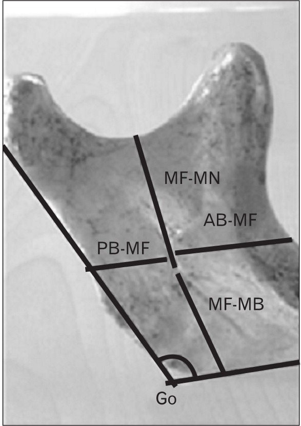
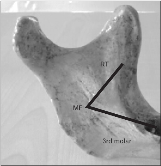
To precisely locate the mandibular foramen, the following parameters were measured on both sides of the mandible with a sliding vernier callipers of 0.1 mm accuracy (Figs. 1, 2).
Fig. 1. Medial surface of mandibular ramus showing measurements taken from various landmarks to determine the position of the mandibular foramen. AB-MF, distance from the midpoint of anterior margin of mandibular foramen to the nearest point on the anterior border of the ramus of mandible; Go, angle of the mandible; MF-MB, sistance from inferior limit of mandibular foramen to the base of the mandible; MF-MN, distance from the lowest point of mandibular notch to the inferior limit of mandibular foramen; PB-MF, distance from the midpoint of posterior margin of mandibular foramen to the nearest point on the posterior border of the ramus of mandible.

Fig. 2. Medial surface of mandibular ramus showing distance of mandibular foramen from apex of retromolar trigone and from midpoint of third molar socket. MF, mandibular foramen; RT, apex of retromolar trigone.

(1) AB-MF: distance from the midpoint of anterior margin of Mandibular foramen to the nearest point on the anterior border of the ramus of mandible (Fig. 1), (2) PB-MF: distance from the midpoint of posterior margin of mandibular foramen to the nearest point on the posterior border of the ramus of mandible (Fig. 1), (3) AB-PB: breadth of the ramus from anterior to posterior border, (4) MF-MN: distance from the lowest point of mandibular notch to the inferior limit of mandibular foramen (Fig. 1), (5) MF-MB: distance from inferior limit of Mandibular foramen to the base of the mandible (Fig. 1), (6) III Molar-MF: distance from the midpoint of third molar tooth or socket to anterior margin of Mandibular foramen (Fig. 2), and (7) RT-MF: distance between the apex of the retromolar trigone and mandibular foramen (Fig. 2).
The angle of the mandible was measured with a goniometer at the junction of inferior and posterior borders of the ramus of the mandible (Fig. 1).
The mandibles were further observed for the presence of accessory mandibular foramen in and around mandibular foramen on the medial surface of mandibular ramus by means of a simple visual observation with the help of a magnifying lens and their prevalence rate was noted and analyzed.
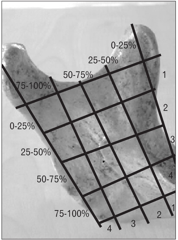
The quadrant where mandibular foramen was located horizontally, was obtained by identifying the distance between the anterior border of the mandibular ramus and the midpoint of the mandibular foramen opening hole, calculated by subtracting, from AB-PB, the sum of the distances AB-MF, and PB-MF. The result yields the width of mandibular foramen. This distance was divided into halves to determine the midpoint of the mandibular foramen opening hole, and added to distance AB-MF. It was then calculated, in percentage, how much the distance between anterior border of the ramus and the midpoint of mandibular foramen represented in relation to the total width of the ramus (AB-PB). This value indicated in which quadrant mandibular foramen was in an anteroposterior location (Fig. 3). Values from 0% to 25% comprised the first quadrant; from 26% to 50%, the second; from 51% to 75%, the third and from 76% to 100%, the fourth. In the vertical direction, the quadrant on which Mandibular foramen was located was identified by calculating how much MF-MN represented, in percentage of the addition of MF-MN and MF-MB distance (Fig. 3).
Fig. 3. Medial surface of mandibular ramus showing the scheme of divisions in anteroposterior and superoinferior axis.

All the above parameters were carefully tabulated and statistically analysed. SPSS version 16.0 (SPSS Inc., Chicago, IL, USA) was used for the statistical analysis of this study. Student's t test was used as test of significance to compare the mean values of right and left sides and a P-value less than 0.05 was taken to be statistically significant. The results of the present study were compared with the results of previous studies done on various ethnic groups.
Results
Distance of mandibular foramen from various landmarks on the right and left sides
A total of 204 adult human mandibles were studied for the position of mandibular foramen. The minimum, maximum, average, and standard deviation values of the various parameters which were studied on either side of the mandible are shown in Table 1. There was no statistically significant difference between the values obtained on the right and left sides (P>0.05).
Table 1. Distance of mandibular foramen (MF) from various mandibular landmarks on the right and left sides.
| Measurement | Right side (mm) | Left side (mm) |
|---|---|---|
| AB-MF | 17.11±2.74 | 17.41±3.05 |
| PB-MF | 10.47±2.11 | 9.68 ±2.03 |
| AB-PB | 31.76±3.83 | 31.49±3.97 |
| Foramen-width | 4.19 ±1.57 | 4.37±1.67 |
| MF-MN | 21.74±2.74 | 21.92 ±3.33 |
| MF-MB | 22.33±3.32 | 25.35±4.5 |
| III Molar-MF | 22.84±3.94 | 23.23±4.21 |
| RT-MF | 12.27±2.13 | 12.13± 2.35 |
Values are presented as mean±SD. AB-MF, distance from midpoint of anterior margin of mandibular foramen to the nearest point on anterior border of ramus; PB-MF, distance from midpoint of posterior margin of mandibular foramen to the nearest point on posterior border of ramus; AB-PB, distance from anterior to posterior border of ramus; foramen width, calculated by subtracting, from AB-PB, the sum of the distances AB-MF and PB-MF; MF-MN, distance from mandibular notch to inferior limit of mandibular foramen; MF-MB, distance from Mandibular Base to inferior limit of mandibular foramen; III Molar-MF, distance from III molar to mandibular foramen; RT-MF, distance from apex of retromolar trigone to mandibular foramen.
Angle of the mandible:
The angle of the mandible- Gonion was 117.47°±4.95° on the right side and 117.47°±5.88° on the left side. There was no statistically significant difference between the angles of the mandible on the right and left sides (P>0.05).
Incidence of symmetrical measurement of different parameters:
The incidence of symmetrical measurement of different parameters on the right and left sides is shown in Table 2. Symmetrical measurement of localization of mandibular foramen on right and left sides was observed only in 13%–20% of the mandibles. The angle of the mandible was symmetrical in 76.7% of the mandibles.
Table 2. Incidence of symmetrical measurement of different parameters on right and left sides.
| Measurement | No. of bones showing symmetrical measurements | Percentage of bones showing symmetrical measurements | No. of bones showing asymmetrical measurements | Percentage of bones showing asymmetrical measurements |
|---|---|---|---|---|
| AB-MF | 28 | 13.7 | 0176 | 86.3 |
| PB-MF | 28 | 13.7 | 176 | 86.3 |
| AB-PB | 36 | 17.6 | 168 | 82.4 |
| Foramen-width | 28 | 13.7 | 176 | 86.3 |
| MF-MN | 48 | 23.5 | 156 | 76.5 |
| MF-MB | 12 | 5.9 | 192 | 94.1 |
| III Molar-MF | 8 | 3.9 | 196 | 96.1 |
| RT-MF | 44 | 20.6 | 160 | 79.4 |
| Gonial angle | 156 | 76.7 | 48 | 23.3 |
AB-MF, distance from midpoint of anterior margin of mandibular foramen to the nearest point on anterior border of ramus; PB-MF, distance from midpoint of posterior margin of mandibular foramen to the nearest point on posterior border of ramus; AB-PB, distance from anterior to posterior border of ramus; foramen width, calculated by subtracting, from AB-PB, the sum of the distances AB-MF and PB-MF; MF-MN, distance from mandibular notch to inferior limit of mandibular foramen; MF-MB, distance from mandibular base to inferior limit of mandibular foramen; III Molar-MF, distance from III Molar to mandibular foramen; RT-MF, distance from apex of retromolar Trigone to mandibular foramen.
Localization of mandibular foramen in anteroposterior and superoinferior axis of the ramus of mandible:
The percentile of the distance from anterior border of ramus to midpoint of mandibular foramen in relation to the distance from anterior to posterior border of ramus (AB-PB) on the right side was 56.73±3.44% and it was localized in the third quadrant in the anteroposterior axis and on the left side it was 62.2±2.32% and it was also localized in the third quadrant in the anteroposterior axis of the mandibular ramus. The percentile of distance MF-MN in relation to MF-MN+MF-MB was 49.68±3.46% on the right side and it was localised at the junction of second and third quadrant in the superoinferior axis and 46.51±5.1% on the left side and it was also localised at the junction of second and third quadrant in the superoinferior axis of the mandibular ramus (Table 3). There was no statistically significant difference in the location of mandibular foramen on the right and left sides (P>0.05) in both the anteroposterior axis and superoinferior axis.
Table 3. Percentile of the distance from anterior border of ramus to midpoint of mandibular foramen in relation to the AB-PB, percentile of distance MF-MN in relation to MF-MN+MF-MB and quadrant of mandibular foramen localization on anteroposterior and superoinferior axis of mandibular ramus on both sides.
| Side | Anteroposterior localization (%) | Quadrant in anteroposterior axis | Superoinferior localization (%) | Quadrant in Superoinferior axis |
|---|---|---|---|---|
| Right | 56.73±3.44 | Third | 49.68±3.46 | Junction of second and third |
| Left | 62.2±2.32 | Third | 46.51±5.1 | Junction of second and third |
Values are presented as mean±SD. AB-PB, distance from anterior to posterior border of ramus; MF-MN, distance from mandibular notch to inferior limit of mandibular foramen; MF-MB, distance from mandibular base to inferior limit of mandibular foramen.
Accessory mandibular foramen:
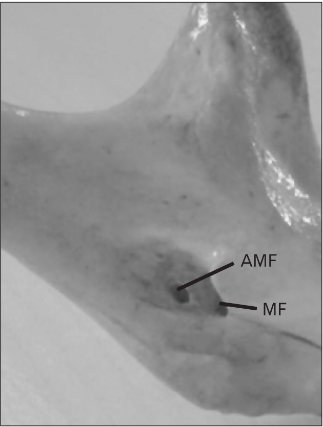
Single accessory mandibular foramen (Fig. 4) was found unilaterally in 28 mandibles (13.72%) and bilaterally in 18 mandibles (8.8%). Double accessory mandibular foramen was found unilaterally in 17 mandibles (8.33%) and bilaterally in 3 mandibles (1.5%) (Table 4). The accessory foramens were located at distance of less than 5 mm from the mandibular foramen in all the specimens showing the accessory mandibular foramen and the width of the accessory foramen was observed to be less than 2 mm.
Fig. 4. Medial surface of mandibular ramus showing an accessory mandibular foramen. AMF, accessory mandibular foramen; MF, mandibular foramen.

Table 4. Incidence of accessory mandibular foramen in 204 dry adult human mandibles.
| Accessory mandibular foramen | No. (%) |
|---|---|
| Right side–single accessory foramen | 17 (8.33) |
| Right side–double accessory foramen | 8 (3.92) |
| Left side–single accessory foramen | 11 (5.4) |
| Left side–double accessory foramen | 9 (4.41) |
| Bilateral single accessory foramen | 18 (8.8) |
| Bilateral double accessory foramen | 3 (1.5) |
| Absent–accessory foramen | 138 (67.64) |
Unilateral single accessory mandibular foramen was statistically predominant on the right side compared to the left side (P<0.05). But there was no statistically significant difference on the occurrence of double accessory mandibular foramen on the right and left sides (P>0.05).
Discussion
The location of the mandibular foramen is essential for mandibular surgeries like vertical ramus osteotomy, inverted L osteotomy and also esthetic surgeries for dentofacial deformities. The inferior alveolar nerve is at a greater risk during these surgical procedures. Daw et al. [5] have reported great variability in the position of mandibular foramen from Non-Asian hemi mandibles. They have also emphasised that the knowledge of the location of the mandibular foramen would assist in performing a proper sagittal split of the mandibular ramus.
During pterygomandibular technique of inferior alveolar nerve blockage long needles of size 33 mm and short needles of size 21.5 mm are used. If a long needle is used in a patient with small mandible, there is a risk of perforating the parotid gland capsule and injuring the branches of facial nerve. If a short needle is used in a patient with big sized mandible, there may be a fracture of the needle when it is completely introduced in the oral tissues [13].
There are significant differences in the localization of mandibular foramen in different ethnic groups. Mbajiorgu in his study [14] on adult black Zimbabweans has reported that the mandibular foramen lies 2.56 mm behind the midpoint of the ramus width on the right side and 2.08 mm behind the midpoint of the ramus width on the left side. The mean distance from anterior border of mandibular ramus to anterior margin of mandibular foramen (AB-MF) was 18.95±0.41 mm, the mean distance from posterior border of ramus to posterior margin of mandibular foramen (PB-MF) was 14.30±0.35 mm, the mean distance from mandibular notch to inferior end of the mandibular foramen (MF-MN) was 22.5±0.5 mm and the mean distance from the inferior end of mandibular foramen to base of the ramus (MF-MB) was 28.44±0.65 mm in his study.
Oguz and Bozkir [4] have tried to localize the mandibular foramen in Turkish mandibles. Ennes and Medeiros [13] and Prado et al. [15] have studied the location of mandibular foramen in Brazilian population. Samanta and Kharb [11] and Raghavendra and Benjamin [16] have tried to localize the mandibular foramen in Indian mandibles. There are variations in the values obtained in each of the studies and the values are compared with the results of the present study in Table 5.
Table 5. Comparison of results of different studies on localization of mandibular foramen on right and left sides.
| Author | Population | Right side AB-MF (mm) | Left side AB-MF (mm) | Right side PB-MF (mm) | Left side PB-MF (mm) | Right side MF-MN (mm) | Left side MF-MN (mm) |
|---|---|---|---|---|---|---|---|
| Oguz and Bozkir [4] | Turkey | 16.9 | 16.78 | 14.09 | 14.37 | 22.37 | 22.17 |
| Ennes and Medeiros [13] | Brazil | 9.4±2.03 | 6.9±2.06 | 8.6±1.2 | 8.4±1.77 | 18.3±3.25 | 17.5±3.37 |
| Prado et al. [15] | Brazil | 19.2±3.6 | 18.8±3.8 | 14.2±2.4 | 13.9±2.6 | 23.6±3.1 | 23.1±3.0 |
| Samanta and Kharb [11] | India | 15.72±2.92 | 16.23±2.88 | 13.29±1.74 | 12.73±2.04 | 22.7±3.0 | 22.27±2.92 |
| Raghavendra and Benjamin [16] | India | 16.21± 2.12 | 16.67±2.34 | 11.08±2.34 | 11.11±2.34 | 21.38±3.91 | 20.95±3.39 |
| Present study | South India | 17.11±2.74 | 17.41±3.05 | 10.47±2.11 | 9.68±2.03 | 21.74±2.74 | 21.92±3.33 |
Values are presented as mean±SD. AB-MF, distance from midpoint of anterior margin of mandibular foramen to the nearest point on the anterior border of ramus of mandible; PB-MF, distance from midpoint of posterior margin of mandibular foramen to the nearest point on the posterior border of ramus of mandible; MF-MN, distance from mandibular notch to inferior aspect of mandibular foramen.
Kilarkaje et al. [17] have reported that mandibular foramen was within 25 mm from the third molar tooth. Varma et al. [18] have reported that the mean distance of mandibular foramen from the third molar tooth was 15 mm on the right side and 18 mm on the left side. Ghorai et al. [19] have reported the distance to be 22.8±4.9 mm on the right side and 21.7±4.7 mm on the left side. The results of the present study are similar to Kilarkaje et al. [17] and Ghorai et al. [19], as the measurements were 22.84±2.13 mm on the right side and 23.23±4.21 mm on the left side.
Valente et al. [20] have measured the distance of mandibular foramen from the apex of retromolar trigone and reported it to be 14.23±2.57 mm on the right side and 14.40±2.48 mm on the left side. In the present study, the measurements were 12.27±2.13 mm on the right side and 12.13±2.35 mm on the left side.
Ennes and Medeiros [13] have reported that the average gonial angle to be 125.6° with a standard deviation between 6.2° and 9.2°. Oguz and Bozkir [4] reported the angle of mandible to be 120.2°±4.7°. In the present study, it was 117.47°±4.95° on the right side and 117.47°±5.88° on the left side. The gonial angle is inversely proportional to the anteroposterior width of the mandibular ramus and the distance between mandibular foramen and base of the mandible (MF-MB). So, in individuals with wide gonial angle, inferior alveolar nerve blockage has to be performed at a site lower than the conventional site and with a short needle and in individuals with small gonial angle, the inferior alveolar nerve block has to be performed at a site higher than the conventional site and with a long needle [13].
Kilarkaje et al. [17] from their study have reported that the mandibular foramen maintains bilateral symmetry in dry mandibles in all ages and the foramen was found to be within 25 mm from the third molar, anterior border of ramus (AB) and mandibular notch. In the present study bilateral symmetry of distance of mandibular foramen from various landmarks of the mandibular ramus ranged between 13% to 20% only.
Ennes and Medeiros [13] have localized the mandibular foramen in the third quadrant in the anteroposterior and superoinferior axis of the mandibular ramus. Padmavathi et al. [21] have localized the mandibular foramen in the third quadrant in the anteroposterior axis and in the junction between second and third quadrant in the superoinferior axis. In the present study also the mandibular foramen was localized in the third quadrant in the anteroposterior axis and junction between second and third quadrants in the superoinferior axis of the mandibular ramus.
The embryological basis for the occurrence of accessory mandibular foramen has been explained in literatures. Chávez-Lomeli et al. [22] have reported that, initially, 3 inferior alveolar nerves, innervating each of the 3 groups of mandibular teeth are formed in the embryo. Later, all the 3 nerves fuse and a single inferior alveolar nerve is formed. The incomplete fusion of the nerves leads to the formation of double mandibular canals. In 60% of the cases, the mandibular canal was found to have the entire inferior alveolar nerve passing through it and in 40% the nerves were found to be scattered. Pancer et al. [23] has reported the incidence of accessory mandibular foramen to vary from 0.88% to 10.66%. In the study by Padmavathi et al. [21] the accessory mandibular foramen was present in 41.5% of the cases and it was present unilaterally in 29.2% of the cases and bilaterally in 12.3% of the cases. Gopalakrishna et al. [24] have reported the incidence of single accessory mandibular foramina to be 18%. Samanta and Kharb [11] have found accessory mandibular foramen in 16.66% of the mandibles of which 10% of the mandibles had single accessory mandibular foramen and 6.66% of the mandibles had double accessory mandibular foramens. Freire et al. [25] have observed a single accessory mandibular foramen in 27.93% of the Brazilian mandibles located below the mandibular foramen and in 43.24% of the Brazilian mandibles, the accessory foramen was located above the mandibular foramen. In the present study, single accessory mandibular foramen was found unilaterally in 13.72% of the mandibles and bilaterally in 8.8% of the mandibles. Double accessory mandibular foramen was found unilaterally in 8.33% of the mandibles and bilaterally in 1.5% of the mandibles (Table 4). The statistically significant predominance of unilateral single accessory mandibular foramen on the right side may be due to chance and there is no literature explanation for it. Hanihara and Ishida [26] have reported that, there is a higher prevalence of accessory mandibular foramen in Asian males, hence there is a higher incidence of accessory mandibular foramen in our study and also in the study by Padmavathi et al. [21] on Indians.
The smaller size of the accessory mandibular foramen limits its visualisation in traditional radiographs of the mandible. Panoramic radiographs of the mandible have limitations like distortion, overlap and magnification, which may lead to false interpretation of important anatomical structures [23]. Haas et al. [27] and Pancer et al. [23] have suggested that cone beam computed tomography (CBCT) is a reliable and accurate three dimensional radiographic technique to visualise the accessory mandibular foramen. The higher cost and level of radiation associated with CBCT limit its usage in dental patients.
The range of distance between the accessory mandibular foramen and mandibular foramen is clinically important, because the spread of local anaesthetic affects the efficiency of the inferior alveolar nerve block. The spread of local anaesthetic in inferior alveolar nerve blocks, depends on the drug used, its concentration and the volume of drug injected [28]. In the present study, the accessory foramens were located at a distance of less than 5 mm from the mandibular foramen, hence if a higher concentration and larger volume of local anaesthetic is used, the accessory nerves can also be anaesthetised.
The precise localization of mandibular foramen is very important to achieve a successful inferior alveolar nerve block, prior to dental surgeries in the lower jaw like osteotomy, orthognathic reconstruction surgeries of the mandible and dental implant procedures, and also to avoid injury to the neurovascular contents passing through it. Accessory mandibular foramina will serve as a route for spread of infection and tumour cells. The present study on the morphometry of the mandibular foramen and the incidence of accessory mandibular foramen will provide useful information to dental surgeons for planning and conducting dental and maxillofacial surgeries in south Indian population. This study will also help radiologist and oncologist in localizing the mandibular foramen in south Indian population.
References
- 1.Collins P. Infratemporal and pterygopalatine fossae and temporomandibular joint. In: Standring S, editor. Gray's Anatomy: The Anatomical Basis of Clinical Practice. 40th ed. Edinburg: Churchill and Livingstone; 2008. pp. 530–532. [Google Scholar]
- 2.Yu SK, Kim S, Kang SG, Kim JH, Lim KO, Hwang SI, Kim HJ. Morphological assessment of the anterior loop of the mandibular canal in Koreans. Anat Cell Biol. 2015;48:75–80. doi: 10.5115/acb.2015.48.1.75. [DOI] [PMC free article] [PubMed] [Google Scholar]
- 3.Shah K, Shah P, Parmar A. Study of the location of the mandibular foramina in Indian dry mandibles. Global Res Anal. 2013;2:128–130. [Google Scholar]
- 4.Oguz O, Bozkir MG. Evaluation of location of mandibular and mental foramina in dry, young, adult human male, dentulous mandibles. West Indian Med J. 2002;51:14–16. [PubMed] [Google Scholar]
- 5.Daw JL, Jr, de la Paz MG, Han H, Aitken ME, Patel PK. The mandibular foramen: an anatomic study and its relevance to the sagittal ramus osteotomy. J Craniofac Surg. 1999;10:475–479. [PubMed] [Google Scholar]
- 6.Clarke J, Holmes G. Local anesthesia of the mandibular molar teeth a new technique. Dent Practit Dent Rec. 1959;36:321. [Google Scholar]
- 7.Nicholson ML. A study of the position of the mandibular foramen in the adult human mandible. Anat Rec. 1985;212:110–112. doi: 10.1002/ar.1092120116. [DOI] [PubMed] [Google Scholar]
- 8.Komar D, Lathrop S. Frequencies of morphological characteristics in two contemporary forensic collections: implications for identification. J Forensic Sci. 2006;51:974–978. doi: 10.1111/j.1556-4029.2006.00210.x. [DOI] [PubMed] [Google Scholar]
- 9.Neiva RF, Gapski R, Wang HL. Morphometric analysis of implant-related anatomy in Caucasian skulls. J Periodontol. 2004;75:1061–1067. doi: 10.1902/jop.2004.75.8.1061. [DOI] [PubMed] [Google Scholar]
- 10.Sutton RN. The practical significance of mandibular accessory foramina. Aust Dent J. 1974;19:167–173. doi: 10.1111/j.1834-7819.1974.tb05034.x. [DOI] [PubMed] [Google Scholar]
- 11.Samanta PP, Kharb P. Morphometric analysis of mandibular foramen and incidence of accessory mandibular foramen in adult human mandibles of an Indian population. Rev Arg Anat Clin. 2013;5:60–66. [Google Scholar]
- 12.Fanibunda K, Matthews JN. Relationship between accessory foramina and tumour spread in the lateral mandibular surface. J Anat. 1999;195(Pt 2):185–190. doi: 10.1046/j.1469-7580.1999.19520185.x. [DOI] [PMC free article] [PubMed] [Google Scholar]
- 13.Ennes JP, Medeiros RM. Localization of mandibular foramen and clinical implications. Int J Morphol. 2009;27:1305–1311. [Google Scholar]
- 14.Mbajiorgu EF. A study of the position of the mandibular foramen in adult black Zimbabwean mandibles. Cent Afr J Med. 2000;46:184–190. doi: 10.4314/cajm.v46i7.8554. [DOI] [PubMed] [Google Scholar]
- 15.Prado FB, Groppo FC, Volpato MC, Caria PH. Morphological changes in the position of the mandibular foramen in dentate and edentate Brazilian subjects. Clin Anat. 2010;23:394–398. doi: 10.1002/ca.20973. [DOI] [PubMed] [Google Scholar]
- 16.Raghavendra VP, Benjamin W. Position of mandibular foramen and incidence of accessory mandibular foramen in dry mandibles. Int J Pharm Bio Sci. 2015;6:282–288. [Google Scholar]
- 17.Kilarkaje N, Nayak SR, Narayan P, Prabhu LV. The location of the mandibular foramen maintains absolute bilateral symmetry in mandibles of different age-groups. Hong Kong Dent J. 2005;2:35–37. [Google Scholar]
- 18.Varma CL, Haq I, Rajeshwari T. Position of mandibular foramen in south Indian mandibles. Anatomica Karnataka. 2011;5:53–56. [Google Scholar]
- 19.Ghorai S, Pal M, Jana S. Distance of mandibular foramen from 3 rd molar tooth in dry adult mandible of West Bengal population. Anat J Afr. 2016;5:640–643. [Google Scholar]
- 20.Valente VB, Arita WM, Garcia Concalves PC, Duarte Bonini Campos JA, de Oliveira Capote TS. Location of the mandibular foramen according to the amount of dental alveoli. Int J Morphol. 2012;30:77–81. [Google Scholar]
- 21.Padmavathi G, Tiwari S, Varalakshmi KL, Roopashree R. An anatomical study of mandibular and accessory mandibular foramen in dry adult human mandibles of south Indian origin. IOSR J Dent Med Sci. 2014;13:83–88. [Google Scholar]
- 22.Chávez-Lomeli ME, Mansilla Lory J, Pompa JA, Kjaer I. The human mandibular canal arises from three separate canals innervating different tooth groups. J Dent Res. 1996;75:1540–1544. doi: 10.1177/00220345960750080401. [DOI] [PubMed] [Google Scholar]
- 23.Pancer B, Garaicoa-Pazmiño C, Bashutski JD. Accessory mandibular foramen during dental implant placement: case report and review of literature. Implant Dent. 2014;23:116–124. doi: 10.1097/ID.0000000000000056. [DOI] [PubMed] [Google Scholar]
- 24.Gopalakrishna K, Deepalaxmi S, Somashekara SC, Rathna BS. An anatomical study on the position of mandibular foramen in 100 dry mandibles. Int J Anat Res. 2016;4:1967–1971. [Google Scholar]
- 25.Freire AR, Rossi AC, Prado FB. Incidence of the mandibular accessory foramina in Brazilian population. J Morphol Sci. 2012;29:171–173. [Google Scholar]
- 26.Hanihara T, Ishida H. Frequency variations of discrete cranial traits in major human populations. IV. Vessel and nerve related variations. J Anat. 2001;199:273–287. doi: 10.1046/j.1469-7580.2001.19930273.x. [DOI] [PMC free article] [PubMed] [Google Scholar]
- 27.Haas LF, Dutra K, Porporatti AL, Mezzomo LA, De Luca Canto G, Flores-Mir C, Corrêa M. Anatomical variations of mandibular canal detected by panoramic radiography and CT: a systematic review and meta-analysis. Dentomaxillofac Radiol. 2016;45:20150310. doi: 10.1259/dmfr.20150310. [DOI] [PMC free article] [PubMed] [Google Scholar]
- 28.Parirokh M, Yosefi MH, Nakhaee N, Abbott PV, Manochehrifar H. The success rate of bupivacaine and lidocaine as anesthetic agents in inferior alveolar nerve block in teeth with irreversible pulpitis without spontaneous pain. Restor Dent Endod. 2015;40:155–160. doi: 10.5395/rde.2015.40.2.155. [DOI] [PMC free article] [PubMed] [Google Scholar]


