SUMMARY
In animal models, inflammation is both a cause and consequence of seizures. Less is known about the role of inflammation in human epilepsy. We performed PET using a radiotracer sensitive to brain inflammation in a patient with frontal epilepsy ~36 hours after a seizure as well as during a seizure-free period. When statistically compared to a group of 12 matched controls, both of the patient’s scans identified a frontal (supplementary motor area) region of increased inflammation corresponding to his clinically-defined seizure focus, but the post-seizure scan showed significantly greater inflammation intensity and spatial extent. These results provide new information about transient and chronic neuroinflammation in human epilepsy and may be relevant to understanding the process of epileptogenesis and guiding therapy.
Keywords: Positron Emission Tomography, microglia, translocator protein, supplementary motor area, frontal lobe epilepsy, focal cortical dysplasia
INTRODUCTION
Understanding the mechanism by which "seizures beget seizures,"4 i.e. how a single seizure might alter the brain to predispose to future seizures, is critical to understanding the process of epileptogenesis and to developing effective antiepileptogenic treatments. There is mounting preclinical evidence that inflammation is important in epileptogenesis, with seizures inducing inflammation via a number of direct18 and indirect mechanisms, and inflammation increasing the likelihood and severity of subsequent seizures.12; 17 In animal studies, inflammation and neural damage at different time points after various inciting epileptogenic events can be quantified directly via tissue analysis. In humans, however, the short and long-term effects of seizures on the brain are difficult to quantify and poorly understood. While a prolonged convulsive seizure can be fatal and is considered a medical emergency, it is unknown whether shorter and/or nonconvulsive seizures cause transient or permanent neural damage, and this is an area of active research.15 Here, using [11C]PK11195 Positron Emission Tomography (PET) – a type of PET scanning sensitive to inflammation,8 we describe a patient with refractory frontal lobe epilepsy who was scanned shortly after a typical seizure as well as during a seizure-free period, providing, to our knowledge, the first estimate of transient seizure-induced inflammation in humans.
METHODS
Subjects consisted of one 35 year old man with a history of refractory epilepsy for >20 years and twelve healthy controls (mean age 35.9, range 26–50; 6 male). The patient took Carbamazepine and Zonisamide for his epilepsy. Controls took no prescription medications. The patient had exclusively nocturnal hypermotor seizures occurring several times per month. Video-EEG showed seizures originating from sleep characterized by flipping onto his left side, right > left arm dystonic posturing and “frog-like” movements of both legs. EEG during events was mostly obscured by muscle artifact. Interictal EEG showed left frontal-temporal slowing and infrequent left temporal epileptiform discharges. Expert interpretation of multisequence high-resolution brain MRI revealed no significant abnormalities. Two FDG PETs showed left anterior frontal hypometabolism. Based primarily on seizure semiology of hypermotor seizures arising from sleep with asymmetric dystonic arm posturing and froglike (similar to bicycling) leg movements, he was considered to have a (nonfamilial) syndrome of nocturnal frontal lobe epilepsy14 with a likely left frontal seizure focus.
As part of an IRB-approved research study, the patient underwent PET using [11C]PK11195, a radiotracer that identifies brain inflammation by binding to the translocator protein (TSPO) expressed by activated microglia.8 TSPO PET has identified inflammation associated with a number of inflammatory brain disease8 as well as in temporal lobe epilepsy3; 5 and focal cortical dysplasia (FCD).2 By experimental design, the patient underwent a post-seizure TSPO PET within 1–4 days (in this case ~36 hours) following a typical, self-recognized nocturnal seizure. This timing was based on animal studies showing that microglial activation is detectable 1–2 hours after a seizure, peaks within 2–4 days, then declines after 5–7 days.13; 17 The patient also underwent volumetric T1 MRI, and a repeat TSPO PET during a seizure-free period of at least two weeks (in this case, the patient had been seizure free for one month.) 58 days separated the two TSPO PET scans, with no medication changes occurring during this time. Control subjects underwent a single TSPO PET scanning session and MRI.
To quantify regional radiotracer brain uptake, Binding Potential (BPND) images reflecting TSPO expression irrespective of tracer delivery/bloodflow were generated from dynamic PET using a multilinear reference tissue model (MRTMo) implemented in PMOD7 (PMOD Technologies, Zurich, Switzerland), with the reference tissue time-activity curve identified via optimized supervised cluster analysis.19 Using SPM 8 software,11 MRI images were coregistered to summed PET images and stereotactically normalized to MNI standard space. MRI-derived normalization parameters were then applied to BPND images to bring them into standard space. The patient’s post-seizure and seizure-free BPND images were statistically compared, separately, to the group of 12 controls, without adjustment for global values, and thresholded at Z=2.5.
RESULTS
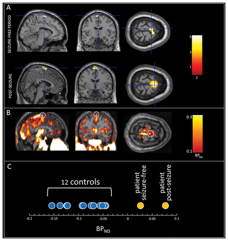
As shown in Figure 1A, during a seizure-free period, the patient had a single region of abnormally increased radiotracer binding (as compared to controls) in bilateral supplementary motor area (SMA; peak coordinates: x=−8, y=−8, z=76; peak Z =3.6) with the left hemisphere showing higher uptake than the right. The post-seizure image revealed an overlapping area of abnormality (peak coordinates: x=4, y=−4, z=76) with a higher peak Z score of 5.2 and larger spatial extent including greater extension to the right SMA. As compared to the image obtained during a seizure-free period, the area of abnormality in the post-seizure image was 61% larger (216mm3 vs 84mm3 at a threshold of Z=2.5) with 66% higher mean BPND (0.077 vs 0.026). Figure 1B is the patient’s post-seizure BPND image which shows radiotracer uptake in the region identified by the Z comparison as well as in multiple other cortical and subcortical regions including thalamus, brainstem, posterior superior sagittal sinus and extracranial tissues, all of which are also present in controls, making the patient’s abnormally increased SMA uptake difficult to appreciate on visual inspection.
Figure 1.

Abnormally increased neuroinflammation (TSPO expression by activated microglial) as assessed using [11C]PK11195 PET in a patient with clinically-defined frontal lobe epilepsy. A: Z-maps (the patient’s PET images statistically compared to 12 age-matched controls) obtained during a seizure-free period (top) and ~36 hours after a typical seizure (bottom.) Note the nearly identical location of abnormally increased TSPO expression in the midline SMA region in both scans, with greater intensity and spatial extent in the post-seizure scan. Crosshairs are at the voxel of peak intensity in each scan. Images are thresholded at Z=2.5 and overlaid onto a template T1 MRI. B: The patient's post-seizure BPND image, reflecting TSPO expression, overlaid on his native T1 MRI, without statistical comparison to controls. Note radiotracer uptake in the region identified in the post-seizure Z-map (white circles) as well as in multiple other cortical and subcortical regions including thalamus, brainstem, posterior superior sagittal sinus and extracranial tissues, all of which are also present in controls, making focally abnormal uptake more difficult to appreciate on visual inspection. C: Average BPND for each subject within the region of abnormality (bilateral SMA) identified in the comparison between the patient’s post-seizure image and the group of controls. Note significantly higher BPND in the patient’s two scan as compared to the twelve controls.
DISCUSSION
These results provide evidence that TSPO PET can identify a clinically (semiologically and FDG-PET) defined seizure focus that has no visible MRI correlate, and that inflammation at this seizure focus, while detectable during a seizure-free period, increases considerably following a seizure. This result has implications both for guiding epilepsy surgery and for understanding and differentiating the effects of seizures and epilepsy on brain inflammation.
We think that this patient, if he elects to proceed with epilepsy surgery, may turn out to have Focal Cortical Dysplasia (FCD) type II, as in a prior report of a patient with normal clinical MRI and a seizure focus detected by TSPO PET.2 FCD refers to a class of neuropathological abnormalities commonly found in patients with refractory epilepsy and normal MRI who undergo epilepsy surgery.1 FCD, especially FCD type II, is characterized by prominent neuroinflammation including activated microglia.6; 17 A prospective study of TSPO PET in presurgical epilepsy patients would be needed to determine whether TSPO PET can identify MRI-invisible FCD and help guide epilepsy surgery.
Our results suggest that inflammation at a seizure focus increases in intensity and spatial extent following a seizure. Interestingly, the region of peak inflammation was in the SMA just left of midline during a seizure-free period, and became more bilateral (with a peak just right of midline) post-seizure, perhaps reflecting seizure spread with bilateral motor semiology in this patient, and in in frontal/SMA seizures more generally.14 This could indicate that a post-seizure TSPO PET scan, while demonstrating greater abnormality, might be less precise in terms of lateralizing/localizing a seizure focus. These results highlight the importance of considering the time since a patient’s last seizure when interpreting TSPO PET results; this information was not considered in prior studies.2; 3; 5
It is important to note that the region of TSPO PET abnormality correlating with this patient’s clinically-defined seizure focus was identified via voxelwise statistical comparison within a standardized stereotactic space to a group of controls, and while detectable on his post-seizure scan (Figure 1B), was not obvious with visual inspection. Unbiased, observer-independent statistical analysis techniques such as we used here are proving superior to traditional expert interpretation in a variety of clinical nuclear medicine applications.10; 16
These results have a number of significant limitations: (1) Control subjects underwent only a single TSPO PET, so we do not have a measure of normal intra-subject variability against which to gauge the magnitude of the difference between the patient’s post-seizure and seizure-free scans. Mitigating somewhat against this limitation are preliminary results of an ongoing study by our group showing approximately 10% intra-subject variability in brain regions such as the thalamus in normal subjects who underwent two [11C]PK11195 PET scans on the same day. (2) These single subject results require replication in additional patients with well-localized seizures. While confidence in our patient’s seizure localization is high based on the distinct semiology of SMA seizures,14 it remains suboptimal given his normal MRI, uninformative scalp EEG and the lack of intracranial monitoring or resective surgery (both of which he has refused.) (3) The detected location of TSPO PET abnormality (bilateral SMA) at the edge of the brain can be prone to artifact associated with spatial normalization, and is also quite close to the superior sagittal sinus, which strongly expresses TSPO.19 However, even if the area of abnormality does correspond in part to the superior sagittal sinus, our finding of post-seizure increased TSPO expression would be of interest given the increasingly recognized role of large draining veins in immune surveillance and response.9
Because TSPO PET was performed only twice, we do not know the true time course of seizure-induced brain inflammatory changes. We have shown that brain inflammation in a clinically-defined seizure focus 36 hours after a typical seizure decreases back down to a “baseline” level but we do not know how stable this baseline is. We suspect, based on strong evidence that uncontrolled epilepsy is associated with slowly progressive cognitive decline and brain atrophy,15 that the transient seizure-induced neuroinflammation we have documented is superimposed on a slowly increasing level of chronic brain inflammation. Brain inflammation and in particular activated microglia are considered “double-edged swords” because of their complex role in neural repair and plasticity and neurodegeneration.8 Transient seizure-induced microglial activation, which we demonstrate in humans for the first time here, epitomizes this role – allowing recovery from any seizure-related neural damage, but also contributing to functional and structural neural changes, and mediating potentially maladaptive neuroplasticity that can lead to future seizures, i.e. epileptogenesis. Future studies in animals and humans are critical to better understanding transient and chronic neuroinflammation associated with seizures, epilepsy, epileptogenesis and epilepsy-related functional and structural neural decline.
Acknowledgments
This research was supported by NINDS K23 NS057579 and CURE
Footnotes
CONFLICTS OF INTEREST
No authors have any financial interests relevant to this report to disclose.
Ethical Publication Statement
We confirm that we have read the Journal’s position on issues involved in ethical publication and affirm that this report is consistent with those guidelines.
References
- 1.Besson P, Andermann F, Dubeau F, et al. Small focal cortical dysplasia lesions are located at the bottom of a deep sulcus. Brain. 2008;131:3246–3255. doi: 10.1093/brain/awn224. [DOI] [PubMed] [Google Scholar]
- 2.Butler T, Ichise M, Teich AF, et al. Imaging inflammation in a patient with epilepsy due to focal cortical dysplasia. J Neuroimaging. 2013;23:129–131. doi: 10.1111/j.1552-6569.2010.00572.x. [DOI] [PMC free article] [PubMed] [Google Scholar]
- 3.Gershen LD, Zanotti-Fregonara P, Dustin IH, et al. Neuroinflammation in temporal lobe epilepsy measured using positron emission tomographic imaging of translocator protein. JAMA neurology. 2015;72:882–888. doi: 10.1001/jamaneurol.2015.0941. [DOI] [PMC free article] [PubMed] [Google Scholar]
- 4.Gowers W. Epilepsy and other chronic convulsive diseases: their causes, symptoms, and treatment. Churchill; London: 1881. [Google Scholar]
- 5.Hirvonen J, Kreisl WC, Fujita M, et al. Increased in vivo expression of an inflammatory marker in temporal lobe epilepsy. J Nucl Med. 2012;53:234–240. doi: 10.2967/jnumed.111.091694. [DOI] [PMC free article] [PubMed] [Google Scholar]
- 6.Iyer A, Zurolo E, Spliet WGM, et al. Evaluation of the innate and adaptive immunity in type I and type II focal cortical dysplasias. Epilepsia. 2010;51:1763–1773. doi: 10.1111/j.1528-1167.2010.02547.x. [DOI] [PubMed] [Google Scholar]
- 7.Kimura Y, Ichise M, Ito H, et al. PET quantification of Tau pathology in human brain with 11C-PBB3. J Nucl Med. 2015;56:1359–1365. doi: 10.2967/jnumed.115.160127. [DOI] [PubMed] [Google Scholar]
- 8.Liu GJ, Middleton RJ, Hatty CR, et al. The 18 kDa translocator protein, microglia and neuroinflammation. Brain Pathol. 2014;24:631–653. doi: 10.1111/bpa.12196. [DOI] [PMC free article] [PubMed] [Google Scholar]
- 9.Louveau A, Smirnov I, Keyes TJ, et al. Structural and functional features of central nervous system lymphatic vessels. Nature. 2015 doi: 10.1038/nature14432. [DOI] [PMC free article] [PubMed] [Google Scholar]
- 10.McNally KA, Paige AL, Varghese G, et al. Localizing value of ictal–interictal SPECT analyzed by SPM (ISAS) Epilepsia. 2005;46:1450–1464. doi: 10.1111/j.1528-1167.2005.06705.x. [DOI] [PubMed] [Google Scholar]
- 11.Penny WD, Friston KJ, Ashburner JT, et al. Statistical parametric mapping: the analysis of functional brain images: the analysis of functional brain images. Academic press; 2011. [Google Scholar]
- 12.Rodgers KM, Hutchinson MR, Northcutt A, et al. The cortical innate immune response increases local neuronal excitability leading to seizures. Brain. 2009;132:2478–2486. doi: 10.1093/brain/awp177. [DOI] [PMC free article] [PubMed] [Google Scholar]
- 13.Rosell DR, Nacher J, Akama KT, et al. Spatiotemporal distribution of gp130 cytokines and their receptors after status epilepticus: comparison with neuronal degeneration and microglial activation. Neuroscience. 2003;122:329–348. doi: 10.1016/s0306-4522(03)00593-1. [DOI] [PubMed] [Google Scholar]
- 14.Salanova V, Morris H, Ness P, et al. Frontal lobe seizures: electroclinical syndromes. Epilepsia. 1995;36:16–24. doi: 10.1111/j.1528-1157.1995.tb01659.x. [DOI] [PubMed] [Google Scholar]
- 15.Sutula TP, Hagen J, Pitkänen A. Do epileptic seizures damage the brain? Curr Opin Neurol. 2003;16:189–195. doi: 10.1097/01.wco.0000063770.15877.bc. [DOI] [PubMed] [Google Scholar]
- 16.Varrone A, Asenbaum S, Vander Borght T, et al. EANM procedure guidelines for PET brain imaging using [18F] FDG, version 2. Eur J Nucl Med Mol Imaging. 2009;36:2103–2110. doi: 10.1007/s00259-009-1264-0. [DOI] [PubMed] [Google Scholar]
- 17.Vezzani A, French J, Bartfai T, et al. The role of inflammation in epilepsy. Nature Reviews Neurology. 2011;7:31–40. doi: 10.1038/nrneurol.2010.178. [DOI] [PMC free article] [PubMed] [Google Scholar]
- 18.Xanthos DN, Sandkühler J. Neurogenic neuroinflammation: inflammatory CNS reactions in response to neuronal activity. Nature Reviews Neuroscience. 2014;15:43–53. doi: 10.1038/nrn3617. [DOI] [PubMed] [Google Scholar]
- 19.Yaqub M, van Berckel BN, Schuitemaker A, et al. Optimization of supervised cluster analysis for extracting reference tissue input curves in (r)-[11c]-pk11195 brain pet studies. J Cereb Blood Flow Metab. 2012;32:1600–1608. doi: 10.1038/jcbfm.2012.59. [DOI] [PMC free article] [PubMed] [Google Scholar]


