

Abstract
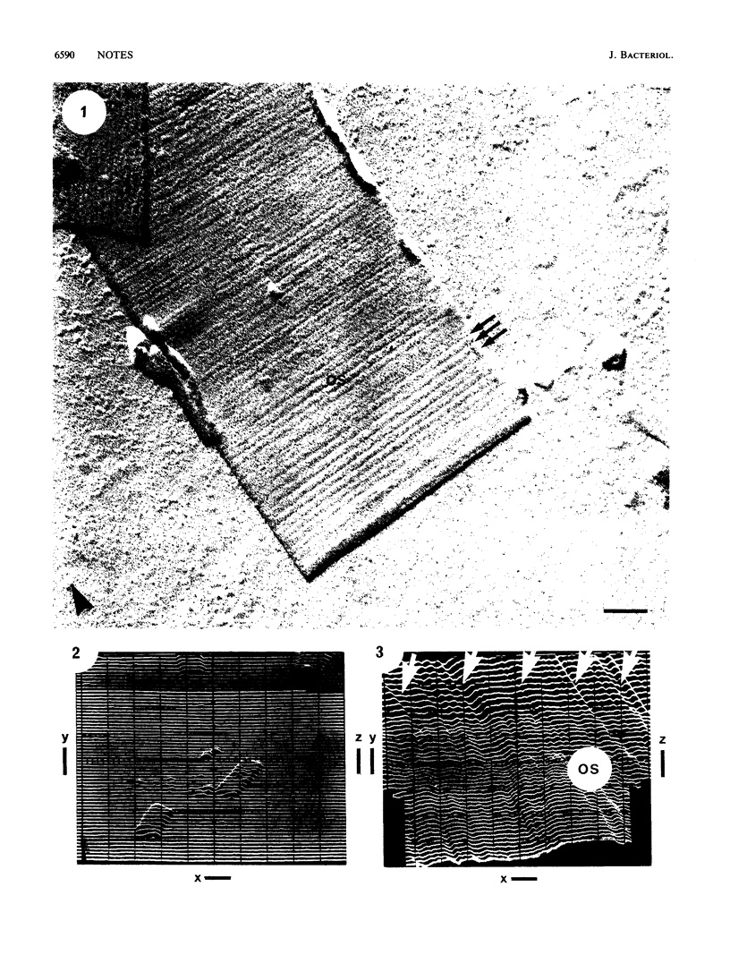
The inner and outer surfaces of the sheath of Methanospirillum hungatei GP1 have been imaged for the first time by using a bimorph scanning tunneling microscope (STM) on platinum-coated or uncoated specimens to a nominal resolution in height of ca. 0.4. nm. Unlike more usual types of microscopy (e.g., transmission electron microscopy), STM provided high-resolution topography of the surfaces, giving good depth detail which confirmed the sheath to be a paracrystalline structure possessing minute pores and therefore impervious to solutes possessing a hydrated radius of greater than 0.3 nm. STM also confirmed that the sheath consisted of a series of stacked hoops approximately 2.5 nm wide which were the remnants of the sheath after treatment with 2% (wt/vol) sodium dodecyl sulfate-2% (vol/vol) beta-mercaptoethanol (pH 9.0). No topographical infrastructure could be seen on the sides of the hoops. This research required the development of a new long-range STM capable of detecting small particles such as bacteria on graphite surfaces as well as a new "hopping" STM mode which did not deform the poorly conducting bacterial surface during high-resolution topographical analysis.
Full text
PDF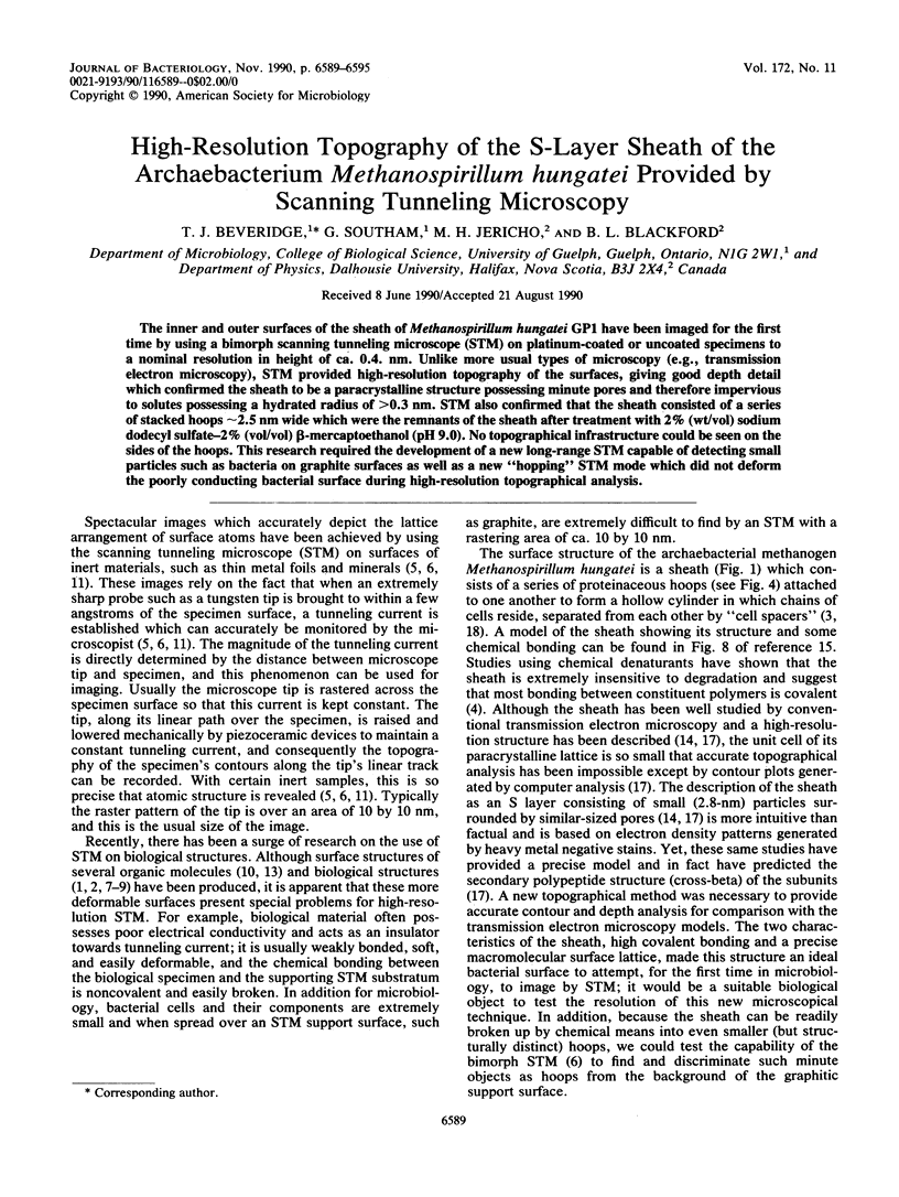


Images in this article


Selected References
These references are in PubMed. This may not be the complete list of references from this article.
- Amrein M., Dürr R., Stasiak A., Gross H., Travaglini G. Scanning tunneling microscopy of uncoated recA-DNA complexes. Science. 1989 Mar 31;243(4899):1708–1711. doi: 10.1126/science.2928803. [DOI] [PubMed] [Google Scholar]
- Amrein M., Stasiak A., Gross H., Stoll E., Travaglini G. Scanning tunneling microscopy of recA-DNA complexes coated with a conducting film. Science. 1988 Apr 22;240(4851):514–516. doi: 10.1126/science.3358130. [DOI] [PubMed] [Google Scholar]
- Beveridge T. J., Stewart M., Doyle R. J., Sprott G. D. Unusual stability of the Methanospirillum hungatei sheath. J Bacteriol. 1985 May;162(2):728–737. doi: 10.1128/jb.162.2.728-737.1985. [DOI] [PMC free article] [PubMed] [Google Scholar]
- Golovchenko J. A. The tunneling microscope: a new look at the atomic world. Science. 1986 Apr 4;232(4746):48–53. doi: 10.1126/science.232.4746.48. [DOI] [PubMed] [Google Scholar]
- Shaw P. J., Hills G. J., Henwood J. A., Harris J. E., Archer D. B. Three-dimensional architecture of the cell sheath and septa of Methanospirillum hungatei. J Bacteriol. 1985 Feb;161(2):750–757. doi: 10.1128/jb.161.2.750-757.1985. [DOI] [PMC free article] [PubMed] [Google Scholar]
- Stewart M., Beveridge T. J., Sprott G. D. Crystalline order to high resolution in the sheath of Methanospirillum hungatei: a cross-beta structure. J Mol Biol. 1985 Jun 5;183(3):509–515. doi: 10.1016/0022-2836(85)90019-1. [DOI] [PubMed] [Google Scholar]
- Zeikus J. G., Bowen V. G. Fine structure of Methanospirillum hungatii. J Bacteriol. 1975 Jan;121(1):373–380. doi: 10.1128/jb.121.1.373-380.1975. [DOI] [PMC free article] [PubMed] [Google Scholar]