Figure 4.
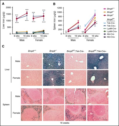
Liver iron is increased and spleen iron is reduced in Bmp6fl/fl;Tek-Cre+ mice, but not Bmp6fl/fl;Alb-Cre+ or Bmp6fl/fl;LysM-Cre+ mice. Eight- and 16-week-old littermate male and female Bmp6+/+, Bmp6+/−, and Bmp6−/− global knockout mice and Bmp6 conditional knockout mice in ECs (Bmp6fl/fl;Tek-Cre+), macrophages (Bmp6fl/fl;LysM-Cre+), and hepatocytes (Bmp6fl/fl;Alb-Cre+) compared with littermate controls (Bmp6fl/fl;Cre–) were analyzed for tissue iron in (A,C) liver and (B,C) spleen by (A-B) biochemical analysis or (C) Perls’ Prussian blue. n = 4-7 mice per group (supplemental Table 2), with tissues from 1 representative mouse shown in (C) (original magnification ×20; scale bar represents 100 μM). *P < .05, **P < .01, ***P < .001 relative to control Bmp6+/+ mice by one-way ANOVA with Dunnett’s post hoc test; +P < .05, ++P < .01, +++P < .001 relative to control Bmp6fl/fl;Cre– mice by Student t test.

