Abstract
Site-directed deuterium NMR spectroscopy is a valuable tool to study the structural dynamics of biomolecules in cases where solution NMR is inapplicable. Solid-state 2H NMR spectral studies of aligned membrane samples of rhodopsin with selectively labeled retinal provide information on structural changes of the chromophore in different protein states. In addition, solid-state 2H NMR relaxation time measurements allow one to study the dynamics of the ligand during the transition from the inactive to the active state. Here we describe the methodological aspects of solid-state 2H NMR spectroscopy for functional studies of rhodopsin, with an emphasis on the dynamics of the retinal cofactor. We provide complete protocols for the preparation of NMR samples of rhodopsin with 11-cis-retinal selectively deuterated at the methyl groups in aligned membranes. In addition, we review optimized conditions for trapping the rhodopsin photointermediates; and lastly we address the challenging problem of trapping the signaling state of rhodopsin in aligned membrane films.
Keywords: G-protein–coupled receptor, Lipids, Membrane, Nuclear magnetic resonance, Protein dynamics, Relaxation, Rhodopsin, Vision
1. Introduction
Rhodopsin is responsible for dim light vision in vertebrates and is representative of a large family of G-protein–coupled receptors (GPCRs) that regulate many important signaling functions in humans (1). Despite progress in recent years in crystallizing GPCRs for X-ray studies (2–4), the available structures of rhodopsin in the signaling state (5–7) are insufficient to provide a complete picture of the protein activation. Alternatively, methods such as NMR spectroscopy can be applied to obtain additional structural information. Yet the most innovative and valuable aspect of such NMR studies is the ability to produce experimental information about the molecular dynamics as a complement to X-ray crystallography (8). Site-directed 2H NMR spectroscopy provides orientational constraints for functional molecular groups, and relaxation times characterize their thermal fluctuations. The results can be further analyzed using a generalized model-free approach (9–11) to obtain novel information about the molecular dynamics (see below). Structural and dynamical modeling allows one to determine both the local conformation and correlation times of the molecular motions at an atomistically resolved level (9–11). In this article we outline the methodology for solid-state 2H NMR studies of the retinal cofactor bound to rhodopsin, including trapping of rhodopsin photointermediates in aligned membrane films. Data reduction and analysis are described at an introductory level appropriate for non-specialists.
2. Materials
Protocols for the preparation and characterization of aligned phospholipid lipid bilayers containing rhodopsin for solid-state 2H NMR studies are presented. Native rod outer segment (ROS) disk membranes (also referred to as RDM, retinal disk membranes) are prepared from bovine retinas following standard procedures (12). Recombinant membranes with a defined lipid composition are prepared by purifying detergent-solubilized rhodopsin on a hydroxyapatite column, followed by recombination with solubilized phospholipids, and subsequent detergent dialysis. Proteolipid membrane bilayers with rhodopsin then are harvested by ultracentrifugation.
2.1 Retinas, Buffers, Detergents, Gels, Phospholipids, Retinals, and Peptides
Retinas: Frozen bovine retinas are purchased from W.L. Lawson, Co. (Omaha, NE, USA) and delivered in 30% (w/w) sucrose solution containing 10 mM Tris-acetate buffer, pH 7.4, 1 mM dithiothreitol (DTT), and 0.001% (v/v) aprotinin (A6279, Sigma-Aldrich, St. Louis, MO, USA), on dry ice. Retinas are stored in lightproof glass containers under nitrogen or argon at −80°C until use.
Buffers and solutions: Homogenizing solution—30% sucrose (w/w), 5 mM Tris-acetate pH 7.4, 65 mM NaCl, 2 mM MgCl2, and 2 mM EDTA. Dilution buffer—10 mM Tris-acetate pH 7.4. Stock detergent buffer—3% (v/v) Ammonyx LO containing 100 mM NH2OH (hydroxylamine) and 10 mM Na phosphate buffer, pH 6.8. Opsin buffer—10 mM 2-[4-(2-hydroxyethyl)piperazin-1-yl]ethanesulfonic acid (HEPES) buffer containing 50 mM NH2OH and 10 mM MgCl2 at pH 6.8. Adjust pH using 1 M NaOH (approximately 7 drops). Regeneration buffer—10 mM HEPES, pH 6.8. Dialysis buffer—5 mM HEPES, 1 mM EDTA, and 1 mM DTT (add fresh). Chromatography detergent buffer—100 mM DTAB, 15 mM Na phosphate, pH 6.8, containing 0.02% (w/w) NaN3 (sodium azide) and 1 mM DTT (optional, for regenerating the column if desired). Storage buffer—15 or 67 mM Na phosphate buffer, pH 7.0. Retinal stock solution—0.02 mM 11-cis-retinal in 99.5% ethanol. Deuterium-depleted buffer—5 mM 2-(N-Morpholino)ethanesulfonic acid (MES) buffer prepared with deuterium-depleted water containing 5 mM NaCl, pH 7.0 (at room temperature for the dark or Meta I state), or pH 5.5 (at 4 °C and pH 5.0 at room temperature for the Meta II state).
Detergents: Ammonyx LO (laurylamine oxide, 29–31%) (Stepan Co., Northfield, IL, USA), dodecyltrimethylammonium bromide (DTAB) (Sigma-Aldrich, St. Louis, MO, USA) (see Note 1).
Column packing materials: Hydroxyapatite (Bio-Gel HTP Gel; Bio-Rad Laboratories, Hercules, CA, USA).
Phospholipids: l-palmitoyl-2-oleoyl-sn-glycero-3-phosphocholine (POPC) and l,2-dioleoyl-sn-glycero-3-phosphoethanolamine (DOPE) are procured from Avanti Polar Lipids (Alabaster, AL, USA).
Density gradient solutions: 1.10, 1.11, 1.13, and 1.15 g/mL sucrose gradient solutions are prepared in 100-mL volumetric flasks by mixing 62.4 g, 68.4 g, 81.4 g, or 93.0 g of a 42% (w/w) sucrose solution, respectively, with 0.1 mL of 1 M Tris-acetate buffer, pH 7.4, containing 0.1 mL of 0.1 M MgCl2, and adding double-distilled water to make the total volume of each solution 100 mL.
Isomerically pure 11-cis-retinal can be procured from the US National Eye Institute (Bethesda, MD, USA).
Selectively deuterated 11-cis-retinals having 2H isotope labels at the C5-, C9-, or C13-methyl groups, denoted as 11-Z-[5-C2H3]-, 11-Z-[9-C2H3]-, and 11-Z-[13-C2H3]-retinal, were synthesized in the laboratory of Prof. K. Nakanishi (Department of Chemistry, Columbia University, New York) as described (13).
High-affinity transducin-derived GαCT2 peptide with the amino-acid sequence ILENLKDVGLF, also denoted as Gαt340–350(K341L, C347V), is obtained from Biomatik (Wilmington, DE, USA).
2.2. Purification of Retinal Disk Membranes Containing Rhodopsin
Preparation of ROS disk membranes: all procedures are performed on ice or at 4 °C under dim red light (11-W Bright Lab™ Universal Red Safelight bulb, CPM Delta1, Inc., Dallas, TX, USA; or a 11-W incandescent white bulb with red filter, Kodak safelight Red filter 1, Eastman Kodak Co., USA). Begin the preparation by thawing the frozen bovine retinas overnight at 4 °C. Transfer the thawed bovine retinas to a loose-fitting Teflon homogenizer (chamber clearance is 0.3 to 0.45 mm). Add 30 mL of homogenizing solution per 50 retinas under a gentle stream of argon gas (see Note 2). Homogenize the retinas by applying about ten strokes of the pestle slowly under a gentle argon gas stream. Centrifuge at 2,600 × g (Sorvall GSA rotor) for 20 min at 4 °C. The centrifuge speed is expressed as maximum relative centrifugal force (× g) throughout the text. Collect the supernatant with a spring-loaded syringe (18-gauge cut off needle), and transfer the pellet to a tight-fitting Teflon homogenizer (chamber clearance is 0.1 to 0.15 mm). Add an equal volume of homogenizing solution, and apply about 6–8 strokes of the pestle under a gentle argon gas stream for the release of additional ROS disk membranes. Centrifuge at 2,600 × g for 20 min at 4 °C, and collect the supernatant.
Combine the supernatants from the two centrifugation steps, and add two volumes of dilution buffer (10 mM Tris-acetate, pH 7.4) (see Note 2). Centrifuge at 8,000 × g (Sorvall GSA rotor) for 50 min at 4 °C. Resuspend the pellet in a small volume of 1.10 g/mL sucrose density gradient solution (to yield 25 mL or less of total volume). Prepare sucrose step gradients on ice or in the cold room in polyallomer 38.5-mL centrifuge tubes. The volumes of the layers are 8, 10, and 10 mL for the 1.15, 1.13, and 1.11 g/mL gradient solutions, respectively. Four tubes are required for 50 retinas, or six tubes for 100 retinas. Pull up the resuspended pellet with an 18-gauge cut-off needle, then switch to a 20-gauge needle, and expel it to fragment the crude rod outer segments. Place an equal amount of the ROS solution on the top of the 1.11 g/mL density gradient solution of each of the centrifuge tubes, and use additional 1.10 g/mL sucrose density gradient solution to balance the tubes. Centrifuge at 113,000 × g in a swinging bucket rotor (Beckman SW 28 rotor) for 1 h at 4 °C. Collect the carpet (band) at the 1.11/1.13 g/mL interface with a spring-loaded syringe, having a cutoff 18-gauge needle tip.
Removing the sucrose: combine the collected bands, and dilute with two volumes of double-distilled deionized water. Add argon gas to the centrifuge bottles, and centrifuge at 48,000 × g (Sorvall SS 34 rotor) for 30 min at 4 °C. After centrifugation, take the pellet, resuspend it in water, and centrifuge for 30 min at 48,000 × g at at 4 °C. Repeat 1–2 times to ensure the removal of sucrose. Note that hypotonic water “washing” produces membrane fragments due to the osmotic shock. Resuspend the ROS disk membranes in either 15 mM or 67 mM (for longer storage) storage buffer. Tansfer to low-temperature freezer storage vials, e.g. Eppendorf safe-lock tubes (Eppendorf North America, Inc., Westbury, NY, USA) or polypropylene leakproof screw cap centrifuge tubes (e.g. Nunc™ 15-mL Graduated Centrifuge Tubes, Thermo Fisher Scientific, Inc., Waltham, USA). Overlay with argon gas, wrap in aluminum foil, and store at −80 °C until use.
2.3. Characterization of Rhodopsin by UV-visible Spectroscopy
The procedure is carried out under red dim light. The UV-visible absorption spectra are acquired using a Cary 4000 dual-beam scanning spectrophotometer (Varian, Inc., USA) from 700 nm to 250 nm with 10-mm pathlength quartz cuvettes at room temperature. Alternatively, a single beam Cary 50 spectrophotometer (Varian, Inc., USA) with Xenon flash lamp technology is used.
First record the baseline using a blank solution containing 980 μL of stock detergent buffer and 20 μL of 15 mM storage buffer.
Dissolve and mix thoroughly 20 μL of a suspension of disk membranes with rhodopsin in 980 μL of stock detergent buffer. Acquire the spectrum of this test sample.
Photobleach the test sample completely using about 6 flashes from a handheld flash lamp (Sunpak auto 555 or equivalent) equipped with the 515-nm long wavelength pass filter (OG515, Schott, Mainz, Germany), and record the spectrum of the bleached sample.
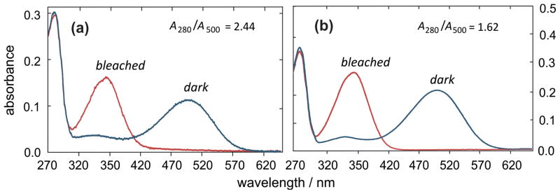
Measure the absorbance at 500 nm of the dark spectrum versus the bleached spectrum (A500) and at 280 nm for the dark spectrum (A280). Generally rhodopsin in purified disk membranes has a A280/A500 ratio of 2.4. For comparison, a representative spectrum is provided in part (a) of Fig. 1 (see Note 3).
Calculate the molar concentration of rhodopsin using the molar absorption coefficient of ε = 40,600 M−1 cm−1 at 500 nm. Alternatively, optical density units (OD) can be used. One OD unit is defined as 1 mL of a solution with absorbance of A500 = 1 in a 10-mm path length cuvette, and affords a convenient measure of the equivalents of retinal. One OD corresponds to 24.6 nmol or 0.958 mg rhodopsin, assuming its relative molecular mass is Mr = 39,000 Da.
Fig. 1.
Representative UV-visible absorption spectra of rhodopsin (a) in ROS disk membranes solubilized in 3% Ammonyx LO, pH 6.8, (Section 2.1), and (b) after column purification in 100 mM DTAB detergent buffer, pH 6.8, in presence of 10 mM hydroxylamine. The 11-cis retinal absorption in the dark-state rhodopsin is shifted to 500 nm due to the protonated Schiff base covalently bound to the protein, and its interaction with surrounding amino acids in the binding pocket. After bleaching in the presence of hydroxylamine, the absorption with maximum around 360 nm is due to free hydrolyzed retinal oxime. The A280/A500 absorption ratio characterizes the spectral purity of rhodopsin (see Note 3).
2.4. Regeneration Assay for Testing Rhodopsin Photochemical Functionality
A regeneration assay is carried out to determine the amount of bleached rhodopsin present in the disk membranes, their regenerability, and the post-regeneration A280/A500 ratio.
Prepare 11-cis-retinal solution in 99.5 % ethanol with a concentration of about 20 mM (see Note 4).
UV-visible characterization of 11-cis-retinal: record the absorption spectrum using 1 μL of the retinal solution in ethanol as follows. Prepare two cuvettes with 1 mL of ethanol (≥ 99.5 % w/w). Add 1 μL of 11-cis-retinal solution into the sample cuvette; the other cuvette is the blank (reference sample). Record the absorption spectrum from 600 to 200 nm. At 380 nm the absorbance should be in the range 0.3–0.8, and the ratio A380/A250 should be in the range of 1.2–1.5. As the 11-cis-retinal isomerizes, this ratio increases considerably up to a value of 8 (14).
Place 200 μL of ROS disk membranes in each of three 1.5-mL Eppendorf tubes. Label them as “control” (C), “bleached+regenerated” (B), and “regenerated” (R). Photobleach the “bleached+regenerated” sample using the flash lamp with the 515-nm long-wavelength pass filter, as described in the previous section.
Add the same amount of ODs at 380 nm of 11-cis-retinal as the amount of rhodopsin ODs in disk membranes to the tubes labeled “bleached+regenerated” and “regenerated”, but not to the “control”. Taking into account that the molar absorption coefficient for retinal in ethanol at 376.5 nm is ε ≈25,000 M−1 cm−1 (15) and for rhodopsin ε = 40,600 M−1 cm−1 at 500 nm, the molar retinal/rhodopsin ratio is about 1.6:1.
Incubate all the three tubes at 37 °C in the water bath for about 1.5 hours. Take the UV-visible absorption spectra after adding 800 μL of stock detergent buffer (Section 2.1), pH 6.8, as described earlier.
The percent regeneration is calculated by the ratio of A500 of sample B to A500 of sample R multiplied by 100. For calculating the percentage of rhodopsin in the membrane sample that is bleached, take the ratio of A500 of sample C to A500 of sample R, and multiply by 100. Subtract this number from 100 to obtain the percentage of rhodopsin bleached. The post-regenerated A280 /A500 ratio is calculated by dividing the A280 absorption of sample C by the A500 absorption of sample R, and multiplying by 100.
Typical results are: 93% regenerability; 3–5% bleached; and 2.1 post-regeneration A280 /A500 ratio. For further details see ref. (16).
2.5 Regeneration of Disk Membranes with Deuterium-Labeled Retinals
All regeneration procedures are carried out in the dark (except opsin handling before regeneration) on ice, or in the cold room (at 4 °C). Always work with and store the disk membrane suspensions under a gentle stream or blanket of argon gas.
Retinal preparation: Synthetic deuterated 11-cis-retinals are stored in benzene at −80°C. Typically each vial contains about 0.5 mg of deuterated 11-cis-retinal. Take the vial from the freezer, and evaporate the benzene under a gentle stream of argon or nitrogen gas. Add 300 μL of ethanol (≥ 99.5% w/w) to the retinal bottle, dissolve the retinal, and measure the UV-visible absorption spectrum as described above (Section 2.4, step 2). Calculate the amount of 11-cis-retinal in OD units at 380 nm.
Calculate the amount of rhodopsin required for regeneration using a 1:1 retinal/rhodopsin ratio in OD units. Because the retinal molar absorption coefficient in ethanol is ε ≈ 25,000 M−1cm−1 at 380 nm (15), and for rhodopsin ε = 40,600 M−1 cm−1 at 500 nm, the molar retinal/rhodopsin ratio is about 1.6:1 (see Note 5).
Check the purity and concentration of rhodopsin in disk membranes as described above (Section 2.3). The purity of the protein is very important: with an A280 /A500 ratio about 2.6 the regeneration gives approximately 75% yield.
Opsin preparation: Centrifuge the exact amount of the disk membranes previously calculated (according to the exact amount of deuterated retinal) for 30 min at 48,000 × g (Sorvall SS-34 rotor). Resuspend the pellet in ca. 50 mL of opsin buffer containing hydroxylamine (Section 2.1). Suspension is a crucial point; first mix everything using a Pasteur pipette, then use a syringe with an 18-gauge needle (the syringe is used to disperse material visible to the naked eye in the protein suspension; try to pass the protein through the syringe only once) to obtain a homogeneous suspension. Bleach the ROS membranes at 4 °C using a 150-W tungsten–halogen light source (Schott-Fostec ACE light source; Fostec, Auburn, NY, USA) fitted with yellow filter (λ > 500 nm) for 30 min. Check that all the protein is in suspension. Take a UV-visible spectrum of the opsin using a 20 μL aliquot of this solution. The color of the suspension changes visibly from pink to slightly pink, then slightly yellow, and finally white. Centrifuge again for 30 min at 48,000 × g at 4 °C. Discard the supernatant; the pellet should be very tight. Then resuspend the pellet in 60 mL of regeneration buffer (Section 2.1) very gently to avoid denaturation. This time do NOT use a syringe when you are manipulating the opsin, use only a Pasteur pipette, and centrifuge again for 30 min at 48,000 × g at 4 °C. Repeat this step 4 times to completely remove the hydroxylamine.
Opsin regeneration: After the last centrifugation, take out the supernatant, and resuspend the pellet completely in ca. 30 mL of regeneration solution (Section 2.1). Again do NOT use a syringe. Add all the ethanol solution of the deuterated 11-cis-retinal to the opsin. More ethanol can be used to rinse the residual 11-cis-retinal from the vial; however, do not exceed 1% for the ethanol concentration in the total volume of the sample (for instance, use less than 350 μL for a Vtot = 35 mL). Put the tube in the water bath at 37 °C for l.5 h in the dark. At the end of the regeneration, check the UV-visible spectra using about 20 μL of the solution. Calculate the amount of regenerated rhodopsin as described above (Section 2.3). Centrifuge for 30 min at 48,000 × g at 4 °C. After centrifugation, follow the rhodopsin purification protocol (Section 2.6) starting with step 2, or resuspend the pellet in 10 mL of storage buffer, and store in the −80 °C freezer.
2.6. Rhodopsin Purification by Hydroxyapatite Column Chromatography
All procedures are carried out at 4 °C under dim red light conditions.
Take the suspension of the ROS disk membranes kept in storage buffer from the −80 °C freezer and thaw. Centrifuge the disk membranes at 48,000 × g for 30 min at 4 °C. After centrifugation, remove the supernatant, and solubilize the rhodopsin and lipids by resuspending the pellet in 10–15 mL of the chromatography detergent buffer. The concentration of rhodopsin should be about 3–5 mg/mL. Incubate for one hour, and then centrifuge at 163, 000 × g (Sorvall T865.1 rotor) for 20 min at 4 °C. The solubilized rhodopsin and lipids should not have any insoluble precipitate. Any pellet observed at this point is discarded (see Note 6).
Prepare a 2.5 cm (diameter) column by weighing 18 g of hydroxyapatite (DNA grade, Bio-Rad, Hercules, CA), which is enough to purify about 50 mg of rhodopsin. The length of the column with this amount of hydroxyapatite is about 8 cm. For more rhodopsin than this amount, the column needs to be proportionately longer.
Equilibrate the column using 120 mL (about 3 times the column volume) of DTAB detergent buffer. Load the detergent-solubilized rhodopsin onto the hydroxyapatite column. Elute the rhodopsin using a gradient mixer with a linear gradient of 0–0.5 M salt (NaCl) in DTAB detergent buffer.
Elute the fractions with a flow rate of 0.6 mL/minute, and collect using a fraction collector. Check the absorption spectrum of the fractions to monitor the fractions containing rhodopsin. Normally after about 3 h the protein starts eluting. The pure rhodopsin has purity (A280/A500 ratio) of 1.60–1.75, see Fig. 1b.
Combine the rhodopsin-containing fractions together, to determine the overall yield and the purity of purified rhodopsin. Generally 70–75% rhodopsin is recovered. For specifics see refs. [8,9].
2.7. Preparation of Rhodopsin–Lipid Recombinant Membranes
All the procedures are performed in the dark or under dim red light conditions at 4 °C. Always work with and store the disk membranes suspension under a stream or blanket of argon gas. Rhodopsin is recombined with POPC at a 1:50 rhodopsin-lipid molar ratio for the dark and the Meta I states. For the Meta II sample, a mixture of POPC with DOPE (3:1 POPC/DOPE ratio) is used, and the rhodopsin-lipid molar ratio is decreased to 1:75.
After combining the eluted rhodopsin fractions from hydroxyapatite column chromatography (typically ≈50 mL) as mentioned above, adjust the DTAB concentration to 300 mM.
Calculate the amount of lipids corresponding to the amount of purified rhodopsin. Dissolve the phospholipids in 3–4 mL of chromatography buffer having a higher DTAB concentration (300 mM DTAB concentration instead of 100 mM).
Mix the rhodopsin solution with the solubilized phospholipids in the desired molar ratio, cover the sample, and vortex. Gently overlay the sample with argon gas, cap the sample tightly with parafilm, cover in aluminum foil, and incubate for 1–2 hours at 4 °C.
In the meantime, prepare the dialysis tubing (molecular weight cut off 12–14 kDa; 1.6 cm diameter; approximately 2.0 mL solution per centimeter of dialysis tubing) (Spectrum Laboratories Inc.; Rancho Dominguez, CA, USA) by immersing the dialysis bag in hot boiling water for a few seconds, then transfer from the hot water to the colder (4 °C) water (repeat three times), and leave it in the refrigerator (4 °C) for 30 minutes, after which it is ready to be used.
Transfer the sample (rhodopsin solution incubated with the lipids) into the dialysis tubing as prepared already. Securely clamp both ends, and place the tubing with the sample in the dialysis buffer (5 mM HEPES and 1 mM EDTA, pH 6.8), with a continuous gentle purging of N2 gas through the buffer using a gas dispersion tube. Dialyze with one exchange of the dialysis buffer every 6–8 hours, giving a total of 8 or more exchanges. The total volume of buffer used for the whole dialysis procedure is about 900 mL of buffer per 1 mL of DTAB solution in the dialysis bag.
Take out the dialysis bag and record the UV–visible absorption spectrum of the recombinant sample to check the yield and the A280/A500 ratio.
Centrifuge the contents of the dialysis bag at 48,000 × g for 1 hour at 4 °C. Resuspend the pellet in a 67 mM sodium phosphate buffer pH 7.0, or 100 mM HEPES buffer pH 7.0 containing 1 mM EDTA. Overlay with argon and freeze it at −80 °C. The recombinant membrane samples can be stored for several years at −80 °C. For further details see refs. (17–19).
3. Methods
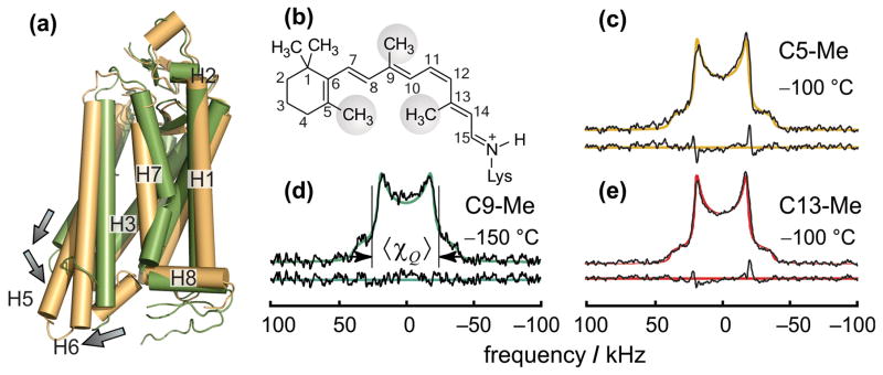
Site-directed deuterium NMR spectroscopy allows one to study the local dynamics and structure of biological systems by specifically 2H-labeling the molecular groups of interest. The deuteron has a spin I = 1, and like other nuclei with spin larger than ½, it has a lower symmetry of the nuclear charge distribution than spin-½ nuclei. The measure of the charge asymmetry is called the quadrupole moment, which can interact with the electric field gradient of the carbon-deuterium bond present in the molecule (20). This interaction leads to two transitions present in the solid-state 2H NMR spectrum (21). Examples are provided in Fig. 2, and are further discussed below. Unlike a solution 2H NMR spectrum, where the angular-dependent interactions are averaged to zero giving a single peak, in the solid-state NMR spectrum more information is available. The quadrupolar splittings carry information regarding the spatial orientation of the carbon–deuterium bonds, including their average orientation and the amplitude of their angular fluctuations. Below we include the protocols for preparation of aligned samples of lipid bilayers containing rhodopsin, and provide a description of structural and relaxation 2H NMR methods for the bound retinal chromophore.
Fig. 2.
Solid-state deuterium NMR spectroscopy of selectively 2H-labeled retinal bound to rhodopsin in randomly oriented membranes. (a) Structure of the Meta II state of rhodopsin (protein databank accession code 4A4M) compared to dark-state rhodopsin (code 1U19) showing activating movement of helices. (b) Numbering scheme of retinal ligand indicating methyl groups studied. (c) Solid-state 2H NMR spectra of deuterated retinal specifically labeled at C5-, C9-, or C13-Me positions bound to rhodopsin in unoriented membranes (10). The spectra are calculated by convolution of the frequency distribution for the randomly oriented quadrupolar coupling tensor and intrinsic line broadening Δν. The spectral fitting indicates that the average quadrupolar coupling constant 〈χQ〉 is reduced by a factor of three due to rapid methyl group spinning, and by another 10 % due to off-axial fluctuations with respect to the average orientation (see text). The line broadening Δν is in the range of 3.2–5.0 kHz. Figure adapted from ref. (10).
3.1 Solid-State Deuterium NMR Spectroscopy for Non-Aligned Samples of Rhodopsin
In so-called powder-type samples, the membranes are randomly oriented (hence the analogy to a random powder). Orientational constraints for the labeled group are not available, because the 2H NMR spectrum lineshape is the same for any orientation (e.g. a methyl group) with respect to the protein. Fig. 2 presents examples of solid-state 2H NMR spectra obtained for randomly oriented samples of rhodopsin with specifically 2H-labeled methyl groups of retinal at the C5, C9, or C13 carbon positions. An important aspect is that experimental information about the molecular dynamics is uniquely available from such solid-state 2H NMR studies (11). Moreover, certain information on the orientational distribution of the labeled molecular groups (distribution versus the average orientation) can be derived from the spectral line shape, as described below.
In the solid-state 2H NMR spectrum the weak shoulders are due to the C2H3 groups oriented parallel (lower relative probability) to the external magnetic field. The two stronger peaks are due to the C2H3 groups oriented perpendicular (higher relative probability) to the external magnetic field.
For rapidly rotating methyl groups, the static quadrupolar coupling constant χQ (= 170 kHz) is reduced by the spinning. The effective quadrupolar coupling is , where θt = 109.47 ° is the tetrahedral angle defining the geometry of the methyl group, and P2(cosθ) is the second order Legendre polynomial, defined as P2(cosθ) = (1/2)(3 cos2 θ − 1). Calculate the effective quadrupolar coupling constant by using ideal tetrahedral angle for rapidly spinning methyl group ( ).
The effective quadrupolar interaction ( ) of the deuterium nuclei is further reduced by the fluctuations of the methyl group symmetry axis (aligned with C–C2H3 bond) about its mean orientation. The average quadrupolar coupling constant 〈χQ 〉 is given by the relation , where θ is the angle between the instantaneous and averaged orientations of the methyl group, and SC3 is the order parameter characterizing the amplitude of the off-axial fluctuations of the three-fold axis of the methyl group. The brackets 〈…〉 indicate the angular average. Calculate the value of the order parameter (SC3) for the methyl three-fold axis by assuming that the value of 〈χQ〉 (see Fig. 2d) is 51 kHz (SC3 = 0.9). Note that for all three labeled methyl groups at carbon positions C5, C9, or C13, the order parameter is found to be practically the same (≈0.9), despite the different environment in the rhodopsin binding pocket and the very different rotational dynamics (see below).
The order parameter (SC3) is related to the angular amplitude (range) of the fluctuations by . It is assumed for simplicity that the probability of the methyl angle is constant over the angular range Δθ, and is zero otherwise (rectangular probability distribution). To establish a physical picture of the fluctuations, calculate the value of Δθ for the methyl group fluctuations from the order parameter (SC3) obtained directly from the solid-state 2H NMR spectrum. (Answer: Δθ = 21 °.)
3.2 Preparation of Aligned Membranes Containing Rhodopsin in the Dark State
All procedures are performed in the dark or under dim red light conditions at 4 °C unless mentioned otherwise. Always work with and store the rhodopsin samples under a gentle steam or blanket of argon gas. The best alignment of proteolipid membranes containing rhodopsin can be achieved by isopotential spin-dry ultracentrifugation (22,23) (see Note 7).
Prepare a suspension of POPC membranes with rhodopsin (rhodopsin/lipid ratio 1:50) using 5 mL of deuterium-depleted 5 mM MES buffer containing 5 mM NaCl, pH 7, at 4 °C. Place an ultrathin glass slide (16 × 6 × 0.07 mm; Marienfeld Laboratory Glassware, Lauda-Königshofen, Germany) on the isopotential surface of the stainless steel plate (see Note 7). Assemble the insert, and place it into the centrifuge tube. Transfer about 150 μL of a membrane suspension containing ca. 1 mg of rhodopsin (1:50 rhodopsin/POPC molar ratio) into the sample compartment.
Centrifuge the membranes at 141,000 × g (Beckman SW 28 rotor) for ca. 2 h at 4 °C.
After slow evaporation, rehydrate the membrane film by isopiestic transfer in a closed container (desiccator), using saturated KCO3 in 2H-depleted water with a relative humidity ≈ 43% at 4 °C. Stack the individual glass plates, each with a membrane film, insert them into a 8 × 22 mm cut-off NMR tube, and tightly seal with a machined Teflon plug.
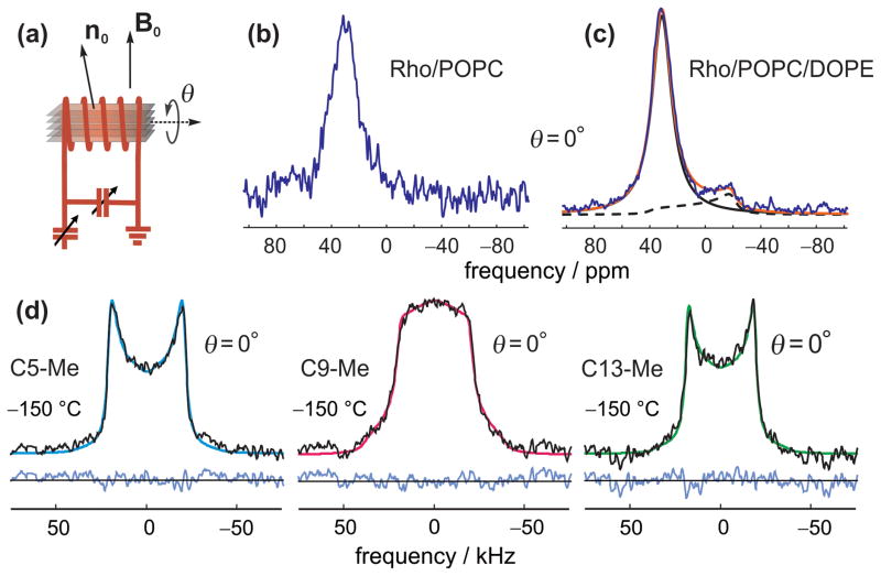
Check the alignment by 31P NMR spectroscopy at room temperature. Well-aligned samples with the director axis (the average membrane normal) parallel to the magnetic field of the NMR spectrometer (θ = 0 °) give a single peak, with a chemical shift of about 30–35 ppm depending on the lipid (Fig. 3a). Unoriented lipid bilayers produce a powder-type spectrum with a maximum around −15 to −20 ppm, corresponding to θ = 90 ° (most probable orientation), Fig. 3b (dashed line). The fraction of aligned bilayers can be readily estimated by the area of a single peak with respect to the total area of the spectrum (Fig. 3b). Knowing the amounts of aligned and unoriented membranes, one can simulate and subtract the contribution from unoriented membranes to the 2H NMR spectrum (see Note 8), and use incompletely aligned samples in structural studies. Examples of solid-state 2H NMR spectra of selectively deuterated retinal bound to rhodopsin in aligned membranes are shown in Fig. 3c.
Fig. 3.
Examples of phosphorus-31 (a,b) and deuterium NMR (c) spectra of aligned membranes containing rhodopsin with 2H-labeled retinal: (a) solid-state 31P NMR spectrum of rhodopsin-POPC (1:50 protein/lipid ratio) recombinant membranes with a largely complete alignment; (b) solid-state 31P NMR spectrum of recombinant membranes (3:1 POPC/DOPE molar ratio) with rhodopsin (1:75 protein/lipid ratio) in 5 mM 2H-depleted MES buffer (pH 5.5 at 4 °C). One can see a noticeable peak at about −20 ppm in the right-hand spectrum, corresponding to the θ = 90 ° contribution of unoriented bilayers. The experimental spectrum (blue) in Fig. 1b is fitted to the theoretical spectrum (red solid line), approximated as a superposition of a Lorenzian (corresponding to aligned fraction, indicated by black solid line) and a powder-type spectrum (dashed line). The fitting parameters are the chemical shift anisotropy (Δσ), line broadening (Δν), and intensities of the aligned and powder-type spectra. The calculated unoriented fraction is 14%. The 31P NMR spectra are measured with a Bruker AMX-500 spectrometer (magnetic field 11.7 T) at room temperature using a locally constructed (“home built”) 31P probe with a transverse solenoid coil (8-mm diameter, 10-mm length). Spectral resolution is high enough to resolve a contribution to the spectrum from unoriented bilayers even without proton decoupling. (c) Experimental and simulated 2H NMR spectra of 11-cis-retinal with selectively 2H-labeled C5-, C9-, or C13-Me groups bound to rhodopsin in aligned membranes. The rhodopsin/POPC molar ratio is 1:50. Experimental 2H NMR spectra are measured on a Bruker AMX-500 spectrometer (see Note 12) at −150 °C with the average bilayer normal inclined at θ = 0 ° to the magnetic field.
3.3. Experimental Protocol for Trapping of Metarhodopsin I in Aligned Membranes
The complete procedure is performed under dim red light conditions at 4 °C unless stated otherwise. Always overlay the rhodopsin samples with a blanket of argon gas.
Prepare a suspension of POPC membranes with rhodopsin (rhodopsin/lipid ratio 1:50) using 5 mL of deuterium-depleted 5 mM MES buffer containing 5 mM NaCl, pH 7, at 4 °C. Align the proteolipid membranes on glass slides by isopotential ultracentrifugation, as described above for the dark state (Section 3.2).
Rehydrate the aligned membrane films by isopiestic transfer using a saturated solution of KCO3 in 2H-depleted water with a relative humidity ≈ 43% at 2 °C.
Irradiate each glass slide with the aligned membranes containing rhodopsin for approximately 30 s at 2 °C using a 150-W Fostec light source (see Section 2.5) fitted with a green bandpass filter (A08071, with maximum transmittance at λ = 530 nm).
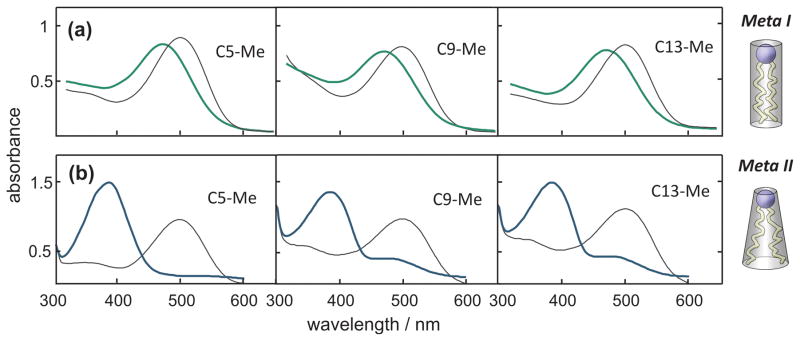
Record the UV-visible absorption spectrum between 700 and 250 nm (the baseline may be recorded with an empty glass slide). The photolyzed sample gives a band with λmax ≈ 479 nm indicative of the Meta I state (Fig. 4a). Freeze the glass slide with the membrane film at −30 °C or below (e.g. by placing it immediately on dry ice).
Collect ca. 25 glass slides with the membrane film containing rhodopsin in the Meta I state and stack them in an 8-mm NMR tube in a thermally insulated styrofoam glove box at −30 °C (see Notes 9–11). Store the sample at −80 °C, and always keep and handle the sample below −30 °C to prevent transformation of the Meta I state into the Meta II state.
Fig. 4.
UV-visible absorption spectra of aligned membrane films containing rhodopsin (on glass slides) in the dark (dashed lines), and trapped Meta I (a) and Meta II (b) states (both states are indicated by solid lines). The Meta I state was obtained by illuminating rhodopsin-POPC recombinant membranes (1:50 protein/lipid ratio) with actinic light at 2 °C, pH 7, and 43% relative humidity. The Meta II state was trapped by bleaching recombinant membranes (3:1 POPC/DOPE molar ratio) with rhodopsin (1:75 protein/lipid ratio) in 5 mM 2H-depleted MES buffer at room temperature, pH 5.0, and 100% relative humidity. The average shape of the DOPE lipid molecule, shown on the right (bottom) corresponds to a negative spontaneous (intrinsic) monolayer curvature, which favors the Meta II state. By contrast POPC, shown on the right (top), favors flat bilayers thus shifting the metarhodopsin equilibrium back to Meta I in accord with a flexible surface model (35).
3.4 Experimental Protocol for Trapping of Metarhodopsin II in Aligned Membranes
All the procedures are performed under dim red light conditions in cold room at 4 °C unless mentioned otherwise. Keep the rhodopsin samples under argon gas at all times.
Centrifuge the proteolipid membranes (3:1 POPC/DOPE ratio) containing rhodopsin (1:75 rhodopsin/lipid ratio) kept in storage buffer (15 or 67 mM Na phosphate, pH 7) at 48,000 × g (Sorvall T865.1 rotor), discard the supernatant, and resuspend the pellet in 5 mL of 5 mM MES buffer prepared with deuterium-depleted water containing 5 mM NaCl, pH 5.5 (at 4 °C). Centrifuge again at the same speed, and resuspend the pellet in the same buffer to make sure the pH is adjusted to 5.5 (very important to trap Meta II state) and the water is replaced with deuterium-depleted water.
Calculate the amount of GαCT2 peptide (see Section 2.1) required to stabilize the Meta II state based on 1:5 protein-to-peptide ratio.
Dissolve the calculated amount of GαCT2 peptide in 300 μL of 5 mM MES buffer prepared with deuterium-depleted water containing 5 mM NaCl, pH 5.5. Add the solution of the GαCT2 peptide to rhodopsin suspension.
Align the proteolipid membranes on glass slides by isopotential ultracentrifugation as described above for the dark state (Section 3.2).
Rehydrate the aligned membrane films at 100% relative humidity by isopiestic transfer using 2H-depleted water at room temperature for about 1 h. Note that the alignment can decrease at this point due to the hydration; however, it can be restored later by dehydrating the sample (see Note 9). At the same time, the pH at room temperature decreases to 5.0.
Bleach the aligned membranes with rhodopsin on a glass slide for about 30 s at room temperature using the 150-W Fostec light source fitted with a green bandpass filter (see Section 3.3).
Record the UV-visible absorption spectrum between 700 and 250 nm (with the baseline measured for an empty glass slide). If the 500-nm peak in the Meta II sample does not disappear completely (incomplete conversion into Meta II), most likely it is because of insufficient hydration (Fig. 4b). The spectrophotometer cuvette must be sealed (e.g. with parafilm) to prevent dehydration of the sample during the measurement.
Dehydrate and cool the membrane film on a glass slide under a stream of nitrogen or argon gas at 4 °C for 20–30 s (see Note 9). This step can be avoided for the preparation of the samples that are used only for the relaxation studies (see Note 10).
Freeze the sample at −60 °C or below (e.g. by placing it in the styrofoam box with dry ice or liquid nitrogen, see Note 11). Because the membrane film on the glass slide is thin, it freezes rapidly to trap the Meta II state.
After collecting 20–30 slides with membranes containing rhodopsin in the Meta II state, stack them in an 8-mm cutoff NMR tube at −60 °C (see Note 11). Store the sample at −80 °C, and from this point on it is recommended to always handle the sample below −60 °C to prevent decay of the Meta II state (see Notes 9, 11).
3.5 Experimental Protocol for Structural Studies of Rhodopsin in Aligned Bilayers using Solid-State Deuterium NMR Spectroscopy
The structural studies are performed under dim red light conditions as described above. All manipulations of the samples in the Meta I state are carried out at temperatures below −30 °C. For the Meta II state all manipulations are done below −60 °C, except for measurements of the 2H NMR spectra and relaxation times at −30 °C.
Acquire solid-state 2H NMR spectra of the rhodopsin in aligned lipid bilayers, Fig. 3c, by applying a quadrupolar-echo pulse sequence (see Note 12). The 2H NMR spectra are recorded with a Bruker AMX-500 spectrometer using a deuterium probe with a transverse radiofrequency coil (see Note 12). The sample with aligned membranes can be rotated in the NMR coil (see Fig. 3a), which allows one to study the angular dependence of the 2H NMR spectra, and increase the accuracy of the method. The presence of unoriented bilayers can be determined from solid-state 31P NMR spectroscopy (typically 10%), and is taken into account by spectral subtraction (see Note 8). If necessary, the 2H NMR signals from the natural 2H abundance of the lipids are also subtracted (see Note 13).
Determine the orientation of the methyl groups in rhodopsin (θB) to the local membrane normal (n) by fitting the experimental spectra for aligned samples to theoretical ones (Fig. 3c). The latter are calculated using a Monte Carlo method (see Note 14) by assuming a static uniaxial distribution of the methyl group orientations about the local membrane normal n (24). The distribution is static because the experimental temperatures are set below −30 °C (lipid gel phase) to prevent rotational diffusion of the proteins in the membrane. In addition the alignment disorder—namely, the distribution of the local membrane normal n with respect to the average normal n0 (mosaic spread)—is taken into account. It is assumed to be uniform in the plane of the glass slide (the directions perpendicular to n0) and Gaussian (with a standard deviation of σ) for the angle θ′ between the n and n0 axes. Apart from the orientation of the 2H-labeled methyl groups (θB), the mosaic spread (σ ) is also determined from the 2H NMR spectral data fitting procedures. Excellent fits of the simulated (color lines) and experimental (black lines) spectra (Fig. 3c), and the sharp minima in the error surfaces (not shown, see ref. (9)) indicate high accuracy and sensitivity of this method for determining orientational constraints (for selectively deuterated methyl groups in this case).
The calculated orientations of the methyl groups can be used for direct comparison with X-ray crystallography data (if available), which is quite valuable considering that NMR is a completely different and independent technique. The 2H NMR results can also be used as orientational constraints in computer simulations of rhodopsin (e.g. in ref. (25) they are used to evaluate the retinal counterion switch mechanism), or in retinal structure modeling in ref. (9) (see Note 15).
3.6 Experimental Protocol for Solid-State Deuterium NMR Relaxation Studies of Rhodopsin Dynamics
Manipulations of the samples for the relaxation experiments are carried out under dim red light at temperatures below 4 °C for the dark state. For the Meta I state, temperatures are kept below −30 °C and generally below −60 °C for the Meta II state. To obtain information on the dynamics of the retinal cofactor, the relaxation measurements are performed using either freely suspended (unoriented, so-called powder-type) or aligned membrane samples. However, for the structural studies only aligned samples can provide the orientational constraints. Aligned samples of rhodopsin also have the advantage of a small membrane film thickness as compared to the unoriented membrane specimens, which makes them easier to bleach with the actinic light. In general, for both relaxation and structural studies it is most desirable to use aligned samples (see Notes 9, 10, 11).
Measure inversion-recovery relaxation spectra, Fig. 5a, in the available temperature range (usually from −30 to −150 °C) using the quadrupolar-echo inversion-recovery pulse sequence (see Note 12). The repetition time t1 for the pulse sequence is adjusted in the course of experiment according to the longitudinal (T1Z) relaxation time (t1 ~ 3–5 T1Z). Integrate the spectra and plot their relative intensities (with respect to equilibrium spectrum) proportional to the magnetization of the 2H nuclei in the sample versus the time t between the 180° and 90° pulses (see Note 12), Fig. 5b.
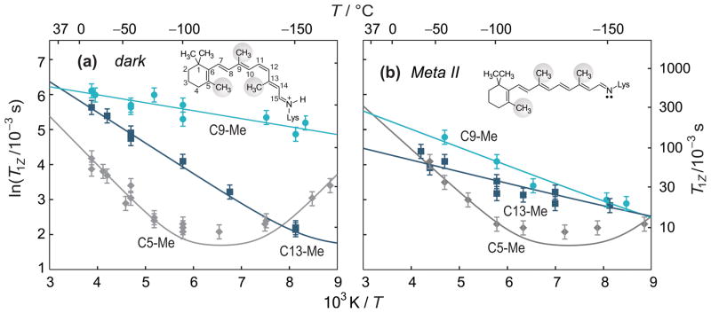
Fit the time dependencies for Zeeman relaxation, Fig. 5b, using a single-exponential function for the recovery of nuclear magnetization: M / M0 = 1 − b exp(−t/T1Z) with three fitting parameters—viz. the equilibrium magnetization, M0, longitudinal relaxation time, T1Z, and the inversion parameter b (≤ 2, characterizing incomplete spectral inversion). Determine the T1Z relaxation times from fitting the inversion recovery plots, Fig. 5b. Examples of the T1Z values plotted against temperature are provided in Fig. 6.
-
According to NMR relaxation theory (26–29) the 2H NMR longitudinal relaxation rates R1Z (or relaxation times T1Z) correspond to the spectral densities Jm(ω) (or power spectra) of the thermal fluctuations of the 2H-labeled group (i.e. the quadrupolar coupling tensor) near the resonance frequency (ω0) by . The spectral densities depend on the orientation of the methyl group in the sample with respect to the magnetic field, together with the mean-square amplitudes and rates of the motions. Analysis of the NMR relaxation times in terms of molecular motions can be performed using our generalized model-free approach (11), which provides the spectral densities Jm(ω) for time-dependent rotational transformations connecting components of quadrupolar tensor in the molecular and laboratory frames. As an alternative, specific motional models can be introduced (10). For methyl groups, two motional models can be considered: 3-fold hops about the methyl axis with a rate constant k, or continuous diffusion within a potential of mean torque, with axial (D||) and off-axial (D⊥) rotational diffusion coefficients. For detailed relationships between spectral densities Jm(ω) and parameters of the molecular motion (k, D||, and D⊥), see refs. (11,27,28).
4. Simulate and fit the T1Z temperature dependences, Fig. 6, for the 2H-labeled methyl groups, e.g. using 3-fold hops or continuous diffusion in a potential of mean torque model (11,27,28). The Arrhenius equation A=A0 exp(−Ea/RT) is introduced for motional parameters k, D||, and D⊥, Fig 6. Simulation is performed using locally written MATLAB script (see Note 14), where the spectral densities Jm(ω) are calculated according to refs. (11,27,28) with activation energies (Ea) and motional parameters (k, D||, and D⊥) as fitting parameters. Determine the pre-exponential factors for reorientational jump rates (k0), or rotational diffusion coefficients (D0), and corresponding activation energies Ea; calculate the spectral densities and correlation times at different temperatures.
5. Information on local molecular dynamics obtained from relaxation data is very important, as the protein functioning is believed to be directly related to mobility. Moreover, this information can be used to study inter- and intramolecular interactions, because the activation energies Ea for molecular motions are determined by the potential barriers. On the other hand, analysis of the dynamical data with regard to molecular interactions requires structural information. Once the structure is available, one might qualitatively analyze the dynamical parameters with respect to steric hindrances (10,30), or apply molecular mechanics or quantum mechanical simulations to calculate potential barriers. The results can be compared with experimentally obtained activation energies (31) and used to improve the force field, or refine the protein structure.
Fig. 5.
Inversion-recovery solid-state 2H NMR spectra (a) for methyl groups of 11-cis-retinal 2H-labeled at the C9- or C13-Me positions bound to rhodopsin (in the dark state), and magnetization-recovery plots (b) used to determine the spin-lattice (T1Z) relaxation time (T = −150 °C). One can observe much faster relaxation for the C13-Me than for C9-Me group, indicating the different environments for the retinylidene methyl groups in the rhodopsin binding pocket (8). Figure adapted from ref. (8).
Fig. 6.
Temperature dependences of the Zeeman (T1Z) relaxation times characterize the molecular dynamics of retinal ligand (8,10) in the dark and Meta II states. The slope of the plots directly provides the activation energy for methyl group spinning. The pre-exponential factors for motional parameters k, D||, and D⊥ (see text) can be determined by fitting the T1Z temperature dependences. Analysis of the relaxation data indicates relatively fast rotation of the C9-Me group, intermediate for the C13-Me group, and slow for the C5-Me group at low temperatures (Fig. 6), The rotational rates correlate with the corresponding activation energies Ea for these groups, and are possibly related to intra-retinal potential barriers (10). It is also shown that both 3-fold hops and rotational diffusion models give good agreement with the experimental T1Z relaxation times (8,10). Figure adapted from ref. (8).
Footnotes
Rhodopsin can also be solubilized in other detergents, including digitonin, n-dodecyl-β-D-maltopyranoside (DDM), DTAB, [(Cholamidopropyl)dimethylammonio]-propanesulfonate (CHAPS), Emulphogene, lauryldimethylamine oxide (LDAO), octaethylene glycol n-dodecyl monoether (C12E8), n-octyl-β-D-glucopyranoside (OG), sodium cholate, nonylglucopyranoside (NG), heptylthioglucoside (HTG), or Tween 80. However, Ammonyx LO (lauryldimethylamine oxide, 30%) is an inexpensive detergent and gives reliable results.
The yield of rhodopsin in the retinal disk membranes can be increased by dissecting and storing the retinas in 10 mM Tris-acetate buffer, pH 7.4 containing 30% sucrose, 1 mM dithiothreitol (DTT), and 0.001% (v/v) aprotinin (instead of only buffer) as obtained from the supplier (W. L. Lawson, Co., Omaha, NE, USA). The volume of the homogenizing buffer added in the second homogenization step is generally about 25 mL. The amount of dilution buffer (10 mM Tris-acetate buffer, pH 7.4) should be added meticulously (Section 2.2, step 2). Excess of will result in higher yield but lower purity, whereas a lower amount of will lead to good purity but lower yields. Generally, the optimum value is 2–3 volumes of the dilution buffer. This ratio should be optimized for each shipment of retinas depending on the purpose. For disk membrane studies the highest purity is required, while for purification of rhodopsin the yield is more important.
The band at 500 nm in the UV-visible rhodopsin spectrum, Fig. 1, is related to the retinal absorption, and consequently it is proportional to the rhodopsin concentration. The maximum at 280 nm is due to the absorption of aromatic amino acids, and it can be used to estimate the overall protein concentration (not just rhodopsin). Hence the A280 /A500 spectral ratio is a reliable indicator of the rhodopsin purity.
The 11-cis-retinal is best stored as dry crystals in a lightproof container at −80 °C. Before opening the jar containing retinal, keep it at room temperature for 1–2 h to make sure the retinal is warmed. Cold crystals may condense water from the air and isomerize if they are wet. Dry crystals can be handled in visible light (not recommended). Take one or two small small crystals, about the size of fine table salt crystals, and under red light dissolve them in approximately 200 μL of 99.5% ethanol.
One can use a smaller molar retinal-to-rhodopsin ratio (down to 1:1) at the expense of extra rhodopsin to obtain more rhodopsin regenerated with 2H-labeled retinal. (Note that deuterated retinal is much more valuable than rhodopsin.) In that case, the absolute yield of regenerated rhodopsin would be higher; but the percentage yield would be lower.
During solubilization of rhodopsin in DTAB (step 1), rhodopsin should be solubilized completely before loading onto the column. The total volume should not exceed 15 mL for the column length specified (8 cm) (Section 2.6). More than this volume will lead to rhodopsin elution in a broader band, or even in two bands with worse quality. Note that DTAB is a denaturing detergent, and thermally bleaches the rhodopsin at temperatures above 4 °C. It is essential that all the procedures with rhodopsin in DTAB should be conducted in the cold room at 4 °C.
Isopotential spin-dry ultracentrifugation (22,23) is carried out using custom-designed inserts for polycarbonate centrifuge tubes. The insert consists of two main parts: a black delrin body with the sample compartment, and a stainless steel plate with the isopotential surface (the surface is curved accordingly to the radius of rotation of the steel plate, providing the same relative centrifugal force on the whole surface during centrifugation). A glass slide serves as a substrate for membrane alignment, and it is placed on the isopotential surface. The bottom of the polycarbonate centrifuge tube is filled with epoxy resin and polymerized to make a flat surface supporting the insert.
Alignment of recombinant membranes with rhodopsin on supporting glass slides is difficult. Evidently the difficulty is related to the fact that substantial portions of the protein are protruding from the both sides of the lipid bilayers. In our hands, near perfect alignment as in Fig. 3a is rarely achieved. That is why estimating the fraction of unoriented bilayers and subtracting the powder-type contribution from the experimental 2H NMR spectrum is very important. However, this subtraction leads to a reduction of the signal-to-noise ratio in the experimental spectrum, and therefore can be used only for relatively small fractions of unoriented bilayers. We would not recommend using samples with an unoriented fraction larger than 20%. The reason is that apart from unoriented bilayers, there is also additional alignment disorder due to the deviation of the local membrane normal from the average membrane normal in the sample (mosaic spread, σ). In lipid bilayers containing rhodopsin, the mosaic spread may be in the range of σ = 10–20 °. It changes the lineshape of the 2H NMR spectra, making it more powder-type like, and additionally reducing the accuracy of the method, because the greater the mosaic spread, the smaller the effect of alignment on the lineshape. At the limit of very large mosaic spread the orientational distribution becomes random.
Trapping of the Meta II state in aligned lipid bilayers is challenging. The reason is that alignment is favored by the sample dehydration, yet Meta II formation requires fully hydrated membranes. These opposite requirements can be overcome by initially preparing the aligned bilayers with rhodopsin in the dark state by isopotential spin-dry ultracentrifugation (22,23), followed by rehydration at 100% relative humidity, and then bleaching with the actinic light. Subsequent dehydration of the thin membrane film under an intensive stream of cold (4 °C) N2 gas allows one to quickly (within 20–30 s) dehydrate the membranes, while rhodopsin still remains in the Meta II state. Stabilization of the Meta II state at low temperature and in dehydrated membranes (both conditions favoring Meta I state) is achieved by low pH 5 and with GαCT2 peptide binding. Another option is to skip the last dehydration step (step 8 in Section 3.4), and to work with a poorly aligned sample by subtracting the powder-type contribution to the 2H NMR spectrum. However, a large fraction of unoriented bilayers (which may increase up to 50% in a fully hydrated sample) will essentially reduce the accuracy of the method. The temperature conditions for storage and handling the samples with rhodopsin in the Meta II state after trapping is stricter than for the Meta I state, because the Meta I state is stabilized by the low hydration of the membranes. The Meta II state is mostly stabilized by the peptide binding, so that additional measures must be taken to freeze the sample, including bound water (water in the vicinity of the protein and the lipid bilayer, which may be in a liquid phase down to −30 °C (13)), to prevent dissociation of the peptide. That is why the recommended temperatures for the storage and manipulation of the Meta I and the Meta II samples are below −30 °C and −60 °C, respectively.
For relaxation measurements, the alignment is less essential (unless one wants to study the angular dependence of relaxation). Hence there may be no need to dehydrate the membrane film with rhodopsin at all (step 8 in Section 3.4). However, it is still logical to prepare the sample on glass slides for more complete bleaching, and fast cooling upon trapping the Meta I and Meta II states. The isopotential ultracentrifugation method (step 4 in Section 3.4) may be avoided in this case. Instead, one may just spread the suspension of disk membranes over the glass slides, and let them dry partially under a stream of N2 gas to reduce the volume of the sample. That allows one to put more rhodopsin into the NMR tube (up to 40 mg in an 8 × 22 mm tube). To be certain that the sample is fully hydrated, it can be rehydrated at 100 % relative humidity.
The Meta I and Meta II states are unstable, and therefore it is recommended to freeze the Meta I and Meta II samples immediately after preparation, and handle them afterwards at temperatures below −30 °C and −60 °C, respectively. Stacking the glass slides with membranes containing rhodopsin in the Meta I and Meta II states at these temperatures is carried out in a custom-designed thermally insulated styrofoam glove box (with window and thermometer), which is cooled by placing dry ice on the sides, and liquid nitrogen at the bottom. A similar styrofoam box is used to place the sample into the NMR probe, and to pre-cool the NMR probe before the experiment. The transfer line which carries the cold N2 gas to the NMR probe to stabilize the temperature should also be pre-cooled by turning on the liquid N2 evaporator 5–10 min before the experiment.
Typically the 2H NMR spectra are acquired using a Bruker AMX-500 spectrometer with a narrow-bore magnet (magnetic field B0 is 11.7 T, corresponding to a deuterium frequency of 76.77 MHz). The 2H NMR probe is locally constructed (“home built”) and has an 8-mm diameter and 12-mm long transverse solenoid as the radiofrequency coil, with high-voltage capacitors (Polyflon company, Norwalk, CT, USA; or Jennings Technology, Vandalia, OH, USA). The membrane tilt angle to the magnetic field is adjusted manually using a protractor by rotating the sample in the radiofrequency coil. The transmitter pulses are amplified with a 2-kW Henry Radio amplifier (Henry Radio Co., USA) or equivalent, providing a 4.2 μs 90° pulse. Acoustical ringing of the radiofrequency coil can be minimized by increasing the delay τ (see pulse sequences below) up to 70 μs if necessary. The repetition time for the pulse sequence is in the range of 0.1–1.5 s depending on the longitudinal (T1Z) relaxation time. The spectra are processed with locally-written data processing software (MATLAB; The MathWorks, Inc., Natick, MA). Echo signals are multiplied by a decaying exponential, representing a linebroadening of 500 Hz in the frequency domain. The number of scans depends on the temperature and the delay τ, and is in the range of ≈ 0.2–1 × 10⁶ (typically 1 d signal acquisition). The solid-state 2H NMR spectra are recorded using a composite-pulse, quadrupolar-echo sequence ( ). Experimental longitudinal (spin-lattice) 2H NMR relaxation rates (T1Z ) (Figs. 5,6) for selectively 2H labeled methyl groups are measured by an inversion-recovery, 180°–t–(90°)ϕ–τ–(90°)ϕ±90º–τ–acquire pulse sequence. The 31P NMR spectra are acquired with a Hahn echo (90°)ϕ–τ–(180°)ϕ–τ–acquire pulse sequence. Appropriate phase (ϕ)-cycling is applied in each case (9,10).
The most straightforward way to subtract the spectrum, which accounts for the natural abundance of deuterium in phospholipids, is to prepare a sample with the same amount of lipids (it can be with natural, unlabeled rhodopsin, or without rhodopsin), and measure the 2H NMR spectrum below the gel to liquid-crystalline phase transition temperature. The spectrum amplitude may be adjusted before subtraction according to the number of scans acquired. The 2H NMR spectrum of water is usually not observed, because the signal is too weak for deuterium-depleted water, and because it is very broad. However, if necessary the subtraction may be performed for the water signal as well.
Monte Carlo simulation is performed using a locally-written program (MATLAB; The MathWorks, Inc., Natick, MA, USA) as follows. For rapidly rotating 2H-labeled methyl groups (assuming an axially symmetric quadrupolar coupling tensor), the 2H NMR spectrum represents a symmetric doublet. The two quadrupolar frequencies are determined by the angle θ̃ between the methyl group symmetry axis (parallel to C–C2H3 bond) and the external magnetic field B0 and are given by , where is the effective quadrupolar coupling constant (see Section 3.1). The angle θ̃ depends on the orientation of the methyl group in rhodopsin θB, the mosaic spread σ, and the orientation of the sample θ (tilt angle) with respect to the magnetic field B0, see Fig. 3a. The 2H NMR spectra are simulated by randomly generating the orientations of methyl group according to their distribution functions (see Section 3.5), and calculating the quadrupolar frequencies. The distribution of the quadrupolar frequencies is then convoluted with the intrinsic line broadening. Finally, the theoretical and experimental spectra can be fitted using the methyl bond angle θB and mosaic spread σ as fitting parameters.
Apart from the 2H-labeled C5-, C9-, or C13-methyl groups, and the electronic transition dipole moment of retinal (9), the two C1-methyl groups can also be explored for structural studies. However, the number of orientational constrains is still limited for detailed conformational analysis. The same is valid for any protein segment. That is why the method of solid-state 2H NMR spectroscopy of selectively 2H-labeled aligned samples is greatly enhanced by complementary 13C NMR rotational resonance distance measurements (32,33). The distance restraints allow one to eliminate the ambiguity in structural models, based on orientational restraints due to the symmetry of NMR interactions (34,9), and provide additional structural parameters that allow one to refine the molecular structure.
References
- 1.Lagerström MC, Schiöth HB. Structural diversity of G protein-coupled receptors and significance for drug discovery. Nature Reviews Drug Discovery. 2008;7:339–357. doi: 10.1038/nrd2518. [DOI] [PubMed] [Google Scholar]
- 2.Teller DC, Okada T, Behnke CA, Palczewski K, Stenkamp RE. Advances in determination of a high-resolution three-dimensional structure of rhodopsin, a model of G-protein-coupled receptors (GPCRs) Biochemistry. 2001;40:7761–7772. doi: 10.1021/bi0155091. [DOI] [PMC free article] [PubMed] [Google Scholar]
- 3.Okada T, Sugihara M, Bondar A-N, Elstner M, Entel P, Buss V. The retinal conformation and its environment in rhodopsin in light of a new 2.2 Å crystal structure. J Mol Biol. 2004;342:571–583. doi: 10.1016/j.jmb.2004.07.044. [DOI] [PubMed] [Google Scholar]
- 4.Li J, Edwards PC, Burghammer M, Villa C, Schertler GFX. Structure of bovine rhodopsin in a trigonal crystal form. J Mol Biol. 2004;343:1409–1438. doi: 10.1016/j.jmb.2004.08.090. [DOI] [PubMed] [Google Scholar]
- 5.Choe H-W, Kim YJ, Park JH, Morizumi T, Pai EF, Krauβ N, Hofmann KP, Scheerer P, Ernst OP. Crystal structure of metarhodopsin II. Nature. 2011;471(7340):651–656. doi: 10.1038/nature09789. [DOI] [PubMed] [Google Scholar]
- 6.Standfuss J, Edwards PC, D’Antona A, Fransen M, Xie G, Oprian DD, Schertler GFX. The structural basis of agonist-induced activation in constitutively active rhodopsin. Nature. 2011;471(7340):656–660. doi: 10.1038/nature09795. [DOI] [PMC free article] [PubMed] [Google Scholar]
- 7.Deupi X, Edwards P, Singhal A, Nickle B, Oprian D, Schertler G, Standfuss J. Stabilized G protein binding site in the structure of constitutively active metarhodopsin-II. Proc Natl Acad Sci U S A. 2012;109(1):119–124. doi: 10.1073/pnas.1114089108. [DOI] [PMC free article] [PubMed] [Google Scholar]
- 8.Struts AV, Salgado GFJ, Martínez-Mayorga K, Brown MF. Retinal dynamics underlie its switch from inverse agonist to agonist during rhodopsin activation. Nat Struct Mol Biol. 2011;18(3):392–394. doi: 10.1038/nsmb.1982. [DOI] [PMC free article] [PubMed] [Google Scholar]
- 9.Struts AV, Salgado GFJ, Tanaka K, Krane S, Nakanishi K, Brown MF. Structural analysis and dynamics of retinal chromophore in dark and Meta I states of rhodopsin from 2H NMR of aligned membranes. J Mol Biol. 2007;372(1):50–66. doi: 10.1016/j.jmb.2007.03.046. [DOI] [PMC free article] [PubMed] [Google Scholar]
- 10.Struts AV, Salgado GFJ, Brown MF. Solid-state 2H NMR relaxation illuminates functional dynamics of retinal cofactor in membrane activation of rhodopsin. Proc Natl Acad Sci U S A. 2011;108(20):8263–8268. doi: 10.1073/pnas.1014692108. [DOI] [PMC free article] [PubMed] [Google Scholar]
- 11.Xu X, Struts AV, Brown MF. Generalized model-free analysis of nuclear spin relaxation experiments. eMagRes. 2014 in press. [Google Scholar]
- 12.Papermaster DS, Dreyer WJ. Rhodopsin content in the outer segment membranes of bovine and frog retinal rods. Biochemistry. 1974;13:2438–2444. doi: 10.1021/bi00708a031. [DOI] [PubMed] [Google Scholar]
- 13.Tanaka K, Struts AV, Krane S, Fujioka N, Salgado GFJ, Martínez-Mayorga K, Brown MF, Nakanishi K. Synthesis of CD3-labeled 11-cis-retinals and applications to solid-state deuterium NMR spectroscopy of rhodopsin. Bull Chem Soc Japan. 2007;80:2177–2184. [Google Scholar]
- 14.Sperling W, Rafferty CN. Relationship between absorption spectrum and molecular conformations of 11-cis-retinal. Nature. 1969;224:591–594. doi: 10.1038/224591a0. [DOI] [PubMed] [Google Scholar]
- 15.Garwin GG, Saari JC. High-performance liquid chromatography analysis of visual cycle retinoids. Methods Enzymol. 2000;316:313–324. doi: 10.1016/s0076-6879(00)16731-x. [DOI] [PubMed] [Google Scholar]
- 16.Raubach RA, Franklin LK, Dratz EA. A rapid method for the purificaton of rod outer segment disk membranes. Vision Res. 1974;14:335–337. doi: 10.1016/0042-6989(74)90092-3. [DOI] [PubMed] [Google Scholar]
- 17.Hong K, PJK, Hubbell WL. Purification of rhodopsin on hydroxyapatite columns, detergent exchange, and recombination with phospholipids. Methods Enzymol. 1982;81:144–150. doi: 10.1016/s0076-6879(82)81024-0. [DOI] [PubMed] [Google Scholar]
- 18.Botelho AV, Gibson NJ, Wang Y, Thurmond RL, Brown MF. Conformational energetics of rhodopsin modulated by nonlamellar forming lipids. Biochemistry. 2002;41:6354–6368. doi: 10.1021/bi011995g. [DOI] [PubMed] [Google Scholar]
- 19.Brown MF. Influence of non-lamellar forming lipids on rhodopsin. Curr Top Membr. 1997;44:285–356. [Google Scholar]
- 20.Brown MF, Chan SI. Bilayer Membranes: Deuterium & Carbon-13 NMR. In: Grant DM, Harris RK, editors. Encyclopedia of Nuclear Magnetic Resonance. Vol. 2. Wiley; New York: 1996. pp. 871–885. [Google Scholar]
- 21.Brown MF. Membrane structure and dynamics studied with NMR spectroscopy. In: Merz K Jr, Roux B, editors. Biological Membranes. A Molecular Perspective from Computation and Experiment. Birkhäuser; Basel: 1996. pp. 175–252. [Google Scholar]
- 22.Gröbner G, Taylor A, Williamson PTF, Choi G, Glaubitz C, Watts JA, deGrip WJ, Watts A. Macroscopic orientation of natural and model membranes for structural studies. Anal Biochem. 1997;254:132–138. doi: 10.1006/abio.1997.2415. [DOI] [PubMed] [Google Scholar]
- 23.Clark NA, Rothschild KJ, Luippold DF, Simon BA. Surface-induced lamellar orientation of multilayer membrane arrays. Biophys J. 1980;31(1):65–96. doi: 10.1016/S0006-3495(80)85041-7. [DOI] [PMC free article] [PubMed] [Google Scholar]
- 24.Nevzorov AA, Moltke S, Heyn MP, Brown MF. Solid-state NMR line shapes of uniaxially oriented immobile systems. J Am Chem Soc. 1999;121:7636–7643. [Google Scholar]
- 25.Martínez-Mayorga K, Pitman MC, Grossfield A, Feller SE, Brown MF. Retinal counterion switch mechanism in vision evaluated by molecular simulations. J Am Chem Soc. 2006;128(51):16502–16503. doi: 10.1021/ja0671971. [DOI] [PubMed] [Google Scholar]
- 26.Brown MF. Theory of spin-lattice relaxation in lipid bilayers and biological membranes. 2H and 14N quadrupolar relaxation. J Chem Phys. 1982;77(3):1576–1599. [Google Scholar]
- 27.Trouard TP, Alam TM, Brown MF. Angular dependence of deuterium spin-lattice relaxation rates of macroscopically oriented dilaurylphosphatidylcholine in the liquid-crystalline state. J Chem Phys. 1994;101:5229–5261. [Google Scholar]
- 28.Nevzorov AA, Brown MF. Dynamics of lipid bilayers from comparative analysis of 2H and 13C nuclear magnetic resonance relaxation data as a function of frequency and temperature. J Chem Phys. 1997;107(23):10288–10310. [Google Scholar]
- 29.Nevzorov AA, Trouard TP, Brown MF. Lipid bilayer dynamics from simultaneous analysis of orientation and frequency dependence of deuterium spin-lattice and quadrupolar order relaxation. Phys Rev E. 1998;58:2259–2281. [Google Scholar]
- 30.Brown MF, Salgado GFJ, Struts AV. Retinal dynamics during light activation of rhodopsin revealed by solid-state NMR spectroscopy. Biochim Biophys Acta. 2010;1798:177–193. doi: 10.1016/j.bbamem.2009.08.013. [DOI] [PMC free article] [PubMed] [Google Scholar]
- 31.Mertz B, Struts AV, Feller SE, Brown MF. Molecular simulations and solid-state NMR investigate dynamical structure in rhodopsin activation. Biochim Biophys Acta. 2012;1818:241–251. doi: 10.1016/j.bbamem.2011.08.003. [DOI] [PMC free article] [PubMed] [Google Scholar]
- 32.Spooner PJR, Sharples JM, Verhoeven MA, Lugtenberg J, Glaubitz C, Watts A. Relative orientation between the β-ionone ring and the polyene chain for the chromophore of rhodopsin in native membranes. Biochemistry. 2002;41:7549–7555. doi: 10.1021/bi020007o. [DOI] [PubMed] [Google Scholar]
- 33.Verdegem PJE, Bovee-Geurts PHM, de Grip WJ, Lugtenburg J, de Groot HJM. Retinylidene ligand structure in bovine rhodopsin, metarhodopsin-I, and 10-methylrhodopsin from internuclear distance measurements using 13C-labeling and 1-D rotational resonance MAS NMR. Biochemistry. 1999;38:11316–11324. doi: 10.1021/bi983014e. [DOI] [PubMed] [Google Scholar]
- 34.Salgado GFJ, Struts AV, Tanaka K, Krane S, Nakanishi K, Brown MF. Solid-state 2H NMR structure of retinal in metarhodopsin I. J Am Chem Soc. 2006;128(34):11067–11071. doi: 10.1021/ja058738+. [DOI] [PubMed] [Google Scholar]
- 35.Brown MF. Curvature Forces in Membrane Lipid-Protein Interactions. Biochemistry. 2012;51(49):9782–9795. doi: 10.1021/bi301332v. [DOI] [PMC free article] [PubMed] [Google Scholar]


