Abstract
An electrostatic electron source design capable of producing sub-20 femtoseconds (rms) multi-electron pulses is presented. The photoelectron gun concept builds upon geometrical electric field enhancement at the cathode surface. Particle tracer simulations indicate the generation of extremely short bunches even beyond 40 cm of propagation. Comparisons with compact electron sources commonly used for femtosecond electron diffraction are made.
INTRODUCTION
The field of ultrafast structural dynamics is quickly growing, as shorter and brighter hard X-ray and electron pulses are being produced and implemented to light up atoms in motion.1–8 The advent of forth generation light sources9,10 has made the production of ultra-bright femtosecond (fs) hard X-ray pulses possible, which have been successfully applied for time-resolved diffraction11–14 and ultrafast coherent imaging.15–17 On the other hand, the use of ultrashort electron bursts has also emerged as a powerful means to atomically resolve dynamical phenomena and structure in the laboratory setting.18–35 In this regard, different approaches for the generation of fs multi-electron bunches have been developed to meet the prerequisite time-resolution to observe the movement of atoms; i.e., sub-picosecond electron pulses and ideally the shorter the better to avoid temporal blurring in stroboscopically recorded images. Compact femtosecond electron diffraction (FED) instruments with electrostatic electron guns, based on quasi-flat cathode and anode electrodes, have enabled a time-resolution of 100 fs (rms, root-mean-square deviation) with bright multi-electron pulses.36,37 For simplicity, electron pulses were assumed to be Gaussian in shape, and therefore a conversion factor of 2.355 has been used to calculate fwhm (full-width-at-half-maximum) from rms values. Recent designs with a minimal cathode-to-sample distance have brought the temporal resolution of these sources closer to the limit imposed by their initial energy spread—or single electron pulse limit.30,38–41 Electron kinetic energies (KE) produced by electrostatic guns typically range from sub 1 keV to 100 keV and are commonly referred to as sub-relativistic. More advanced electron sources based on radio frequency (RF) photo-injectors are known to generate ultrashort bright pulses of relativistic electrons (KE > 1 MeV). This technology is relatively mature within the accelerator community due to its use in synchrotron and free electron laser facilities and has become popular for monitoring ultrafast structural dynamics.42–49 As its energy spread gets under control, laser-driven electron acceleration is also arising as a low-cost alternative for the generation of ultrashort and ultrabright electron pulses with KE in the 200 keV–1 GeV range.50–53 In addition, different active and passive electron pulse compression schemes have been proposed and/or demonstrated.27–29,53–64 One of the most successfully applied methods in recent FED experiments with ultrabright electron bursts relies on the use of an RF (or microwave) cavity that acts as a temporal lens.27–29,55,56,60–63 This methodology was found to compress dense sub-relativistic multi-electron pulses down to 67 fs (rms).60 Shorter multi-electron pulses are expected from this approach for which synchronization noise has limited the instrument response to about 80–150 fs (rms).60–62 However, a recent phase-lock scheme based on passive optical enhancement has reduced this timing jitter to only 5 fs (rms).65–67 Therefore, RF pill-lens electron pulse rebunching still holds great promise in providing sub-20 fs (rms) temporal resolution with bright multi-electron bunches.56 Furthermore, all-optical electron pulse compression throughout the use of a single cycle THz resonator has recently shown to bring the duration of multi-electron pulses from 395 fs (rms) to 32 fs (rms) [930 fs (fwhm) to 75 fs (fwhm)] with minimal long-term timing drift 4 fs (rms).68,69 This method is expected to generate even shorter multi-electron bursts.68
RESULTS AND DISCUSSION
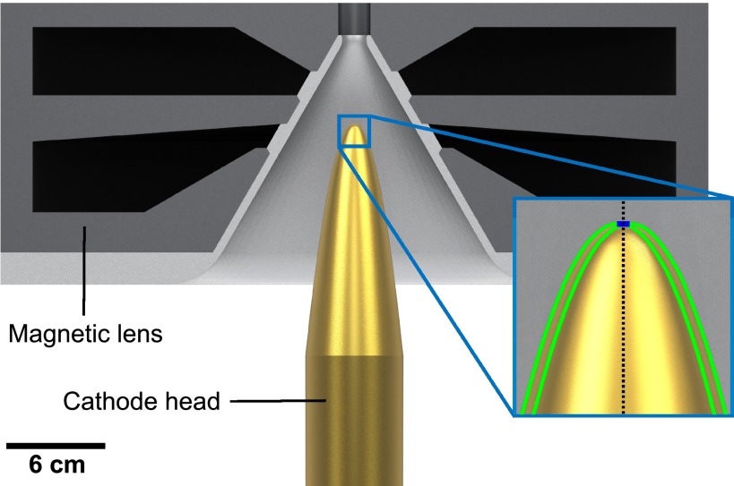
Here, we introduce a rather simple all-electrostatic electron gun design that delivers multi-electron bursts as short as 12 fs (rms) [28 fs (fwhm)] at a relatively long electron propagation distance of 10 cm (sample position in our instrument) without the need of electron pulse temporal rebunching. A 300 kV FED setup based on this source concept is under construction at the University of Waterloo. The electron gun exploits the advantage of strong on-axis electric field acceleration at the electron birth. Figure 1 shows a computer-aided design (CAD) of the key electron source components alongside a geometrical depiction of the photocathode head. The cathode surface has a parabolic shape with a small flat circular area of 1 mm in diameter centered at the symmetry axis or electron propagation axis defined as . This flat region is necessary to avoid an excessive kick in the transverse direction acting on off-axis electrons that greatly deteriorates the transverse and longitudinal properties of the electron bunch. The cathode is positioned inside a double magnetic lens (in-lens system). The magnetic fields generated by each lens point in the opposite direction along the z-axis in order to provide a resultant field Bz = 0 at the cathode surface (0, 0, 0). This is a necessary condition to null emittance growth caused by magnetic fields at the electron pulse birth.70 This in-lens source design yields a lateral spot size 190 μm (rms) for a quasi-parallel electron beam at the sample's plane. A normalized transverse emittance of 0.025 mm mrad (or transverse coherence length of about 3 nm) was obtained. This value suffices for the study of most inorganic and organic crystalline materials composed of small molecules. The lens system has been optimized to operate under the assumption of a core material with a relative magnetic permeability of 104 and a saturation magnetic field of 1.5 T. Such values are easily attainable by various soft magnetic iron alloys.71 The required total power was estimated to be only 200 W.
FIG. 1.
CAD of the electron source concept. The main components are a parabolically shaped photocathode head with a small flat region and a double magnetic lens system with a conical inner form. This cathode shape has been carefully selected in order to confer an on-axis geometrical surface electric field magnitude of 50 MV/m without exceeding a maximum of 60 MV/m in other parts of the head. The in-lens system ensures Bz = 0 at the electron birth to avoid magnetic emittance growth. The conical anode shape helps to maintain the surface electric field at the anode and brings the magnetic poles closer to the cathode head. The magnetic lens system has been optimized in order to obtain, within practical constraints, a reasonable electron spot size at the sample position and low power consumption to avoid water-cooling. Inlet: black dotted line corresponds to the symmetry z-axis; green curves depict two parabolas displaced from the propagation axis by 0.5 mm in the radial direction and which follow the equation ; blue segment highlights the flat region of 1 mm in diameter.
Local electric field enhancement by several orders of magnitude (1 GV/m) is a well-known effect in field emitters and single electron to a few electrons photo-triggered tip sources.72–78 Recently, such nanoemitters have been successfully applied to monitor photocurrents in nanostructures77 as well as ultrafast structural dynamics.78 The introduced cathode head exploits the use of moderate geometrical field enhancement while permitting the generation of multi-electron bunches.
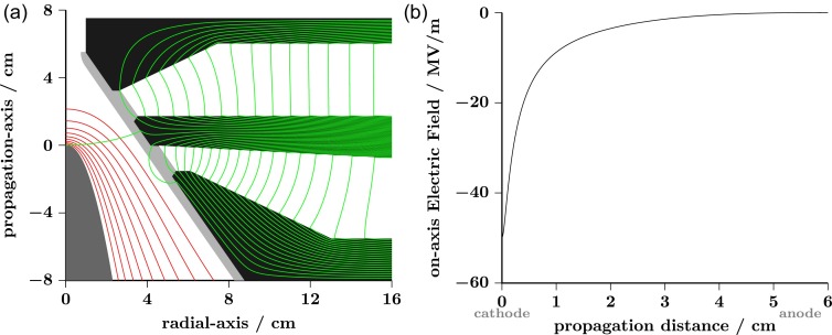
Electrons in simulations were generated at the cathode surface considering a temporal (longitudinal) Gaussian profile of 6 fs (rms) [14 fs (fwhm)], an initial energy spread of 0.2 eV, and a lateral (transverse) Gaussian spot size of 50 μm (rms). Such initial electron pulse parameters can be obtained via single-photon photoemission using the second harmonic from the output of a non-collinear optical parametric amplifier (NOPA).79–81 A NOPA provides the frequency tunability necessary to match the work function of various metal candidates such as Ti, stainless steel, Mo, and W. The photocathode is held at a potential V = 300 kV with respect to ground. The cathode-anode separation distance along the propagation axis, dz 5 cm, confers an average on-axis electric field . Equipotential lines, calculated using Poisson Superfish,82 are shown in panel A of Fig. 2 (red traces). A large increase in the magnitude of the on-axis electric field can be observed as we approach the cathode head reaching a maximum value of at the surface, see Fig. 2(b).
FIG. 2.
(a) Schematic of the electron source concept. Equipotential and magnetic field lines are shown in red and green, respectively. (b) Electric field values obtained along the centrosymmetric axis (0, 0, z) for a cathode held at 300 kV. Calculations were done using Poisson Superfish.82
One of the major concerns of the current design is vacuum breakdown that can compromise the stability of our electron source. Critical surface vacuum breakdown fields, , have been measured and found to be for refractory metals.83–85 Such critical threshold is commonly expressed as , where and are microscopic and geometrical field enhancement factors, respectively, and is the critical applied potential drop over a given separation distance, d, between two electrodes. Thus, equals the magnitude of the average critical applied field , and becomes what we refer to as the “geometrical critical surface electric field” , which therefore results in . Typical values of for polished surfaces lie in the range of 100–300 (with 100 corresponding to mirror-like surface finishing85). On the other hand, the magnitude of the maximum geometrical surface electric field in previous compact electron gun designs was calculated to be about 20 MV/m and therefore satisfies the condition . Note that we cannot modify but and within certain limits with for a roughness free surface. Surface conditioning is known to reduce dark current and greatly increase 86–88 by bringing from several hundreds to about 20–50 for refractory metals.84 The use of a solid cathode head made of Ti, for instance, is therefore essential to allow for proper surface processing. This is difficult to achieve in back illuminated electron guns due to the implementation of ultrathin film photocathodes that can be easily damaged by arcing. On this subject, the cathode shape was optimized to obtain a maximum geometrical surface electric field in order to maintain below . In addition, the source design ensures low surface electric fields at the anode electrode (), a fact that will greatly mitigate anode-initiated vacuum breakdown.88
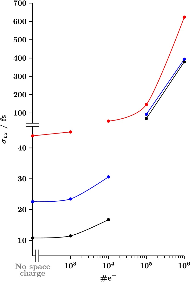
Fig. 3 shows the electron pulse duration (rms) obtained from ASTRA simulations89 for different electron source geometries as a function of the number of electrons per bunch. Black trace corresponds to our 300 kV FED electron gun concept for a total electron propagation distance dT = 10 cm. Blue and red traces refer to the results obtained for conventional 100 kV compact FED setups with flat parallel electrodes, dT = 2 cm, and constant on-axis electric fields of = 20 MV/m and 10 MV/m, respectively. Note that despite the relatively long propagation distance, the proposed design provides < 20 fs for bunches containing 104 electrons, and only 12 fs in the limit of low space charge effects. It should be mentioned, however, that the main disadvantage of the proposed electron source is its relatively larger spot size, 190 μm (rms), when compared with that of a compact FED setup, 55 μm (rms), with the same initial electron beam parameters.
FIG. 3.
Standard deviation or root-mean-square (rms) electron pulse duration (, in fs) as a function of the number of electrons per bunch (#e−) obtained from ASTRA particle tracer simulations.89 Black trace corresponds to our 300 kV FED design and dT = 10 cm. Blue and red traces correspond to compact 100 kV FED setups (i.e., large flat cathodes) with dT = 2 cm and constant electrostatic fields of Ez 20 MV and 10 MV/m, respectively.
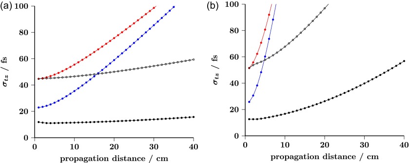
The most noteworthy feature of this new source design is its ability for delivering ultrashort multi-electron bursts after significantly long propagation distances. As can be seen in Fig. 4(a) by direct comparison against conventional FED setups, geometrical field enhancement plays a key role in minimizing the temporal broadening caused by energy spread at photoemission. Temporal broadening is known to be dominant at the initial stage of electron propagation90 and found to be, approximately, inversely proportional to the geometrical surface electric field at the electron birth. Hence, geometrical surface field enhancement appears as a simple means to reduce the temporal broadening introduced by the initial momentum spread.
FIG. 4.
Electron pulse duration (rms) as a function of propagation distance from the cathode surface for bunches containing 103 and 104 electrons, panels (a) and (b), respectively. Black closed dots: our 300 kV FED design. Blue and red dots: compact 100 kV FED setups with constant electric fields of Ez = 20 MV/m and Ez = 10 MV/m, respectively. Black open dots: compact 300 kV FED setup with constant electric field Ez = 10 MV/m.
Therefore, the proposed electrostatic electron source approach lessens the need of using microwave cavities to compensate for such temporal spreading.67 Moreover, we can also observe in Fig. 4 an important decrease of the electron pulse expansion rates when KE is increased from 100 keV to 300 keV (red and blue dots versus closed and open black dots). This is a consequence of relativistic effects that diminish space-charge repulsive forces by a factor of .91 We find remarkable (see black closed dots in Fig. 4(b)) that is below 60 fs even for a pulse containing 104 electrons and after 40 cm of propagation. Longer propagation distances may be advantageous for improving transverse electron beam properties owing to the use of additional electron optics and apertures between the electron source and the sample.
Given the extremely short length of the produced electron bursts, we decided to explore the effect of instabilities of the power source on the instrument response time. Fluctuations of the electron gun voltage, , result in variations of the arrival time (t0) of each electron pulse to the sample (or time zero jitter, ). We found for our 300 kV FED design . Hence, voltage drifts of 10 ppm ( 3 eV) and dT = 10 cm translate into 1.2 fs. A state-of-the-art high-voltage power supply is therefore necessary to guarantee that the overall temporal instrument response is not limited by fluctuations of the voltage source.92
CONCLUSIONS
We have presented an electron source that builds solely on electrostatic fields and that is capable of generating ultrashort and bright multi-electron pulses with minimal temporal degradation over long propagation distances. The main two ingredients of this electron source design are: (i) geometrical field enhancement that increases the strength of the electric field at the electron birth and therefore reduces the temporal broadening caused by initial energy spread; and (ii) higher KE that helps to diminish the detrimental effects of space charge and leads to a decrease of the electron pulse expansion rate. With a time resolution down to 12 fs (rms), FED instruments based on this new electron gun concept hold great promise in resolving even high-frequency vibrational modes without the necessity of implementing RF-electron pulse rebunching or all-optical electron pulse compression schemes.
ACKNOWLEDGMENTS
We would like to dedicate this work in honor to the memory of Professor Ahmed Zewail, who greatly contributed to our community with pioneering experiments. We thank Majed Chergui (EPFL) for providing us with the opportunity to contribute to this special issue. We acknowledge initial discussions about ASTRA code with Gustavo Moriena (University of Toronto). Funding for this project was provided by the Natural Science and Engineering Research Council of Canada and the Canada Foundation for Innovation. G.S. would like to acknowledge the support provided by the Canada Research Chair program.
References
- 1. Zewail A. H., “ 4D ultrafast electron diffraction, crystallography, and microscopy,” Annu. Rev. Phys. Chem. , 65–103 (2006). 10.1146/annurev.physchem.57.032905.104748 [DOI] [PubMed] [Google Scholar]
- 2. Gaffney K. J. and Chapman H. N., “ Imaging atomic structure and dynamics with ultrafast x-ray scattering,” Science , 1444–1448 (2007). 10.1126/science.1135923 [DOI] [PubMed] [Google Scholar]
- 3. Chergui M. and Zewail A. H., “ Electron and x-ray methods of ultrafast structural dynamics: Advances and applications,” ChemPhysChem , 28–43 (2009). 10.1002/cphc.200800667 [DOI] [PubMed] [Google Scholar]
- 4. Sciaini G. and Miller R. J. D., “ Femtosecond electron diffraction: Heralding the era of atomically resolved dynamics,” Rep. Prog. Phys. , 096101 (2011). 10.1088/0034-4885/74/9/096101 [DOI] [Google Scholar]
- 5. Carbone F., Musumeci P., Luiten O. J., and Hebert C., “ A perspective on novel sources of ultrashort electron and X-ray pulses,” Chem. Phys. , 1–9 (2012). 10.1016/j.chemphys.2011.10.010 [DOI] [Google Scholar]
- 6. Hada M., Pichugin K., and Sciaini G., “ Ultrafast structural dynamics with table top femtosecond hard X-ray and electron diffraction setups,” Eur. Phys. J. Spec. Top. , 1093–1123 (2013). 10.1140/epjst/e2013-01909-9 [DOI] [Google Scholar]
- 7. Miller R. J. D., “ Femtosecond crystallography with ultrabright electrons and x-rays: Capturing chemistry in action,” Science , 1108–1116 (2014). 10.1126/science.1248488 [DOI] [PubMed] [Google Scholar]
- 8. Elsaesser T. and Woerner M., “ Perspective: Structural dynamics in condensed matter mapped by femtosecond x-ray diffraction,” J. Chem. Phys. , 020901 (2014). 10.1063/1.4855115 [DOI] [PubMed] [Google Scholar]
- 9. Arthur J., Materlik G., Tatchyn R., and Winick H., “ The LCLS: A fourth generation light source using the SLAC linac,” Rev. Sci. Instrum. , 1987–1989 (1995). 10.1063/1.1145778 [DOI] [Google Scholar]
- 10. Emma P. et al. , “ First lasing and operation of an Ångstrom-wavelength free-electron laser,” Nat. Photonics , 641–647 (2010). 10.1038/nphoton.2010.176 [DOI] [Google Scholar]
- 11. Lindenberg A. M. et al. , “ Atomic-scale visualization of inertial dynamics,” Science , 392–395 (2005). 10.1126/science.1107996 [DOI] [PubMed] [Google Scholar]
- 12. Fritz D. M. et al. , “ Ultrafast bond softening in bismuth: Mapping a solid's interatomic potential with x-rays,” Science , 633–636 (2007). 10.1126/science.1135009 [DOI] [PubMed] [Google Scholar]
- 13. Tenboer J. et al. , “ Time-resolved serial crystallography captures high-resolution intermediates of photoactive yellow protein,” Science , 1242–1246 (2014). 10.1126/science.1259357 [DOI] [PMC free article] [PubMed] [Google Scholar]
- 14. Barends T. R. M. et al. , “ Direct observation of ultrafast collective motions in CO myoglobin upon ligand dissociation,” Science , 445–450 (2015). 10.1126/science.aac5492 [DOI] [PubMed] [Google Scholar]
- 15. Chapman H. N. et al. , “ Femtosecond X-ray protein nanocrystallography,” Nature , 73–77 (2011). 10.1038/nature09750 [DOI] [PMC free article] [PubMed] [Google Scholar]
- 16. Barty A. et al. , “ Self-terminating diffraction gates femtosecond X-ray nanocrystallography measurements,” Nat. Photonics , 35–40 (2012). 10.1038/nphoton.2011.297 [DOI] [PMC free article] [PubMed] [Google Scholar]
- 17. Redecke L. et al. , “ Natively inhibited Trypanosoma brucei cathepsin B structure determined by using an x-ray laser,” Science , 227–230 (2013). 10.1126/science.1229663 [DOI] [PMC free article] [PubMed] [Google Scholar]
- 18. Williamson J. C., Cao J., Ihee H., Frey H., and Zewail A. H., “ Clocking transient chemical changes by ultrafast electron diffraction,” Nature , 159–162 (1997). 10.1038/386159a09296492 [DOI] [Google Scholar]
- 19. Ihee H. et al. , “ Ultrafast x-ray diffraction of transient molecular structures in solution,” Science , 1223–1227 (2005). 10.1126/science.1114782 [DOI] [PubMed] [Google Scholar]
- 20. Siwick B. J., Dwyer J. R., Jordan R. E., and Miller R. J. D., “ An atomic-level view of melting using femtosecond electron diffraction,” Science , 1382–1385 (2003). 10.1126/science.1090052 [DOI] [PubMed] [Google Scholar]
- 21. Baum P., Yang D.-S., and Zewail A. H., “ 4D visualization of transitional structures in phase transformations by electron diffraction,” Science , 788–792 (2007). 10.1126/science.1147724 [DOI] [PubMed] [Google Scholar]
- 22. Sciaini G. et al. , “ Electronic acceleration of atomic motions and disordering in bismuth,” Nature , 56–59 (2009). 10.1038/nature07788 [DOI] [PubMed] [Google Scholar]
- 23. Ernstorfer R. et al. , “ The formation of warm dense matter: Experimental evidence for electronic bond hardening in gold,” Science , 1033–1037 (2009). 10.1126/science.1162697 [DOI] [PubMed] [Google Scholar]
- 24. Eichberger M. et al. , “ Snapshots of cooperative atomic motions in the optical suppression of charge density waves,” Nature , 799–802 (2010). 10.1038/nature09539 [DOI] [PubMed] [Google Scholar]
- 25. Reckenthaeler P. et al. , “ Time-resolved electron diffraction from selectively aligned molecules,” Phys. Rev. Lett. , 213001 (2009). 10.1103/PhysRevLett.102.213001 [DOI] [PubMed] [Google Scholar]
- 26. Hensley C. J., Yang J., and Centurion M., “ Imaging of isolated molecules with ultrafast electron pulses,” Phys. Rev. Lett. , 133202 (2012). 10.1103/PhysRevLett.109.133202 [DOI] [PubMed] [Google Scholar]
- 27. Gao M. et al. , “ Mapping molecular motions leading to charge delocalization with ultrabright electrons,” Nature , 343–346 (2013). 10.1038/nature12044 [DOI] [PubMed] [Google Scholar]
- 28. Morrison V. R. et al. , “ A photoinduced metal-like phase of monoclinic VO2 revealed by ultrafast electron diffraction,” Science , 445–448 (2014). 10.1126/science.1253779 [DOI] [PubMed] [Google Scholar]
- 29. Chatelain R. P., Morrison V. R., Klarenaar B. L. M., and Siwick B. J., “ Coherent and incoherent electron-phonon coupling in graphite observed with radio-frequency compressed ultrafast electron diffraction,” Phys. Rev. Lett. , 235502 (2014). 10.1103/PhysRevLett.113.235502 [DOI] [PubMed] [Google Scholar]
- 30. Ishikawa T. et al. , “ Direct observation of collective modes coupled to molecular orbital-driven charge transfer,” Science , 1501–1505 (2015). 10.1126/science.aab3480 [DOI] [PubMed] [Google Scholar]
- 31. Ruan C.-Y., Lobastov V. A., Vigliotti F., Chen S., and Zewail A. H., “ Ultrafast electron crystallography of interfacial water,” Science , 80–84 (2004). 10.1126/science.1094818 [DOI] [PubMed] [Google Scholar]
- 32. Raman R. K. et al. , “ Direct observation of optically induced transient structures in graphite using ultrafast electron crystallography,” Phys. Rev. Lett. , 077401 (2008). 10.1103/PhysRevLett.101.077401 [DOI] [PubMed] [Google Scholar]
- 33. Erasmus N. et al. , “ Ultrafast dynamics of charge density waves in 4Hb TaSe2 probed by femtosecond electron diffraction,” Phys. Rev. Lett. , 167402 (2012). 10.1103/PhysRevLett.109.167402 [DOI] [PubMed] [Google Scholar]
- 34. Nie S., Wang X., Park H., Clinite R., and Cao J., “ Measurement of the electronic Grüneisen constant using femtosecond electron diffraction,” Phys. Rev. Lett. , 025901 (2006). 10.1103/PhysRevLett.96.025901 [DOI] [PubMed] [Google Scholar]
- 35. Yang D.-S., Baum P., and Zewail A. H., “ Ultrafast electron crystallography of the cooperative reaction path in vanadium dioxide,” Struct. Dyn. , 034304 (2016). 10.1063/1.4953370 [DOI] [PMC free article] [PubMed] [Google Scholar]
- 36. Siwick B. J., Dwyer J. R., Jordan R. E., and Miller R. J. D., “ Ultrafast electron optics: Propagation dynamics of femtosecond electron packets,” J. Appl. Phys. , 1643–1648 (2002). 10.1063/1.1487437 [DOI] [Google Scholar]
- 37. Hebeisen C. T. et al. , “ Grating enhanced ponderomotive scattering for visualization and full characterization of femtosecond electron pulses,” Opt. Express , 3334 (2008). 10.1364/OE.16.003334 [DOI] [PubMed] [Google Scholar]
- 38. Aidelsburger M., Kirchner F. O., Krausz F., and Baum P., “ Single-electron pulses for ultrafast diffraction,” PNAS , 19714–19719 (2010). 10.1073/pnas.1010165107 [DOI] [PMC free article] [PubMed] [Google Scholar]
- 39. Waldecker L., Bertoni R., and Ernstorfer R., “ Compact femtosecond electron diffractometer with 100 keV electron bunches approaching the single-electron pulse duration limit,” J. Appl. Phys. , 044903 (2015). 10.1063/1.4906786 [DOI] [Google Scholar]
- 40. Gerbig C., Morgenstern S., Sarpe C., Wollenhaupt M., and Baumert T., “ Femtosecond transmission electron diffraction on single crystalline graphite,” in International Conference on Ultrafast Structural Dynamics (2012), p. IT3D.3. [Google Scholar]
- 41. Gerbig C., Senftleben A., Morgenstern S., Sarpe C., and Baumert T., “ Spatio-temporal resolution studies on a highly compact ultrafast electron diffractometer,” New J. Phys. , 043050 (2015). 10.1088/1367-2630/17/4/043050 [DOI] [Google Scholar]
- 42. Yang J. et al. , “ Diffractive imaging of coherent nuclear motion in isolated molecules,” Phys. Rev. Lett. , 153002 (2016). 10.1103/PhysRevLett.117.153002 [DOI] [PubMed] [Google Scholar]
- 43. Hastings J. B. et al. , “ Ultrafast time-resolved electron diffraction with megavolt electron beams,” Appl. Phys. Lett. , 184109 (2006). 10.1063/1.2372697 [DOI] [Google Scholar]
- 44. Musumeci P. et al. , “ Multiphoton photoemission from a copper cathode illuminated by ultrashort laser pulses in an RF photoinjector,” Phys. Rev. Lett. , 084801 (2010). 10.1103/PhysRevLett.104.084801 [DOI] [PubMed] [Google Scholar]
- 45. Musumeci P., Moody J. T., Scoby C. M., Gutierrez M. S., and Westfall M., “ Laser-induced melting of a single crystal gold sample by time-resolved ultrafast relativistic electron diffraction,” Appl. Phys. Lett. , 063502 (2010). 10.1063/1.3478005 [DOI] [Google Scholar]
- 46. Murooka Y. et al. , “ Transmission-electron diffraction by MeV electron pulses,” Appl. Phys. Lett. , 251903 (2011). 10.1063/1.3602314 [DOI] [Google Scholar]
- 47. Daraszewicz S. L. et al. , “ Structural dynamics of laser-irradiated gold nanofilms,” Phys. Rev. B , 184101 (2013). 10.1103/PhysRevB.88.184101 [DOI] [Google Scholar]
- 48. Zhu P. et al. , “ Femtosecond time-resolved MeV electron diffraction,” New J. Phys. , 063004 (2015). 10.1088/1367-2630/17/6/063004 [DOI] [Google Scholar]
- 49. Weathersby S. P. et al. , “ Mega-electron-volt ultrafast electron diffraction at SLAC National Accelerator Laboratory,” Rev. Sci. Instrum. , 073702 (2015). 10.1063/1.4926994 [DOI] [PubMed] [Google Scholar]
- 50. Faure J. et al. , “ A laser–plasma accelerator producing monoenergetic electron beams,” Nature , 541–544 (2004). 10.1038/nature02963 [DOI] [PubMed] [Google Scholar]
- 51. Malka V. et al. , “ Principles and applications of compact laser–plasma accelerators,” Nat. Phys. , 447–453 (2008). 10.1038/nphys966 [DOI] [Google Scholar]
- 52. Marceau V., Varin C., Brabec T., and Piché M., “ Femtosecond 240-keV electron pulses from direct laser acceleration in a low-density gas,” Phys. Rev. Lett. , 224801 (2013). 10.1103/PhysRevLett.111.224801 [DOI] [PubMed] [Google Scholar]
- 53. Tokita S. et al. , “ Single-shot femtosecond electron diffraction with laser-accelerated electrons: Experimental demonstration of electron pulse compression,” Phys. Rev. Lett. , 215004 (2010). 10.1103/PhysRevLett.105.215004 [DOI] [PubMed] [Google Scholar]
- 54. Weber P. M., Carpenter S. D., and Lucza T., “ Reflectron design for femtosecond electron guns,” Proc. SPIE , 23–30 (1995). 10.1117/12.218364 [DOI] [Google Scholar]
- 55. Veisz L. et al. , “ Hybrid dc–ac electron gun for fs-electron pulse generation,” New J. Phys. , 451 (2007). 10.1088/1367-2630/9/12/451 [DOI] [Google Scholar]
- 56. van Oudheusden T. et al. , “ Electron source concept for single-shot sub-100 fs electron diffraction in the 100 keV range,” J. Appl. Phys. , 093501 (2007). 10.1063/1.2801027 [DOI] [Google Scholar]
- 57. Kassier G. H., Haupt K., Erasmus N., Rohwer E. G., and Schwoerer H., “ Achromatic reflectron compressor design for bright pulses in femtosecond electron diffraction,” J. Appl. Phys. , 113111 (2009). 10.1063/1.3132834 [DOI] [Google Scholar]
- 58. Wang Y. and Gedik N., “ Electron pulse compression with a practical reflectron design for ultrafast electron diffraction,” IEEE J. Sel. Top. Quantum Electron. , 140–147 (2012). 10.1109/JSTQE.2011.2112339 [DOI] [Google Scholar]
- 59. Grzelakowski K. P. and Tromp R. M., “ Temporal and lateral electron pulse compression by a compact spherical electrostatic capacitor,” Ultramicroscopy , 36–43 (2013). 10.1016/j.ultramic.2013.03.022 [DOI] [PubMed] [Google Scholar]
- 60. van Oudheusden T. et al. , “ Compression of subrelativistic space-charge-dominated electron bunches for single-shot femtosecond electron diffraction,” Phys. Rev. Lett. , 264801 (2010). 10.1103/PhysRevLett.105.264801 [DOI] [PubMed] [Google Scholar]
- 61. Gao M. et al. , “ Full characterization of RF compressed femtosecond electron pulses using ponderomotive scattering,” Opt. Express , 12048 (2012). 10.1364/OE.20.012048 [DOI] [PubMed] [Google Scholar]
- 62. Chatelain R. P., Morrison V. R., Godbout C., and Siwick B. J., “ Ultrafast electron diffraction with radio-frequency compressed electron pulses,” Appl. Phys. Lett. , 081901 (2012). 10.1063/1.4747155 [DOI] [PubMed] [Google Scholar]
- 63. Gliserin A., Apolonski A., Krausz F., and Baum P., “ Compression of single-electron pulses with a microwave cavity,” New J. Phys. , 073055 (2012). 10.1088/1367-2630/14/7/073055 [DOI] [Google Scholar]
- 64. Kassier G. H. et al. , “ Photo-triggered pulsed cavity compressor for bright electron bunches in ultrafast electron diffraction,” Appl. Phys. B , 249–257 (2012). 10.1007/s00340-012-5207-2 [DOI] [Google Scholar]
- 65. Gliserin A., Walbran M., and Baum P., “ Passive optical enhancement of laser-microwave synchronization,” Appl. Phys. Lett. , 031113 (2013). 10.1063/1.4815929 [DOI] [Google Scholar]
- 66. Walbran M., Gliserin A., Jung K., Kim J., and Baum P., “ 5-femtosecond laser-electron synchronization for pump-probe crystallography and diffraction,” Phys. Rev. Appl. , 044013 (2015). 10.1103/PhysRevApplied.4.044013 [DOI] [Google Scholar]
- 67. Gliserin A., Walbran M., Krausz F., and Baum P., “ Sub-phonon-period compression of electron pulses for atomic diffraction,” Nat. Commun. , 8723 (2015). 10.1038/ncomms9723 [DOI] [PMC free article] [PubMed] [Google Scholar]
- 68. Kealhofer C. et al. , “ All-optical control and metrology of electron pulses,” Science , 429–433 (2016). 10.1126/science.aae0003 [DOI] [PubMed] [Google Scholar]
- 69. Ryabov A. and Baum P., “ Electron microscopy of electromagnetic waveforms,” Science , 374–377 (2016). 10.1126/science.aaf8589 [DOI] [PubMed] [Google Scholar]
- 70. Reiser M., Theory and Design of Charged Particle Beams, 2nd ed. ( Wiley-VCH, 2008). [Google Scholar]
- 71. Jiles D. C., Introduction to Magnetism and Magnetic Materials, 2nd ed. ( CRC Press, 1998). [Google Scholar]
- 72. Hommelhoff P., Sortais Y., Aghajani-Talesh A., and Kasevich M. A., “ Field emission tip as a nanometer source of free electron femtosecond pulses,” Phys. Rev. Lett. , 077401 (2006). 10.1103/PhysRevLett.96.077401 [DOI] [PubMed] [Google Scholar]
- 73. Ropers C., Solli D. R., Schulz C. P., Lienau C., and Elsaesser T., “ Localized multiphoton emission of femtosecond electron pulses from metal nanotips,” Phys. Rev. Lett. , 043907 (2007). 10.1103/PhysRevLett.98.043907 [DOI] [PubMed] [Google Scholar]
- 74. Bormann R., Gulde M., Weismann A., Yalunin S. V., and Ropers C., “ Tip-enhanced strong-field photoemission,” Phys. Rev. Lett. , 147601 (2010). 10.1103/PhysRevLett.105.147601 [DOI] [PubMed] [Google Scholar]
- 75. Paarmann A. et al. , “ Coherent femtosecond low-energy single-electron pulses for time-resolved diffraction and imaging: A numerical study,” J. Appl. Phys. , 113109 (2012). 10.1063/1.4768204 [DOI] [Google Scholar]
- 76. Hoffrogge J. et al. , “ Tip-based source of femtosecond electron pulses at 30 keV,” J. Appl. Phys. , 094506 (2014). 10.1063/1.4867185 [DOI] [Google Scholar]
- 77. Müller M., Paarmann A., and Ernstorfer R., “ Femtosecond electrons probing currents and atomic structure in nanomaterials,” Nat. Commun. , 5292 (2014). 10.1038/ncomms6292 [DOI] [PubMed] [Google Scholar]
- 78. Gulde M. et al. , “ Ultrafast low-energy electron diffraction in transmission resolves polymer/graphene superstructure dynamics,” Science , 200–204 (2014). 10.1126/science.1250658 [DOI] [PubMed] [Google Scholar]
- 79. Gale G. M., Cavallari M., Driscoll T. J., and Hache F., “ Sub-20-fs tunable pulses in the visible from an 82-MHz optical parametric oscillator,” Opt. Lett. , 1562 (1995). 10.1364/OL.20.001562 [DOI] [PubMed] [Google Scholar]
- 80. Cerullo G., Nisoli M., Stagira S., and De Silvestri S., “ Sub-8-fs pulses from an ultrabroadband optical parametric amplifier in the visible,” Opt. Lett. , 1283 (1998). 10.1364/OL.23.001283 [DOI] [PubMed] [Google Scholar]
- 81. Shirakawa A., Sakane I., Takasaka M., and Kobayashi T., “ Sub-5-fs visible pulse generation by pulse-front-matched noncollinear optical parametric amplification,” Appl. Phys. Lett. , 2268–2270 (1999). 10.1063/1.123820 [DOI] [Google Scholar]
- 82.See http://laacg.lanl.gov/laacg/services/serv_codes.phtml for Poisson Superfish.
- 83. Alpert D., “ Initiation of electrical breakdown in ultrahigh vacuum,” J. Vac. Sci. Technol. , 35 (1964). 10.1116/1.1491722 [DOI] [Google Scholar]
- 84. Kildemo M., Calatroni S., and Taborelli M., “ Breakdown and field emission conditioning of Cu, Mo, and W,” Phys. Rev. Accel. Beams , 092003 (2004). 10.1103/PhysRevSTAB.7.092003 [DOI] [Google Scholar]
- 85. Slade P. G., The Vacuum Interrupter: Theory, Design, and Application ( CRC Press, 2007). [Google Scholar]
- 86. Ito Y., Yamano Y., Kobayashi S., and Saito Y., “ Vacuum electrical breakdown characteristics and surface condition of Ti electrodes with oxidation conditions,” IEEE Trans. Dielectr. Electr. Insul. , 98–104 (2006). 10.1109/TDEI.2006.1593407 [DOI] [Google Scholar]
- 87. Le Pimpec F. et al. , “ Vacuum breakdown limit and quantum efficiency obtained for various technical metals using dc and pulsed voltage sources,” J. Vac. Sci. Technol., A , 1191 (2010). 10.1116/1.3478300 [DOI] [Google Scholar]
- 88. Nishimori N. et al. , “ Experimental investigation of an optimum configuration for a high-voltage photoemission gun for operation at ≥500 kV,” Phys. Rev. Spec. Top.–Accel. Beams , 053401 (2014). 10.1103/PhysRevSTAB.17.053401 [DOI] [Google Scholar]
- 89.See http://www.desy.de/~mpyflo/ for ASTRA. A space charge tracking algorithm.
- 90. Korobkin V. V., Stepanov B. M., Fanchenko S. D., and Schelev M. Y., “ Pico-femtosecond electron optical photography,” Opt. Quantum Electron. , 367–381 (1978). 10.1007/BF00620303 [DOI] [Google Scholar]
- 91. Wangler T. P., RF Linear Accelerators, 2nd ed. ( Wiley-VCH, 2008). [Google Scholar]
- 92.See http://www.heinzinger.com, Heinzinger-Electronics.


