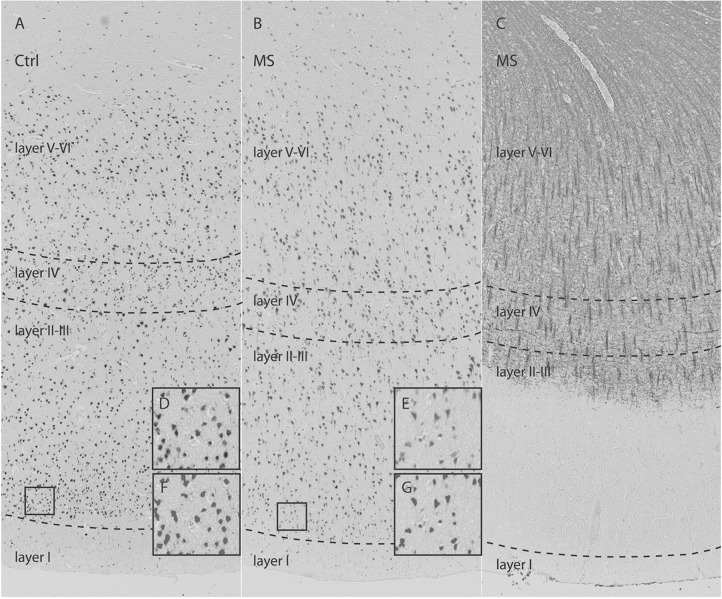
Figure 1. Neuronal loss in type III cerebral cortical lesions in multiple sclerosis (MS).
Control (Ctrl) (A) and MS (B) sections immunostained for NeuN demonstrate overall neuronal loss in MS cortex. (C) Antimyelin PLP-immunostained section from a patient with MS (B) shows subpial demyelination. High-magnification images from cortical layer II (D, E) with matching pictures (F, G) demonstrate the quality of segmentation scripts. The frontal cortex was selected to investigate possible differences in neuron density and axon density between MS (normal-appearing gray matter and type III lesions) and control cortex. Neuron density (−25.4%; p = 0.001) and axon density (−31.4%; p = 0.001) were significantly reduced in type III lesions compared with control cortex. There was no significant difference in neuron sizes between type III lesions and matched control cortex. From Klaver R, Popescu V, Voorn P, et al. Neuronal and axonal loss in normal-appearing gray matter and subpial lesions in multiple sclerosis. J Neuropathol Exp Neurol 2015;74:453–458,11 by permission of the American Association of Neuropathologists, Inc., Copyright © 2015.

