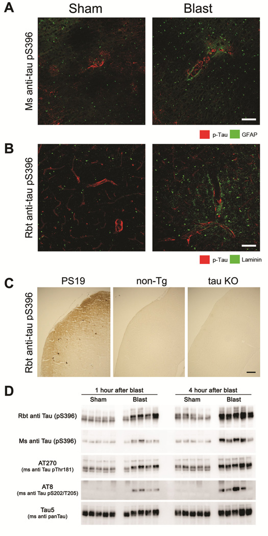
Figure 6. Mild blast exposure causes transient perivascular phospho-tau accumulation.
(A) One hour following a single BOP, phospho-tau (detected by a well-characterized mouse monoclonal anti-phospho-tau S396 antibody, green) aberrantly accumulated around cortical microvessels highlighted by a sheath of endfoot GFAP immunoreactivity (red). (B) Confirmatory immunostaining with a rabbit anti-tau pS396 antibody also revealed altered perivascular phospho-tau expression (green) following blast, with the microvascular space delineated by laminin immunoreactivity (red). (C) Validating the specificity of the less-well characterized rabbit tau-pS396 antibody, phospho-tau deposits in transgenic PS19 mice expressing mutant (P301S) tau were immunopositive, whereas cortical tissue from non-transgenic (non-Tg) mice and tau-deficient (tau KO) mice were immunonegative. (D). Western blots show that in cortex, a single mild blast exposure caused markedly increased expression of multiple indicated phospho-tau species at both 1 and 4 hours, while total tau levels (Tau5 in lower panel) were comparable among shams and BOP-exposed animals (each lane indicates results from one mouse, N=5 and 5, sham and blast-exposed, respectively). Scale bars: (A, B) 20 μm, (C) 100 μm.

