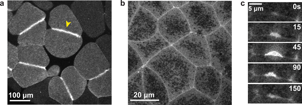
Figure 3. Rho zones and Rho flares in X. laevis embryos.
a. GFP-rGBD (GBD probe for active Rho) in the large blastomeres of the early X. laevis embryo. A distinct zone of active Rho specifies the position of the contractile ring (yellow arrowhead).
b. GFP-rGBD in epithelial cells of the gastrula-stage X. laevis embryo. Zones of active Rho encircle the perimeter of each epithelial cell.
c. A montage depicting a Rho flare over time. These transient accumulations of active Rho at cell-cell junctions were first observed in the X. laevis embryo (Reyes et al. 2014).

