MEDICAL SCIENCES Correction for “NF-κB–driven suppression of FOXO3a contributes to EGFR mutation-independent gefitinib resistance,” by Ching-Feng Chiu, Yi-Wen Chang, Kuang-Tai Kuo, Yu-Shiuan Shen, Chien-Ying Liu, Yang-Hao Yu, Ching-Chia Cheng, Kang-Yun Lee, Feng-Chi Chen, Min-Kung Hsu, Tsang-Chih Kuo, Jui-Ti Ma, and Jen-Liang Su, which appeared in issue 18, May 3, 2016, of Proc Natl Acad Sci USA (113:E2526–E2535; first published April 18, 2016; 10.1073/pnas.1522612113).
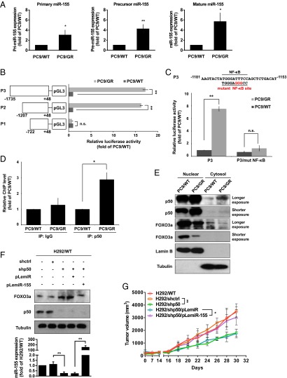
The authors note that Fig. 5 appeared incorrectly. The corrected figure and its legend appear below. This error does not affect the conclusions of the article.
Fig. 5.
miR-155 is transcriptionally regulated by NF-κB of gefitinib-resistant lung cancer cells. (A) Real-time RT-PCR analysis of pri–miR-155, pre–miR-155, and miR-155 expression in PC9/WT and PC9/GR cells. (B) Schematic description of serial deletion reporter constructs of the miR-155 promoter cloned into the pGL3-Basic vector (Left). PC9/WT and PC9/GR cells were transfected with various miR-155 promoter reporters, and the luciferase activity was measured by the dual-luciferase reporter assay (Right). (C) Schematic diagram showing that the NF-κB binding sequences or mutated versions in the miR-155 promoter (Upper), and luciferase activity was measured using the dual-luciferase reporter assay (Lower). (D) ChIP analysis of chromatin extracted from PC9/WT and PC9/GR using polyclonal antibodies directed against the p50, a subunit of NF-κB, followed by real-time RT-PCR analysis to confirm p50 occupancy at the miR-155 promoter. (E) Cytoplasmic and nuclear fractions from PC9/WT and PC9/GR cells were assessed for the presence of FOXO3a, p50, Lamin B (nuclear marker), and Tubulin (cytosolic marker) by Western blot analysis. (F) The FOXO3a and p50 protein expression of H292 cells with stable expression of the indicated transfectants were measured by Western blot (Upper), and miR-155 expression by real-time RT-PCR (Lower), respectively. **P < 0.01 (two-tailed Student's t test). (G) In vivo tumor growth of H292 cells with stable expression of the indicated transfectants. Each point represents the mean ± SEM of tumor volumes of six mice in each group. Tumor volume was calculated as indicated in Materials and Methods. *P < 0.05 and **P < 0.01 (two-tailed Student's t test).


