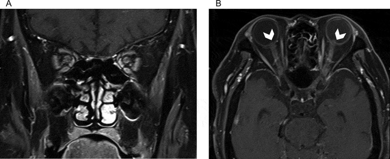
Figure 3.

Gadolinium-enhanced T1-weighted coronal (A) and axial (B) magnetic resonance images. Enhancement of the bilateral optic nerve sheaths is evident in (A) and (B). Protrusion of the optic disc into the vitreous cavity (arrowheads) is also observed in (B).
