Abstract
Duplications of Methyl CpG binding protein 2 (MECP2) -containing segments lead to the MECP2 duplication syndrome, in which severe autistic symptoms were identified. Whether adult neurogenesis may play a role in pathogenesis of autism and the role of MECP2 on state determination of adult neural stem cells (NSCs) remain largely unclear. Using a MECP2 transgenic (TG) mouse model for the MECP2 duplication syndrome, we found that adult hippocampal quiescent NSCs were significantly accumulated in TG mice comparing to wild type (WT) mice, the neural progenitor cells (NPCs) were reduced and the neuroblasts were increased in adult hippocampi of MECP2 TG mice. Interestingly, we found that parvalbumin (PV) positive interneurons were significantly decreased in MECP2 TG mice, which were critical for determining fates of adult hippocampal NSCs between the quiescence and activation. In summary, we found that MeCP2 plays a critical role in regulating fate determination of adult NSCs. These evidences further suggest that abnormal development of NSCs may play a role in the pathogenesis of the MECP2 duplication syndrome.
Methyl CpG-binding protein 2 (MeCP2) is a critical transcriptional regulator and also plays an important role in regulating microRNA biogenesis1,2. Mutations of MECP2 gene were considered as the major cause of the Rett syndrome, a severe neurodevelopmental disorders3. Duplications of genomic segments containing the MECP2 gene and hence increased MeCP2 protein dosage led to a severe neurological disorder, the MECP2 duplication syndrome, which is 100% penetrant in affected males and is associated with infantile hypotonia, severe autistic features, poor speech development, recurrent infections, progressive spasticity, and developmental regression4,5. MECP2 transgenic mice also exhibit various neurological symptoms, including increased anxiety and fear learning, hypoactive, and early death6. Whether adult neurogenesis deficits might be involved in the MECP2 duplication syndrome remains unclear.
Neural stem cells (NSCs) in the hippocampus generate new neurons throughout adulthood7,8. Most of the adult NSCs are relatively quiescent. They will be activated or maintain quiescent in response to extrinsic stimuli or intrinsic signals9,10. Dysregulation of NSCs may contribute to pathogenesis of various neuropsychiatric disorders such as depression and schizophrenia. Previous work identified local parvalbumin (PV) positive interneurons play a critical role in controlling quiescent state of adult NSCs11. Interestingly, numbers of PV positive interneurons were found to increase in Mecp2 null mice12. However, whether MeCP2 may have direct influence on fate determination of adult NSCs remains unknown.
In this work, we showed that adult hippocampal quiescent NSCs were accumulated, the neural progenitor cells (NPCs) were reduced and the neuroblasts were increased in hippocampus of MECP2 TG mice. Furthermore, we found that PV positive interneurons, which were identified as a critical niche component that dictates the adult hippocampal NSCs between the quiescence and activation11, were significantly decreased in dentate gyrus (DG) of MECP2 TG mice. These findings directly link MeCP2 to fate determination of adult NSCs in hippocampus and further suggest that adult neurogenesis may play a critical role in pathogenesis of autism spectrum disorders.
Results
Overexpression of MeCP2 expands the adult NSC pool in vivo
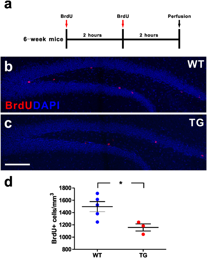
To determine whether MeCP2 overexpression has an impact on adult hippocampal neurogenesis in vivo, we first assessed the dividing cells of hippocampus in both WT and MECP2 TG mice. The dividing cells were identified by the incorporation of BrdU administered through intraperitoneal injections into adult mice twice with a two-hour interval, and analyzed at two hours after the last BrdU injections (Fig. 1a). Quantitative histological ananlysis showed that MECP2 TG mice had fewer BrdU+ cells in the DG, compared with WT mice (Fig. 1b–d). There are at least two types of proliferating cells that can be labeled by BrdU, such as GFAP+/Nestin+ radial glia-like NSCs (RGLs) and GFAP−/Nestin+ NPCs13.
Figure 1. Reduced adult hippocampal dividing cells in MECP2 transgenic mice.
(a) Experimental scheme for assessing new dividing cells in the adult hippocampus. (b,c) Confocal microscopy images of the adult hippocampus showing dividing cells in the subgranular zone (SGZ) of wild type (WT) and MECP2 transgenic (TG) mice, displaying BrdU staining (red). The nuclear label DAPI is shown in blue. Scale bar: 200 μm. (d) Quantitative analysis of BrdU label density in granule cell layer. Values are Mean ± S.E.M (n = 5 WT; n = 3 TG; *P < 0.05, student’s t-test).
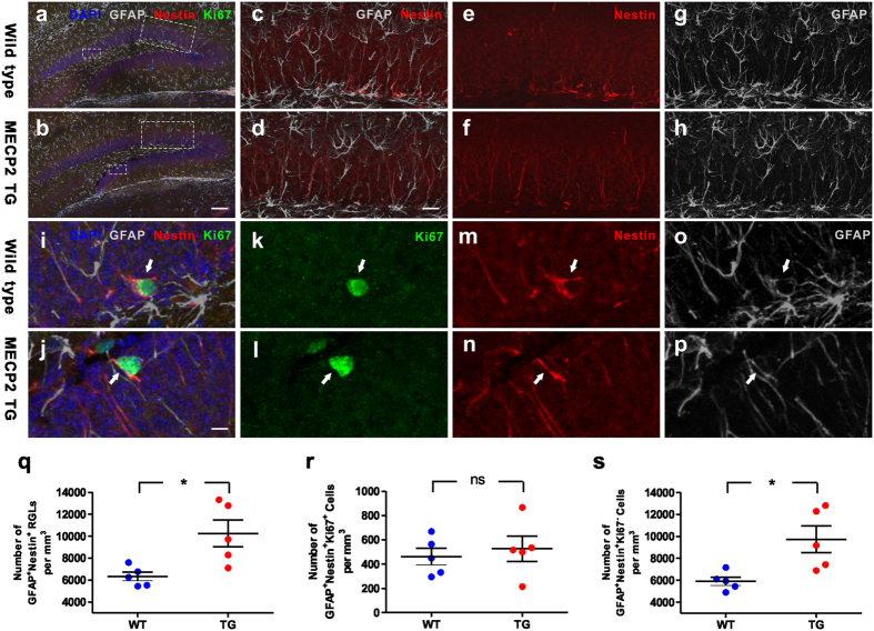
To distinguish which type of cells were affected in MECP2 TG mice, we stained the brain sections with antibodies against GFAP, Nestin and ki67. We found that RGLs (GFAP+/Nestin+) were increased in DG of TG mice, in comparison to WT mice (Fig. 2a–h,q), and proliferating RGLs (GFAP+/Nestin+/Ki67+) appeared not to be different between WT and MECP2 TG mice (Fig. 2i–p,r). Consequently, adult quiescent RGLs (GFAP+/Nestin+/Ki67−) were significantly increased in MECP2 TG mice (Fig. 2s). These data suggest that the adult NSC pool is expanded in hippocampi of MECP2 TG mice.
Figure 2. Increased adult quiescent NSCs in the hippocampal dentate gyrus of MECP2 transgenic mice.
(a,b) Sample confocal images of immunostaining of DAPI, GFAP, Nestin and Ki67. (c–h) High-magnification images of the large boxes in picture A and B showed the adult NSCs (GFAP+/Nestin+) in wild type mice and MECP2 transgenic (TG) mice. (i–p) High-magnification images of the small boxes in picture A and B showed the proliferating RGLs (GFAP+/Nestin+/Ki67+; arrows) in wild type mice and MECP2 transgenic mice. Scale bar: (a,b) 100 μm; (c–h) 25 μm; (i–p) 5 μm. (q–s) Quantitative analysis of GFAP+/Nestin+/RGLs, GFAP+/Nestin+/Ki67+ proliferating RGLs, and GFAP+/Nestin+/Ki67− quiescent RGLs density in granule cell layer respectively. Values are Mean ± S.E.M (n = 5 animals for each genotype; *P < 0.05, student’s t-test; ns: non-significant).
The neural progenitor cells are reduced and the neuroblasts are increased in MECP2 transgenic mice
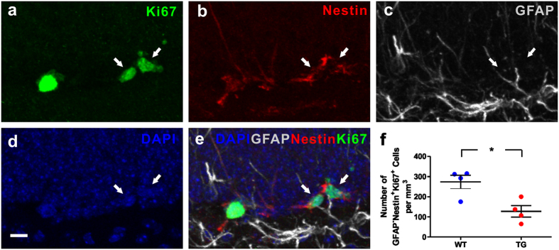
To further characterize the fewer dividing cells in DG of MECP2 TG mice, we examined the NPCs in WT and TG mice. We found that the NPCs (GFAP−/Nestin+/Ki67+) were reduced in DG of TG mice, in comparison to WT mice (Fig. 3). These results suggest that the differentiation of RGL into NPCs or the proliferation of NPCs might be hampered in adult MECP2 TG mice.
Figure 3. Decreased adult neural progenitor cells in the hippocampal dentate gyrus of MECP2 transgenic mice.
(a–e) Sample confocal images of immunostaining of DAPI, GFAP, Nestin and Ki67. Arrows indicate GFAP−/Nestin+/Ki67+ progenitor cells. Scale bar: 10 μm. (f) Quantitative analysis of GFAP−/Nestin+/Ki67+ progenitor cells density in granule cell layer. Values are Mean ± S.E.M (n = 4 animals for each genotype; *P < 0.05, student’s t-test).
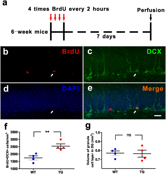
To identify whether neuronal differentiation is affected in the adult hippocampus of TG mice, we gave four doses of BrdU injections with two-hour intervals to label the dividing cells in the DG and analyzed the proliferating neuroblasts at 7 days after the last BrdU injection (Fig. 4a). We counted neuroblasts (BrdU+/DCX+) and found that the neuroblasts were increased in DG of TG mice, in comparison to WT mice (Fig. 4b–f). In addition, WT and MECP2 TG mice exhibited similar volume of granule cell layer in DG (Fig. 4g). These results suggest that MeCP2 overexpression may play an important role in the neuronal differentiation of NPCs into neuroblasts or the proliferation of neuroblasts in adult MECP2 TG mice.
Figure 4. Increased neuroblasts in the hippocampal dentate gyrus of MECP2 transgenic mice.
(a) Experimental scheme for assessing new neuroblasts in the adult hippocampus. (b–e) Sample confocal images of immunostaining of DAPI, MCM2 and DCX. Arrows indicate BrdU+/DCX+ neuroblasts. Scale bar: (b–e), 20 μm. (f) The dentate gyrus of MECP2 TG mice exhibited increased BrdU+/DCX+ neuroblasts analyzed at 7 days after 4 doses of BrdU injections. (g) MECP2 TG mice had similar DG volume with WT mice. Values are Mean ± S.E.M (n = 4 animals for each genotype; **P < 0.01, ns: non-significant, student’s t-test).
MeCP2 overexpression has no impacts on proliferation or differentiation of adult NPCs in vitro
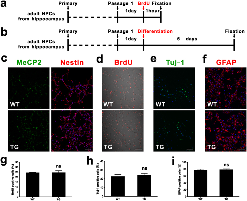
To elucidate the functions of MeCP2 overexpression in adult NPCs, we isolated adult NPCs from the DG of MECP2 TG mice and WT littermates. We found that nearly all cultured cells were positive for the progenitor marker Nestin (Fig. 5c), and MeCP2 was expressed in both WT and TG NPCs (Fig. 5c). We pulsed the cells with BrdU for one hour to assess the proliferation of these NPCs (Fig. 5a) and found that NPCs from MECP2 TG mice exhibited as much BrdU incorporation as WT NPCs (Fig. 5d,g). To assess the effect of MeCP2 overexpression on adult NSC differentiation, both WT and MECP2 TG NPCs were differentiated for five days (Fig. 5b), differentiated cells were stained with Tuj-1 (Fig. 5e) for neurons and GFAP for astrocytes (Fig. 5f). We found that adult NPCs from MECP2 TG mice exhibited similar results in neuronal and astrocytic differentiation compared with adult NPCs from WT mice (Fig. 5h,i). These results suggest that MeCP2 overexpression do not alter the proliferation and differentiation of adult NPCs in vitro. These in vitro results along with in vivio data indicate that there may be non-cell autonomous influence of niche cells on fate decision of adult NPCs in MECP2 TG mice.
Figure 5. Proliferation and differentiation of adult hippocampal NPCs in vitro.
(a) Experimental scheme for assessing proliferation of NPCs in vitro. (b) Experimental scheme for assessing differentiation of NPCs in vitro. (c) Adult hippocampal NPCs culture under proliferating conditions expressed the neural progenitor cell marker Nestin (cytoplasmic, red); MeCP2 in green; Dapi in blue. Scale bar: 100 μm. (d) Both WT and TG NSCs incorporate the thymidine analog, BrdU, under proliferating conditions (BrdU, red). Scale bar: 100 μm. (e) Differentiation of adult NSCs into neurons (Tuj-1, green) by 1 μM RA and 5 μM forskolin. Scale bar: 100 μm. (f) Differentiation of adult NSCs into astrocytes (GFAP, red) by 1% FBS. Scale bar: 100 μm. (g–i) Quantitative analysis showing the percentages of BrdU+, Tuj-1+, and GFAP+ cells in WT and TG individuals respectively. Values are Mean ± S.E.M (ns: non-significant, student’s t-test).
MeCP2 regulates parvalbumin interneurons involved in neurogenesis
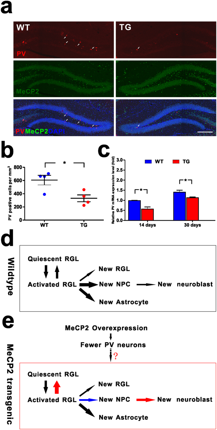
Pavalbumin (PV) interneurons were identified as a critical niche component for adult hippocampal neurogenesis, and was shown to suppress adult quiescent NSCs activation in the adult DG11. To examine the expression of PV in MECP2 TG mice, we stained the brain sections with antibody against PV and found that the density of PV+ neurons was significantly decreased in DG of MECP2 TG mice compared with WT mice (Fig. 6a,b). Consistently, mRNA expression of PV decreased in MECP2 TG mice at different stages before adulthood compared with WT mice (Fig. 6c). These results indicate that decreased expression of PV as a result of MeCP2 overexpression could be responsible for the altered neurogenesis in MECP2 TG mice.
Figure 6. MeCP2 regulates parvalbumin interneurons involved in adult neurogenesis.
(a) Density of parvalbumin interneurons in the DG significantly decreased in MECP2 transgenic mice. Representative images showing the parvalbumin interneurons (red, arrows) in DG were along with the border of the granule cell layer (MeCP2 in green, Dapi in blue). Scale bar: 200 μm. (b) Quantitative analysis of parvalbumin interneurons density in granule cell layer. Values are Mean ± S.E.M (n = 4 animals for each genotype; *P < 0.05, student’s t-test). (c) Expression of PV mRNA decreased in MECP2 transgenic mice (n = 3 animals for each genotype; *P < 0.05, student’s t-test). (d) A model of RGLs/NPCs behaviors in the adult WT mouse hippocampus. an activated RGL usually have four choice points: (1) to be quiescent; (2) symmetric self-renewal to expand the RGL pool; (3) neurogenic asymmetric self-renewal to generate a NPC; (4) astrogliogenic asymmetric self-renewal to generate an astrocyte (modified from Bonaguidi, 2011). The thickness of the arrow indicates the relative probability of each choice. (e) A model on the role of MeCP2 overexpression in regulating RGLs/NPCs in the adult MECP2 transgenic mouse hippocampus. MeCP2 overexpression promotes activated RGLs return to quiescence; and inhibits neurogenic asymmetric self-renewal to generate a NPC or the proliferation of NPCs; also, MeCP2 overexpression promotes the differentiation of NPCs into neuroblasts or the proliferation of neuroblasts. MeCP2 overexpression reduced parvalbumin interneurons. Low activity of parvalbumin interneurons as a result of MeCP2 overexpression could be responsible for the altered neurogenesis in MECP2 TG mice. Red and blue arrows indicate increased and decreased probability, respectively.
Discussion
Previous study showed there was no significant difference in cell proliferation and neuronal differentiation between Mecp2 KO and WT NSCs in vitro14. Moreover, Mecp2 KO mice exhibited no deficits in the number of BrdU+ cells at either 1 day post-BrdU injection or 4 weeks post-BrdU injection14. BrdU+ cells in Mecp2 KO mice differentiated into similar numbers of new neurons compared to WT mice. It was suggested that Mecp2 knockout had no impact on proliferation and differentiation of adult hippocampus NSCs in vivo and in vitro14. Another research showed ectopically expressed MeCP2 in cultured NPCs was able to enhance neuronal differentiation and suppress astrocytic differentiation compared to the control after virus infection, suggesting a possible function of MeCP2 overexpression in NSCs differentiation15. Interestingly, there is no reported effect of MeCP2 overexpression on adult NSCs in vivo. We now provide the first study showing the function of MeCP2 overexpression in the quiescence and activation of NSCs in the adult brain of MECP2 transgenic mice. In the present study, we have uncovered a role for MeCP2 overexpression in the accumulation of adult quiescent NSCs, decreased NPCs and increased neuroblasts in the adult hippocampus, further suggesting the potential involvement of adult neurogenesis in MECP2-related autism spectrum disorders.
Since we found no differences in proliferation and differentiation between MeCP2 overexpression and wildtype adult NPCs in vitro, we considered that there is non-cell autonomous influence of MeCP2 overexpression niche cells on adult NPCs fate decision in MECP2 TG mice. Adult neurogenesis arises from neural stem cells within specialized niches. The local niche is associated with regulating multiple stages of adult neurogenesis, from neural progenitor proliferation to new neuron maturation, synaptic integration and survival16. It was identified that PV interneurons, one of GABA-releasing niche cells among multiple interneuron subtypes in adult DG, interact functionally with adult NSCs11. A study showed that relative PV mRNA expression level was significantly increased in the primary visual cortex of adult Mecp2 KO mice as determined by RT-qPCR12. Another recent study showed decreased number of parvalbumin-expressing interneurons in the medial prefrontal cortex in autism17. We detected significant decrease in the number of PV interneurons in DG of MECP2 TG mice. MeCP2 overexpression may not affect the total number of cells in DG of adult hippocampus, but it can contribute to the decreased parvalbuming interneurons linked to altered neurogenesis11,18.
Our data showed that in MECP2 TG mice, adult hippocampal quiescent NSCs were significantly accumulated. NPCs were reduced and neuroblasts were increased. We also found that parvalbumin positive interneurons were significantly decreased in MECP2 TG mice, which were critical for determining fates of adult hippocampal NSCs between the quiescence and activation. These evidences further suggest that abnormal development of NSCs may play a role in the pathogenesis of the MECP2 duplication syndrome. Low activity of parvalbumin interneurons as a result of MeCP2 overexpression could be responsible for the altered neurogenesis in MECP2 TG mice (Fig. 6d,e).
Methods
All experiments were performed in accordance with the guidelines and under the approval of the Animal Care and Use Committee of the Shanghai Institute for Biological Science of the Chinese Academy of Sciences.
Animals
The MECP2 transgenic (TG) line on FVB background was generated from Dr. Huda Zoghbi laboratory and purchased from the Jackson Laboratory (#008679)6. Heterozygous male was crossed with WT female to maintain a live colony.
BrdU incorporation
For in vivo analysis of adult NSCs, adult (6 weeks old) male mice were injected with BrdU twice (50 mg/kg in saline, every 2 hours, Sigma, flu/Ald, B5002) intraperitoneally, and then were sacrificed at 2 hours post-last injection.
For in vivo analysis of newborn neurons, adult (6 weeks old) male mice were injected with BrdU for 4 times (50 mg/kg in saline, every 2 hours, Sigma, flu/Ald, B5002) intraperitoneally, and then were sacrificed at 7 days post-last injection.
For in vitro analysis of adult NSCs proliferation, BrdU was incorporated into the medium at a concentration of 100 μg/ml for one hour at 37 °C.
Adult NSCs culture, proliferation and differentiation assay
Adult NSCs were prepared as described previously19. For proliferation assay, neurospheres were dissociated into single cells, and seeded onto coverslips at a density of 50,000/ml. The next day, BrdU was incorporated into the medium at a concentration of 100 μg/ml for one hour at 37 °C. Coverslips were fixed with 4% PFA for 10 min at room temperature, washed three times for 5 min each in 0.01 M PBS. For differentiation assay, neurospheres were dissociated into single cells, and seeded onto coverslips at a density of 100,000/ml. The next day, the medium was replaced with 1 μM RA (Sigma) and 5 μM forskolin (Sigma) (differentiated into neuron) or 1% FBS (differentiate into astrocyte). Five days later, coverslips were fixed with 4% PFA for 10 min at room temperature, washed three times for 5 min each in 0.01 M PBS and were further performed immunostaining work.
Tissue preparation and Immunostaining
Mice were euthanized by intraperitoneal injection of sodium pentobarbital, and then transcardially perfused with saline followed by 4% paraformaldehyde (PFA). Brains were dissected out, post-fixed overnight in 4% PFA, and then dehydrated in 30% sucrose.
Coronal sections of 40-μm thickness were cut on the freezing microtome (LEICA CM1950) and stored in PBS.
For Brdu immunostaining, free-floating sections were incubated in 2 N HCl for 15 min at 37 degree then neutralized in 0.1 M boric acid solution (pH 8.5) for 10 minutes and washed by PBS for 3 times (each time for 5 minutes). Primary antibodies used were as follow: Mouse BrdU (1/5000, Millipore), Chicken GFAP (1/1000, Chemicon), Mouse Nestin (1/800, R&D), Rabbit Ki67 (1/400, Abcam), Rabbit MCM2 (1/500, Abcam), Goat DCX (1/200, Santa Cruz), Rabbit MeCP2 (1/1000, Cell signaling), Rabbit Tuj-1 (1/200, Abcam), Mouse PV (1/1000, Millipore). Fluorescently conjugated secondary antibodies (Invitrogen; 1/1000) were used. Nuclei were stained with 4′,6-Diamidino-2-phenylindole (DAPI; Sigma).
Confocal imaging and cell quantification
Confocal z-stack images were acquired on a Nikon A1 confocal laser microscope system.
For in vivo experiments, NIH Image J with NeuronJ plugin was used to count the immunoreactive cell number at the subgranular zone (SGZ) in the dentate gyrus and to quantify the area of granule cell layer in the dentate gyrus. GFAP+ Nestin+ indicates RGL, GFAP+ Nestin+ Ki67+ indicates proliferating RGL, GFAP+ Nestin+ Ki67− indicates quiescent RGL, GFAP−Nestin+ Ki67+ indicates neural progenitor cell, and BrdU+ DCX+ indicates neuroblast. The density of cells was determined by dividing the total number of immunoreactive cells by the corresponding volume (the area * 40 μm) of granule cell layer.
For the analysis of in vitro proliferating adult NSCs or differentiated neurons or astrocytes, Image J was used to count the immuoreactive cell numbers. The percentage of immunoreactive cells was determined by dividing the number of immunoreactive cells by the total number of cells. A minimum of 1000 cells were analyzed for each culture condition.
Quantitative real time PCR
Total RNA was collected from fresh hippocampus dentate gyrus tissue for reverse transcription (Bio-Rad, 1708891). 2× SYBR Green Master Mix (TOYOBO, QPK201) AND QIAGEN Rotor-Gene Q machine were used in real time PCR experiments. Mouse PV real time primers were used as before20.
Statistical analysis
The results were expressed as Mean ± S.E.M. The two-tailed Student’s t-test was carried out for the analysis of animal-related in vivo and in vitro results. Significance was set as follows: *P < 0.05, **P < 0.01.
Additional Information
How to cite this article: Chen, Z. et al. Accumulated quiescent neural stem cells in adult hippocampus of the mouse model for the MECP2 duplication syndrome. Sci. Rep. 7, 41701; doi: 10.1038/srep41701 (2017).
Publisher's note: Springer Nature remains neutral with regard to jurisdictional claims in published maps and institutional affiliations.
Acknowledgments
This study was supported by the CAS Strategic Priority Research Program (XDB02050400), NSFC Grants (#91432111) to Z.Q., and the Knowledge Innovation Program of Shanghai Institutes for Biological Sciences, Chinese Academy of Sciences (2013KIP204 to Z.C.). We thank Dr. Zhengliang Gao for helpful discussions on this work. We thank the Optical Imaging Facility at the Institute of Neuroscience for support with optical imaging.
Footnotes
The authors declare no competing financial interests.
Author Contributions Z.Q. conceived and supervised the project; Z.C. and X.L. performed most experiments, collection and assembly of data; J.Z., B.Y., B.Y. and D.T. contribute to animal experiments; C.C., Y.S., and S.X. contribute to Quantitative real-time PCR experiments; R.Z., J.L., X.Y. and C.D. contribute to data analysis; Z.C., W.-H.Z. and Z.Q. wrote the manuscript.
References
- Lyst M. J. & Bird A. Rett syndrome: a complex disorder with simple roots. Nat Rev Genet 16, 261–75 (2015). [DOI] [PubMed] [Google Scholar]
- Cheng T. L. et al. MeCP2 suppresses nuclear microRNA processing and dendritic growth by regulating the DGCR8/Drosha complex. Dev Cell 28, 547–60 (2014). [DOI] [PubMed] [Google Scholar]
- Amir R. E. et al. Rett syndrome is caused by mutations in X-linked MECP2, encoding methyl-CpG-binding protein 2. Nat Genet 23, 185–8 (1999). [DOI] [PubMed] [Google Scholar]
- Ramocki M. B., Tavyev Y. J. & Peters S. U. The MECP2 duplication syndrome. Am J Med Genet A 152A, 1079–88 (2010). [DOI] [PMC free article] [PubMed] [Google Scholar]
- Van Esch H. MECP2 Duplication Syndrome. Mol Syndromol 2, 128–136 (2011). [DOI] [PMC free article] [PubMed] [Google Scholar]
- Collins A. L. et al. Mild overexpression of MeCP2 causes a progressive neurological disorder in mice. Hum Mol Genet 13, 2679–89 (2004). [DOI] [PubMed] [Google Scholar]
- Spalding K. L. et al. Dynamics of hippocampal neurogenesis in adult humans. Cell 153, 1219–27 (2013). [DOI] [PMC free article] [PubMed] [Google Scholar]
- Bonaguidi M. A. et al. In vivo clonal analysis reveals self-renewing and multipotent adult neural stem cell characteristics. Cell 145, 1142–55 (2011). [DOI] [PMC free article] [PubMed] [Google Scholar]
- Ahn S. & Joyner A. L. In vivo analysis of quiescent adult neural stem cells responding to Sonic hedgehog. Nature 437, 894–7 (2005). [DOI] [PubMed] [Google Scholar]
- Lugert S. et al. Quiescent and active hippocampal neural stem cells with distinct morphologies respond selectively to physiological and pathological stimuli and aging. Cell Stem Cell 6, 445–56 (2010). [DOI] [PubMed] [Google Scholar]
- Song J. et al. Neuronal circuitry mechanism regulating adult quiescent neural stem-cell fate decision. Nature 489, 150–4 (2012). [DOI] [PMC free article] [PubMed] [Google Scholar]
- Durand S. et al. NMDA receptor regulation prevents regression of visual cortical function in the absence of Mecp2. Neuron 76, 1078–90 (2012). [DOI] [PMC free article] [PubMed] [Google Scholar]
- Kempermann G., Jessberger S., Steiner B. & Kronenberg G. Milestones of neuronal development in the adult hippocampus. Trends Neurosci 27, 447–52 (2004). [DOI] [PubMed] [Google Scholar]
- Smrt R. D. et al. Mecp2 deficiency leads to delayed maturation and altered gene expression in hippocampal neurons. Neurobiol Dis 27, 77–89 (2007). [DOI] [PMC free article] [PubMed] [Google Scholar]
- Tsujimura K., Abematsu M., Kohyama J., Namihira M. & Nakashima K. Neuronal differentiation of neural precursor cells is promoted by the methyl-CpG-binding protein MeCP2. Exp Neurol 219, 104–11 (2009). [DOI] [PubMed] [Google Scholar]
- Ming G. L. & Song H. Adult neurogenesis in the mammalian brain: significant answers and significant questions. Neuron 70, 687–702 (2011). [DOI] [PMC free article] [PubMed] [Google Scholar]
- Hashemi E., Ariza J., Rogers H., Noctor S. C. & Martinez-Cerdeno V. The Number of Parvalbumin-Expressing Interneurons Is Decreased in the Medial Prefrontal Cortex in Autism. Cereb Cortex (2016). [Google Scholar]
- Song J. et al. Parvalbumin interneurons mediate neuronal circuitry-neurogenesis coupling in the adult hippocampus. Nat Neurosci 16, 1728–30 (2013). [DOI] [PMC free article] [PubMed] [Google Scholar]
- Brewer G. J. & Torricelli J. R. Isolation and culture of adult neurons and neurospheres. Nat Protoc 2, 1490–8 (2007). [DOI] [PubMed] [Google Scholar]
- He L. J. et al. Conditional deletion of Mecp2 in parvalbumin-expressing GABAergic cells results in the absence of critical period plasticity. Nat Commun 5, 5036 (2014). [DOI] [PubMed] [Google Scholar]


