. 2017 Jan 10;6:e19032. doi: 10.7554/eLife.19032

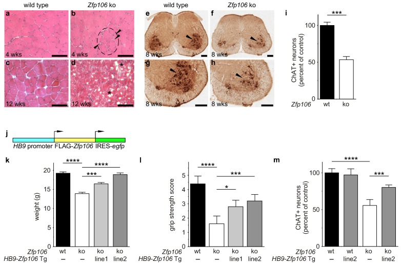
Figure 2. Zfp106 loss-of-function causes a severe neuromuscular phenotype in mice.

(ah) Evidence of profound neuromuscular pathology in Zfp106-null mice. H&E stained sections of quadriceps muscles (ad) from 4-week old and 12-week old wild type (a, c) and Zfp106 knockout (b, d) mice are shown. Note the presence of grouped small angular fibers in knockout mice (dashed circle), already clearly evident by 4-weeks of age (b, arrowheads) compared to the rounder, more evenly-shaped fibers in controls (a). By 12-weeks (end stage disease), Zfp106 knockout mice have profound pathology and the presence of atrophic and degenerated muscle fibers with numerous inclusions (d, asterisks). Scale bar, 100 μm. ChAT immunohistochemical staining (eh) of lumbar spinal cord sections from 8-week old wild type (e, g) and Zfp106 knockout (f, h) mice show a drastic reduction in the number of ChAT+ motor neurons. Arrowheads mark ChAT-expressing motor neurons in the ventral horn. Scale bar, 200 μm in all panels. (i) Quantification of ChAT-positive neurons in the lumbar spinal cord of wild type and Zfp106 knockout mice at 8 weeks of age shows a ~ 47% loss of motor neurons in Zfp106 knockout mice. n = 5 mice for each genotype. Data are expressed as percent of control ± SD and were analyzed by t-test. ***p<0.001. (j) Schematic of the HB9-3×FLAG-2×STREP-Zfp106-IRES-gfp (HB9-Zfp106) transgene. (k, l, m) Transgenic expression of Zfp106 in motor neurons suppresses the wasting phenotype (k), the reduction in grip strength (l) and the loss of lumbar spinal cord motor neurons (m) in Zfp106 knockout mice. Wasting data are expressed as the mean weight ± SEM. Grip strength data are expressed as the mean grip score ± SD; grip score: 1, 1–10 s; 2, 11–25 s; 3, 26–60 s; 4, 61–90 s; 5, > 90 s. ChAT-positive neuron counts are expressed as percent of control ± SD. n = 5 female mice at 8 weeks of age for each genotype. Data were analyzed by one-way ANOVA and Bonferroni's Multiple Comparison Test. *p<0.05; ***p<0.001, ****p<0.0001.

DOI: http://dx.doi.org/10.7554/eLife.19032.005

Figure 2.

Figure 2—figure supplement 1. Zfp106 knockout mice exhibit progressive and severe weight and grip strength loss over time.

Figure 2—figure supplement 1.

(a) Dorsal view of skinned wild type and Zfp106 knockout mice at 12-weeks of age. Zfp106 knockout mice show severe muscle wasting and kyphosis compared to wild type mice. (b) Female mice were weighed at the indicated ages, n = 5 for each group. Body weight of Zfp106 knockout mice was significantly reduced at 2 months of age compared to control and further reduced at 3 months of age. (c) Zfp106-null mice exhibit profound loss of grip strength; grip score: 1, 1–10 s; 2, 11–25 s; 3, 26–60 s; 4, 61–90 s; 5, > 90 s. n = 5 age-matched female mice for each group tested at the indicated ages. Graphical data are presented as the mean ± SD. Data were analyzed by one-way ANOVA and Bonferroni's Multiple Comparison Test. ns, not significant; ***p<0.001, ****p<0.0001.

Figure 2—figure supplement 2. Zfp106 is expressed in skeletal muscle and motor neurons.

Figure 2—figure supplement 2.

Expression of Zfp106 as detected by β-galactosidase activity in quadriceps (a) and spinal cord (b) of 8-week-old Zfp106laczflox/+ mice. β-galactosidase is expressed from a lacZ knock-in at the Zfp106 locus. A section of the quadriceps muscle from an 8-week old Zfp106laczflox/+ mouse stained with X-gal is shown in (a). Arrowheads show β-galactosidase-expressing muscle fibers, weakly detectable by X-gal staining; scale bar, 100 μm. A section through the ventral horn of the spinal cord (lumbar region) of a Zfp106laczflox/+ mouse is shown in (b). The same section is shown in all three panels in (b); the green channel shows expression of the neuronal marker NeuN; the red channel shows expression of β-galactosidase expressed from the Zfp106 locus. Arrows denote co-expression of β-galactosidase and NeuN in large motor neurons in the ventral horn of the spinal cord; scale bar, 50 μm.

Figure 2—figure supplement 3. Expression of Zfp106 in wild type, knockout and HB9-Zfp106 transgenic mice.
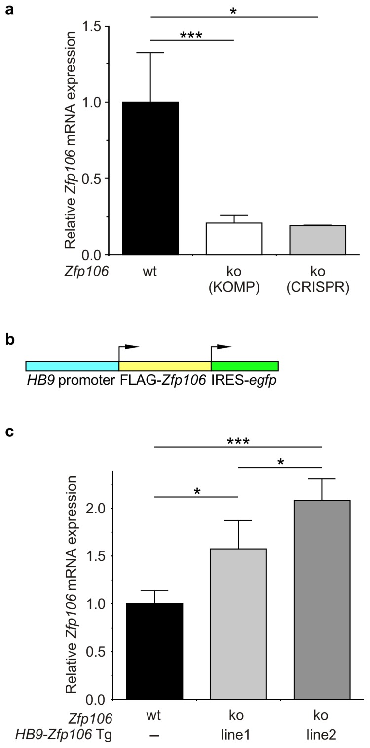
Figure 2—figure supplement 3.

(a) Evaluation of Zfp106 expression by RT-qPCR in lumbar spinal cord of wild type, KOMP Zfp106 knockout and CRISPR Zfp106 knockout mice. Expression of Zfp106 mRNA is drastically and significantly reduced in lumbar spinal cord of both KOMP and CRISPR knockout mice. (b) Schematic of the transgenic construct used for generation of the motor neuron specific Zfp106 mice. The HB9 promoter drives expression of a 3xFLAG-2xSTREP N-terminally tagged Zfp106 and IRES-EGFP. (c) Evaluation of transgene expression by RT-qPCR in lumbar spinal cord at P10. Expression of Zfp106 mRNA ranged from ~1.5 to 2 fold of wild type expression in two independent transgenic mouse lines, as assessed by qPCR with primers amplifying both endogenous and transgenic Zfp106. Data in a, c were analyzed by one-way ANOVA and Bonferroni's Multiple Comparison Test. *p<0.05; ***p<0.001.

Figure 2—figure supplement 4. Rescue of ChAT+ motor neuron loss by restoration of Zfp106 to motor neurons in Zfp106 knockout mice.

Figure 2—figure supplement 4.

Representative images of transverse sections of lumbar spinal cord from wild type (a), motor neuron-specific Zfp106 transgenic (b), Zfp106 knockout (c), and Zfp106 knockout/motor neuron-specific Zfp106 transgenic mice (d), stained by anti-ChAT antibody. Arrowheads mark ChAT+ motor neurons. Scale bar, 200 µm.