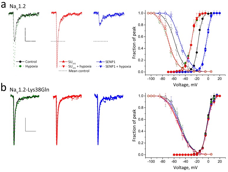
Figure 5. Acute hypoxia induces SUMOylation of NaV1.2 on Lys38.
Rat NaV1.2 was expressed in CHO cells with the β1 subunit and studied under normoxic and hypoxic conditions with control solution (black), 100 pm SUMO1 (red) or 250 pm SENP1 (blue) in the recording pipette, as indicated. Data are mean ± S.E.M. for 10 to 15 cells per group. Measured values are noted in the text and listed in Table 1. Scale bars are 5 ms and 50 pA/pF in a and 10 pA/pF in b. (a) Left, example traces show hypoxia (green dashes) increased NaV1.2 channel current from control conditions (black). SUMO1 in the pipette (red) increased the current to the same level and precluded further augmentation by hypoxia (red dashes). In contrast, SENP1 (blue) decreased the current by ~75% and suppressed sensitivity to hypoxia (blue dashes). Dotted black lines represent mean peak current under control conditions. Right, both Act (solid) and SSI (open) for NaV1.2 were left shifted when cells with hypoxia (green) or SUMO1 in the pipette (red) and were right-shifted with SENP in the pipette (blue). Hypoxia caused no further change to Act or SSI with SUMO1 or SENP1 in the pipette (dashed lines). (b) Left, NaV1.2-Lys38Gln channels passed smaller currents (black) that were not increased by hypoxia (green) or by SUMO1 in the pipette (red) or decreased by SENP1 (blue) in the pipette. Right, the normalized Act and SSI relationships for NaV1.2-Lys38Gln channels (black) are right-shifted compared to wild type NaV1.2 channels and do not change when cells are exposed to hypoxia (green) or are studied with SUMO1 (red) or SENP (blue) in the pipette.

