



Traditionally, large-animal models are used to analyze the impact of clinical ventilators on lung cellular function. In our dual-chamber model, we interface high-frequency oscillatory ventilation (HFOV) directly with airway cells to study the effects of HFOV independently and combined with hyperoxia. Therefore, it is possible to study the preclinical impact of interventional factors without the high cost of animal models, thus reducing staff, time, as well as animal sparing.
Keywords: HFOV, human airway cells, modified chamber
Abstract
High-frequency oscillatory ventilation (HFOV) has been proposed as gentle ventilation strategy to prevent lung injury in the preterm infant. High-frequency jet ventilation leads to dimensional and mechanical airway deformation in animal airway models, which is consistent with translational studies demonstrating the impact of oxygen and biophysical stresses on normal airway cellular function. There is an overall paucity of clinical and cellular data on the impact of HFOV on the conducting airway. We developed an innovative method to test the impact of the clinical HFO Ventilator (SensorMedics 3100A) on human epithelial cell function. In this translational model, we were able to study the differential effects of biophysical stress due to HFOV independently and in combination with hyperoxia on a direct cellular level of the conducting airway system. Additionally, we could demonstrate that hyperoxia and pressure by HFOV independently resulted in significant cell dysfunction and inflammation, while the combination of HFOV and hyperoxia had a synergistic effect, resulting in greater cell death.
NEW & NOTEWORTHY Traditionally, large-animal models are used to analyze the impact of clinical ventilators on lung cellular function. In our dual-chamber model, we interface high-frequency oscillatory ventilation (HFOV) directly with airway cells to study the effects of HFOV independently and combined with hyperoxia. Therefore, it is possible to study the preclinical impact of interventional factors without the high cost of animal models, thus reducing staff, time, as well as animal sparing.
increased awareness about oxygen and pressure-related lung injury in the neonate has resulted in a trend toward more gentle ventilation strategies. Despite advances in critical neonatal care, ventilator-induced lung injury remains an unresolved contributor toward neonatal morbidity and mortality (14). Although survival rates have improved among extremely preterm infants, the proportion of surviving infants with chronic lung disease (CLD) remained unchanged between 1995 and 2006 (13).
Supplemental oxygen in premature infants contributes to the development of CLD due to their immature antioxidant defenses (31). A proposed mechanism by which oxygen causes alveolar injury is through the formation of free radicals and peroxidation of membrane lipids. Also, the secretory upper airway cells are prone to cell dysfunction and inflammation when exposed to progressive oxygen, as shown in a concentration- and time-dependent model (34).
High-frequency oscillatory ventilation (HFOV) has been proposed as a means of decreasing the risk of CLD among infants receiving ventilatory support. Over the last 30 yr, its role has been studied in laboratory and clinical settings, but the application of HFOV remains a topic of controversy in the neonatal intensive care unit with a broad spectrum of indications. While it is used as a primary mode of ventilation in some units, others handle it as a rescue mode of therapy only, when conventional mechanical ventilation (CMV) has been proven unsuccessful (22). Due to the specific characteristics of neonatal airways, the potential complications of tracheal gas flow in neonates need to be differentiated from the mature upper airway. In the rabbit preterm and term isolated airway model, it has been shown that high-frequency jet ventilation leads to dimensional and mechanical airway deformation similar to the sequelae from conventional ventilator-induced airway injury and tracheomalacia in the neonate (7–9). In contrast, there is an overall paucity of clinical and cellular data on the impact of HFOV on the conducting airway.
Furthermore, based on previous cell model work, we and others have shown that oxygen and biophysical stresses can impact normal airway cellular function (25). Building on these airway cellular studies, we developed an innovative model for testing the impact of the clinical HFO Ventilator (SensorMedics 3100A High Frequency Oscillatory Ventilator, CareFusion) on human epithelial cell (EC) function. Our approach parallels the microfabrication techniques with modern tissue engineering, lung-on-a-chip model, which offers a new in vitro approach to mimic the complicated mechanical and biochemical behaviors of a human lung; however, in our modified modulator incubator chamber, we have the capability of interfacing the clinical ventilator with our airway cell model (26). Utilizing this translational model, we sought to develop a method to study the differential effects of biophysical stress due to HFOV on human airway ECs independently and in combination with hyperoxia. Our model, together with the provided data on cell damage due to HFOV, may be used for further research on the effects of HFOV on airway, cartilage, alveolar, and pulmonary vascular cellular function.
MATERIALS AND METHODS
Calu-3 Cell Culture
Calu-3 cells (American Type Culture Collection, ATCC HTB-55) were cultured on transwell plates with the apical surface exposed to gas with 5% CO2 and the basolateral surface bathed in a 50%:50% mixture of Dulbecco’s modified Eagle culture medium/Ham’s F-12 supplemented with 15% fetal calf serum, 500 U/ml penicillin, and 50 μg/ml streptomycin. The cultures were incubated at a constant 37°C in 75-cm2 tissue culture flasks and split when 90% to 95% confluent. Cell culture reagents were acquired from Invitrogen (Carlsbad, CA), and Transwell Permeable Supports were bought from Corning Incorporated Life Sciences (Acton, MA). Calu-3 cells were plated at 2 × 10⁶ cells/cm2 onto Costar transwell inserts (0.4-µm pore size, 12-mm diameter, clear polycarbonate membrane; costar plate; Corning) primed with human placental collagen (Cohesion Technologies, Palo Alto, CA). Approximately 48 h after plating, the apical side of the insert was cleared from culture medium, and the cells were fed from the basolateral side with DMEM/F-12 with 15% fetal calf serum that was changed every other day. The apical side was exposed to air, hereby growing monolayers at an air-liquid interface.1
After 11 days of transwell culture, full confluence was confirmed by measuring transepithelial electrical resistance (TER) with STX2 electrodes and an epithelial volt ohm meter (World Precision Instruments, Sarasota, FL). Calu-3 cell monolayers that exhibited TER values of at least 800-1,000 Ω/cm2 were used for the experiments.
Setup of the Modified Modulator Incubator Chamber for HFOV with Normoxia and Hyperoxia
Cell culture plates were placed into a sterile modular incubator chamber (MIC-101, Billups-Rothenberg, Del mar, CA). A thermometer was placed next to the transwells on the bottom of the chamber, visible from the outside, and temperature was checked every 15 min during the experiment to be kept at a constant 37.0°C. A Mylar membrane was placed 1.5 cm above the culture plates, allowing us to change the gas concentrations on the cells independent of gas concentration (room air) driving the ventilator. In addition, the Mylar sheet allowed us to observe the high-frequency oscillations (HFO) (“wiggle factor”) during HFOV (16). The chamber was then sealed with Dow Corning high-vacuum grease (Dow Corning, Midland, MI) (32). Once the chamber was sealed airtight, the inlet of the modified modular chamber was connected via endotracheal tube (3.0 mm ETT, portex) connection port to a HFO Ventilator at the following study settings: mean airway pressure (MAP) 20 cmH2O; amplitude (Amp) 24 cmH2O, frequency (Freq) 10 Hz (simultaneously pressures were monitored on the opposite side of the Mylar sheet) (see Fig. 1). Low-flow O2/CO2 (95%:5% concentration) or low-flow O2 (21% O2, 5% CO2, balance nitrogen) was infused through an inlet on the level of the culture plates to maintain the cells at the specific group gas concentration, as well as adjust the MAP in combination with the HFO bias flow. A three-way stopcock was attached to the outlet of the chamber on the level of the cell. The connected oxygen analyzer validated the oxygen concentration on the cells and the connected Ruesch pressure manometer (Teleflex Medical, Duluth, GA) measured the MAP pressure throughout the entire time of the experiment. As shown in Fig. 1, ventilator pressures were set to develop pressures on the opposite side of the Mylar sheet, where the cell pressure was monitored.
Fig. 1.

Schematic of experimental modulator incubator chamber.
Setup of the Modified Modulator Incubator Chamber for Hyperoxia without HFOV
The inlet and outlet ports of the chamber were connected to a Servo O2-air 960 mixer (Siemens-Elema, Sweden), a 95% oxygen tank, 5% CO2, and air balance tank. The blended oxygen of 95% was flushed through the airtight chamber for 20 min at a flow rate of 3 l/min. To avoid hypothermia, the blended gas was warmed and humidified with an MR730 respiratory humidifier (Fisher & Paykel Healthcare, Laguna Hills, CA). The oxygen concentration in the chamber was validated by an oxygen analyzer (OM25AE: Maxtec, Salt Lake City, UT). After saturating the chamber with the desired oxygen concentration, the chamber was sealed and placed into a continuous-flow CO2 incubator at 37°C (32).
Study Design
The study is a complete 2 × 2 factorial design with the two factors, oxygen and pressure, each with two levels [oxygen: 21% inspired O2 fraction ()/95% and pressure atmospheric pressure (atm)/20 cmH2O], resulting in four conditions as the combination of the levels of two interactive factors.
Confluent Calu-3 human airway EC monolayers were exposed to one of the four following conditions.
Group 1 (GR1) (control group).
The cultured human airway ECs were exposed to a of 21% under atm during 8 h.
Group 2 (GR2).
The cultured human airway ECs were exposed to a of 95% under atm. The duration of the exposure was 8 h.
Group 3 (GR3).
The cultured human airway ECs were exposed to a of 21% and biophysical stress in the form of HFOV with the following settings: (MAP) 20 cmH2O, Amp 24 cmH2O, and Freq 10 Hz. The duration of the exposure was 8 h.
Group 4 (GR4).
The cultured human airway ECs were exposed to a of 95% and biophysical stress in the form of HFOV with the following settings: MAP 20 cmH2O, Amp 24 cmH2O, and Freq 10 Hz. The duration of the exposure was 8 h.
The duration of HFOV and oxygen targets in this study were based on a retrospective analysis of extracorporeal membrane oxygenation runs initiated for pulmonary reasons at <28 days of life from the extracorporeal life support registry during 2008–2013 (1) and previous cellular exposure studies with increasing doses of oxygen and pressure exposure (32, 34).
At 8 h, monolayer integrity and cell viability were assessed. Apical surface wash fluid (ASF) samples were retrieved from additional wells for analysis of secreted inflammatory mediators. Interleukin (IL)-6 and IL-8 were determined after correction for cell viability.
Measurement of TER
TER measurement was performed with STX2 electrodes and an epithelial volt ohm meter. Medium (0.5 ml) was pipetted into the apical side of the transwell insert, and 1 ml of medium into the basolateral side before the electrodes were inserted into each respective pool of medium and demonstrated resistance to electrical conduction. After the experiment, the medium was suctioned out from the apical transwell side and was exchanged only on the basolateral side. This method has been previously used and validated (34).
Measurement of Cell Viability by Trypan Blue Exclusion Assay
Cells were harvested by trypsinization for single-cell suspensions. Ten microliters of the cell suspension were mixed with 10 µl of 0.4% Trypan blue solution (Sigma Chemical Aldrich, St. Louis, MO). Cell count was done with the hemocytometer, and the ratio of viable (unstained) to total (stained and unstained) cells was calculated to determine cell viability.
Collection of Calu-3 Apical Surface Fluid Washings
Calu-3 ASF was collected from six transwell inserts for each condition after 8 h of exposure. The apical surface was washed twice with 140 μl of sterile deionized water. Samples were centrifuged for 15 min at 13.000 relative centrifugal force and 4°C to remove the cellular debris, and the supernatant was stored in aliquots at −70°C for subsequent IL-6 and IL-8 assays.
Measurement of IL-6 and IL-8 Levels
The ASF levels of IL-6 and IL-8 were measured using quantitative enzyme-linked immunosorbent assay (ELISA) using human IL-6 and IL-8 Quantikine ELISA kits (R&D Systems, Minneapolis, MD). The test sensitivity for respective immunoassays was 0.039 pg/ml for IL-6 and 10 pg/ml for IL-8. Data were corrected for cell viability by calculating enzyme concentration in picograms per milliliter, divided by cell viability, to define whether results were attributable to cell damage or death. Dead cells would not produce enzyme or mediator protein. Thus, as previously reported, mediator production was normalized to live cells by normalizing mediator production to cell viability (10).
Measurement of Total Protein
A DC protein assay kit (Bio-Rad, Hercules, CA) was used. Bovine serum albumin was selected as a reference standard. Absorbance was measured at 655 nm.
Histology and Cytomorphology of Calu-3 Cells
Cytospin slides were prepared using 100 μl of single-cell suspensions and stained for cell differentiation using Kwik-Diff solutions (Thermo Electron, Pittsburgh, PA). Cytomorphology was assessed in a semiquantitative manner as the ratio of abnormal cells to total cells. Abnormal cells were defined by the following parameters: 1) altered cellular appearance; 2) swollen nuclei; 3) intracellular and nuclear vacuoles, or 4) diffuse cytoplasm and cellular debris. All cytospins were examined by light microscopy and photographed at ×40 magnification.
Statistical Analysis
A two-way ANOVA was performed using two factors: HFO (atm/20 cmH2O) and (21% vs. 95%), as well as the interaction in the model for each outcome variable. The omnibus test of the interaction of two factors was highly significant for each parameter. Thus we performed a post hoc analysis of the interaction groups. The four interaction groups are identified above in materials and methods. The Student-Newman-Keuls method of multiple comparisons was used for this purpose. All data were summarized as mean ± 95% confidence interval (CI) for the four groups. Models assumptions were checked before the analysis. All tests were two tailed at the overall level of significance of 0.05. The statistical software SPSS (version 19.0 Chicago, IL) was used for analysis.
RESULTS
The experimental modulator incubator chamber allowed a direct interface between the clinical SensorMedics HFOV with respect to biophysical stress (pressure), while providing independent control of gas concentration exposure on the cell culture plates. In addition, throughout the studies, it was possible to note the HFO on the Mylar surface and monitor the MAP and gas oxygen concentration, while allowing independent control of ventilator driving pressures and gas concentrations from the Calu-3 cell culture plates. After 9 days of culture, Calu-3 cells developed a baseline TER of 923.75 Ω/cm2 for all monolayers before exposure to the four different study conditions. The TER in Calu-3 cell monolayers exposed to hyperoxia and atm (GR2) and normoxia + HFOV (GR3) were decreased to 658.3 Ω/cm2, 95% CI (623.9, 692.8), and 705.2 Ω/cm2, 95% CI (670.7, 739.6), respectively. TER in hyperoxia + HFOV (GR4) was 189.5 ± 6.8 Ω/cm2, 95% CI (155.0, 224.0). In all groups, TER decreased significantly compared with the control group GR1 [TER 984.7 Ω/cm2, 95% CI (950.2, 1,019.1); P < 0.001]. TER in GR4 was significantly lower compared with GR2 and GR3 (P < 0.05) (Fig. 2).
Fig. 2.

Transepithelial electrical resistance (TER) was less than control in all treated groups. GR4 had a significant decrease of TER compared with the other experimental groups. Values are means ± 95% CI; n = 6 for each condition. *Different from control group (P < 0.001). #GR4 different from GR2 and GR3 (P < 0.05).
Cell viability in the control group (GR1) was 92%, 95% CI (86.5, 97.6). Cell viability decreased in all three intervention groups [GR2: 56.4%, 95% CI (51.3, 61.5); GR3: 63.9%, 95% CI (58.8, 69.0); GR4: 20.7%, 95% CI (15.1, 26.2); P < 0.001]. Cell viability in GR4 was significantly diminished compared with the other experimental conditions (P < 0.05) (Fig. 3).
Fig. 3.

Cell viability of Calu-3 monolayers decreased for all experimental conditions compared with control. GR4 had a significant decrease of cell viability compared with GR2 and GR3. Values are means ± 95% CI; n = 6 for each condition. *Different from control group (P < 0.001). #GR4 different from GR2 and GR3 (P < 0.05).
Total protein measured 271.8 µl/ml, 95% CI (225.1, 318.6) in GR1. Protein content in the apical washes increased by 98% to 538.4 µl/ml, 95% CI (491.7, 585.2) in GR2; by 79.8% to 487.3 µl/ml, 95% CI (440.5, 534.1) in GR3; and by 101.5% to 547.7 µl/ml, 95% CI (500.9, 594.5) in GR4 compared with the control group (P < 0.001). There was no statistically significant difference in the total protein content among the three experimental conditions (Fig. 4).
Fig. 4.

Total protein concentration in apical surface wash fluid from Calu-3 monolayers increased in GR2, GR3, and GR4 compared with GR1 (P < 0.001). There was no difference in the total protein secretion among the three experimental conditions. Values are means ± 95% CI; n = 6 for each condition. *Different from control group (P < 0.001).
Levels of the inflammatory mediators IL-6 (Fig. 5) were increased in GR2 [1,549.7 pg/dl, 95% CI (1,423.8, 1,675.7)] and GR3 [1,429.1 pg/ml, 95% CI (1,303.1, 1,555.0)] compared with control [794.7 pg/ml, 95% CI (656.7, 932.7)]. However, levels of IL-6 decreased in GR4 [386.0 pg/ml, 95% CI (248.1, 524.0)] compared with GR2 and GR3 (P < 0.05) and in GR4 compared with GR1 (P < 0.05). The same trends were observed for the inflammatory mediator IL-8 (Fig. 6): the level of IL-8 in the control group measured 13,437.9 pg/ml, 95% CI (10,713.9, 16,162.0). The levels in GR2 increased to 23,422.8 pg/ml, 95% CI (20,936.1, 25,909.5) and to 20,263.4 pg/ml, 95% CI (17,776.7, 22,750.2) in GR3, respectively. In GR4, the level of IL-8 [8,940.9 pg/ml, 95% CI (5,895.3, 11,986.6)] was significantly decreased compared with GR1, GR2, and GR3 (P < 0.05).
Fig. 5.
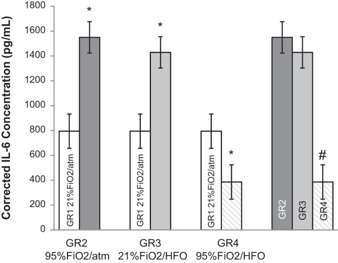
Level of the inflammatory mediator IL-6 corrected for viability was increased in GR2 and GR3 compared with control. Level of IL-6 was decreased in GR4 compared with GR1 and decreased compared with GR2 and GR3. Values are means ± 95% CI; n = 6 for each condition. *Different from control group (P < 0.05). #GR4 different from GR2 and GR3 (P < 0.05).
Fig. 6.

Level of the inflammatory mediator IL-8 corrected for viability was increased in GR2 and GR3 compared with control. Level of IL-8 was decreased in GR4 compared with GR1 and decreased compared with GR2 and GR3. Values are means ± 95% CI; n = 6 for each condition. *Different from control group (P < 0.05). #GR4 different from GR2 and GR3 (P < 0.05).
Semiquantitative assessment of cell morphology showed a greater number of abnormal cells to normal cells in all experimental groups compared with control [4.4%, 95% CI (3.1, 5.8); P < 0.001]. Among the experimental conditions, GR4 [82.1%, 95% CI (80.6, 83.6)] showed an increase of abnormal-to-normal cell ratio compared with GR2 with 24.3%, 95% CI (22.7, 26.0) and GR3 with 22.1%, 95% CI (20.5, 23.8) of abnormal cells (P < 0.05) (Fig. 7).
Fig. 7.

Semiquantitative assessment of cell morphology as the ratio of abnormal cells to total cells showed a higher ratio in GR2, GR3, and GR4 compared with GR1 (P < 0.001). Among the experimental conditions, GR4 showed a higher ratio compared with GR2 and GR3 (P < 0.001). Values are means ± 95% CI; n = 16 for each condition. *Different from control group (P < 0.001). #GR4 different from GR2 and GR3 (P < 0.05).
Abnormal cells are characterized by swollen nuclei, vacuoles, cellular debris, and diffuse cytoplasm. As demonstrated in the representative histological photomicrographs in Fig. 8, cell injury is most pronounced in GR4, exhibiting the characteristics of severe cell damage in almost all cells, while individual exposure to hyperoxia (GR2) and pressure (GR3), respectively, led to a moderate amount of damaged cells.
Fig. 8.

Representative histology. Cytospin preparation of Calu-3 cells after exposure to normoxia or hyperoxia with or without pressure by HFOV is shown. Control showed normal cellular histology. Exposure to hyperoxia (GR2) and pressure by HFOV (GR3) exhibited swollen nuclei and intracellular vacuoles as signs of cell damage. Exposure to hyperoxia and HFOV pressure combined (GR4) demonstrated the highest level of cell damage compared with other experimental conditions. All slides were examined by light microscopy at ×40 magnification (scale bar = 20 µm).
DISCUSSION
As physical and biological sciences come together in medical practice, the boundary between nonliving and living systems requires new engineering models to understand the relationship between these systems. The Biomimetic Microsystems Platform (26) uses an approach to engineer tiny devices containing human cells, mimicking the blood vessels and tissues of living organs. Platform scientists are using these “organs-on-a-chip” to accelerate development of new pharmaceuticals, identify toxins in the environment, and treat life-threatening diseases. In contrast to these microsystems, there is an unmet need for cellular models that can interface with biomedical instrumentation, such as respiratory care devices. In previous studies, our laboratory demonstrated the impact of respiratory gas concentrations and constant pressure, such as continuous positive airway pressure (CPAP), on human airway cell function, structure, and inflammation (32, 34).
Herein, we have developed an airway cellular model, which can interface with a clinical HFOV to provide a better understanding of its impact on cellular function.
Historically, there is a belief that HFOV in the clinical setting may be a more gentle form of ventilation for infants requiring respiratory support in neonatal intensive care units (11, 18). A strategy to decrease ventilator-induced lung injury is to have adequate gas exchange with extremely small tidal volumes, often less than anatomic dead space. In addition, during HFOV, it is possible to maintain a relatively high end-expiratory volume, without inducing overdistension. A Cochrane Review Update published in 2013 revealed that there was no clear evidence that elective HFOV offers important advantages over conventional ventilation when used as the initial ventilation strategy to treat preterm infants with acute pulmonary dysfunction. There may be a small reduction in CLD with HFOV use, but the evidence is weakened by overall borderline significance (3).
Most studies involving HFOV as ventilator mode focus on its effect on the level of the respiratory zone. Data on the effects on cells of the conducting zone remain sparse, despite the fact that bronchial submucosal gland cells have an important immunological function by producing mucus that entraps microorganisms, prevents their replication, and finally removes them from the airway (21), and despite the observation that the oscillatory movements may lead to significant pressure swings in the trachea and at the bronchial level, while being attenuated in the alveoli (19).
For our study, we chose to use a model of Calu-3 cells. The Calu-3 EC line is a well-characterized human airway EC culture that forms high-resistance monolayers when grown on permeable supports. Calu-3 cells are of fast and continuous propagation. The cells have a cuboidal and polygonal shape and grow densely. Confluent monolayers are usually formed after ~3 days postseeding, and they maintain their monolayer condition in transwells for up to 23 days of culture (17). The Calu-3 EC line is derived from bronchial submucosal glands (27) and characterized by its ability to produce airway surface liquid and immunologically active substances (15). As an air-liquid interface culture, it simulates the role of bronchiolar epithelium that functions as a barrier between inspired gas and tissue and protects the upper airway tissue from noxious agents and biophysical injury (29). Calu-3 cells have been used in previous studies to assess cell function (5), the influence of positive pressure in form of CPAP and hyperoxia (25, 34), as well as therapeutic interventions like administration of surfactants and drugs (4, 6, 33) on conducting airway cellular pathophysiology.
In our study, we combined the advantage of using a well-established cell line with a novel model to apply pressure in the form of HFOV, in combination with normoxia or hyperoxia. As such, four groups were studied that allowed us to expose the apical surface of the cells to normoxia and atm (control), hyperoxia and atm, normoxia and HFOV pressure conditions, and a combination of hyperoxia and HFOV pressure. It was shown that hyperoxia and pressure by HFOV individually causes cell damage. In combination, pressure and hyperoxia seem to have a synergistic effect, causing significant cell injury and cell death, as confirmed by an increase in IL-6 and IL-8 concentration, a decrease in cell viability, and in TER as assessment of the integrity of the cell monolayers and the formation of intercellular tight junctions. We also demonstrated an increase of total protein and an abnormal-to-total cell ratio across the three different experimental conditions as markers of cell injury. Our histological photomicrographs exhibit the characteristics of cell damage when exposed to HFOV, with or without hyperoxia. This is contrary to an animal model that examined histopathological tissue changes of cricoid, midtrachea, carina, and left bronchus tissue in the kitten after exposure for 24 h to HFOV vs. conventional ventilation, and that found only minimal airway damage in both groups (23). However, the MAP in the animal study was set at 5 cmH2O, which is much lower than in our study. It might also not reflect common clinical practice for ventilator settings in neonates. MAP on HFOV is often set 2–4 cmH2O higher than on conventional ventilation at 8–10 cmH2O and gets adjusted as needed. In a previous time-dependent study in our laboratory measuring damage of Calu-3 cell exposure to hyperoxia, pressure, or a combination of both factors, it was shown that CPAP of 20 cmH2O applied over 24 and 72 h, respectively, caused less of decrease in cell viability (85% and 87% vs. 63%). Viability due to pressure-related damage seems to be independent from time of exposure, as shown in the time-dependent study (32), In both studies, a MAP of 20 cmH2O was applied and verified by persistent manometer reading at the cell level. Therefore, static pressure was equal in both studies. We speculate that the difference in cell viability might be explained by additional forces during HFOV. During HFOV, the tidal volume oscillates around the constant distending pressure (MAP). Gas is pushed into the lung during inspiration and then pulled out during expiration, causing rapidly changing flow conditions. While tidal volumes are small, usually less than dead space, they are delivered at a high frequency, which might lead to additional dynamic pressure stress and subsequent cell damage.
IL-6 and IL-8 are essential mediators in the cascade of early inflammatory response. Their release is stimulated by hyperoxia and mechanical ventilation, and their levels are found to be persistently high in preterm infants with developing bronchopulmonary dysplasia (3a, 20, 30). The proinflammatory mediators of IL-6 and IL-8 had an increase in the study GR2 and GR3, but a steady decrease in GR4. We speculate that the cells that receive normoxia and pressure by HFOV, as well as the cells that receive hyperoxia without pressure, have significant enough cell damage that causes the IL-6 and IL-8 to seep out of the cell and thus increase in concentration in the ASF. This is consistent with a study on 76 CMV- and HFO-ventilated preterm infants that collected tracheal aspirates for Il-8 over a 10-day period. While the levels of IL-8 were elevated in both ventilation modes, there was no significant difference between CMV and HFOV (28). In previous studies, we have shown that it was necessary to correct mediators expression for cell viability (10). However, in the present study, the combined condition of hyperoxia and positive pressure by HFOV caused a decrease in the concentration of IL-6 and IL-8, even after correcting mediators for viable cells. Therefore, the results cannot be explained with the number of dead cells that are unable to express biological markers. It may be possible that massive cell damage might cause a rapid destruction of Calu-3 cells in GR4 such that the peak IL-6 and -8 expression levels were missed after 8 h of experimentation.
There are several limitations to our study. We should point out that we did not perform a time course study. However, with only 8 h of experimental exposure to pressure, we believe that we chose a minimal time that infants usually spend on HFOV, even when used as a rescue therapy. Additionally, time course exposure to hyperoxia has been performed in previous experiments with similar results (32). Our cells in the present and previous studies were not cultured in a deformable substrate that could quantify the percentage of distension, and, therefore, stretch related injury could not be measured independently. However, applied tidal volumes during HFOV are extremely small, making injury related to stretch less likely than other mechanisms of stress, as mentioned above. Also, pressure was measured at cell level and should accurately measure the pressure applied to the cells.
Calu-3 air-liquid interface cell cultures are a recognized model to simulate airway damage and to assess the effects of new respiratory therapies for humans. However, as in most in vitro experiments with isolated cell lines, systemic interactions like potential repair mechanisms cannot be described. The lack of potential systemic influences in our study has to be taken into consideration.
In summary, this study describes a tissue model that interfaces with a commercial HFO ventilator to demonstrate the effects of HFOV with and without hyperoxia on a direct cellular level of the conducting airway system. Additionally, we showed that hyperoxia and pressure by HFOV independently resulted in significant cell dysfunction and inflammation. The combination of HFOV and hyperoxia had a synergistic effect, resulting in overwhelming cell death. Like the above-mentioned “organ-on-a-chip” technology, our cellular model can also be used to identify the impact of respiratory drugs, toxins, and other forms of respiratory support on airway cellular function (6, 10, 24, 33). Furthermore, more recently, we have established that other lung cell types can be interfaced within this same modified modulator incubator chamber (2). Classically, investigators have used cell cultures in petri dishes and animal models to understand how the respiratory system may react to a medical ventilator, drug, or toxin. Using the technology presented herein, it is possible to study the preclinical impact of these interventional factors without the high cost and need for animal models, thus reducing cost and years of time.
GRANTS
This study was funded by the Nemours Foundation and National Institute of General Medical Sciences COBRE Grants P20 GM-103464 and P30 GM-114736.
DISCLOSURES
No conflicts of interest, financial or otherwise, are declared by the author(s).
AUTHOR CONTRIBUTIONS
A.K.M., B.E.d.J., T.C., and Y.Z. performed experiments; A.K.M. and Y.Z. analyzed data; A.K.M., Y.Z., and T.H.S. interpreted results of experiments; A.K.M. prepared figures; A.K.M. and B.E.d.J. drafted manuscript; A.K.M. and T.H.S. edited and revised manuscript; A.K.M. and T.H.S. approved final version of manuscript.
ACKNOWLEDGMENTS
We thank Paul O’Brian, Respiratory Care Department, A. I. duPont Hospital for Children, for technical assistance during this research effort. We also thank Dr. Jobayer Hossein, Department of Biomedical Research, Nemours, for support with the statistics.
Footnotes
IRB approval and informed consent were not obtained from IRB (Delaware Valley) for our study conducted at Nemours Research Lung Center, Alfred I. duPont Children’s Hospital, Wilmington, DE. Our study did not conduct research on human subjects. The researchers did not obtain data or tissue material through intervention or interaction with human individuals, or obtain any identifiable private information. Animals were not used in the study.
REFERENCES
- 1.Alapati D, Aghai Z, Hossain J, Dirnberger DR, Ogino MT, Shaffer TH. Lung rest during ECMO for neonatal respiratory failure (Abstract). In: Proceedings of the Eastern Society for Pediatric Research Annual Meeting.ESPR, 2016. https://www.aps-spr.org/regions/ESPR/meetings/2016_ESPR_Abstracts.pdf. [Google Scholar]
- 2.Alapati D, Balasubramaniam SL, Hossain J, Soundar S, Fawcett P, Shaffer TH. Effects of inhaled nitric oxide on pulmonary artery smooth muscle cells (PASMC) exposed to positive pressure ventilation and hyperoxia (Abstract). In: Proceedings of the American Thoracic Society 2016 International Conference. New York: ATS, 2016. [Google Scholar]
- 3.[Anon]. Elective high-frequency oscillatory ventilation versus conventional ventilation for acute pulmonary dysfunction in preterm infants. Neonatology 103: 7–9, 2013. doi: 10.1159/000338553. [DOI] [PubMed] [Google Scholar]
- 3a.Babu PB, Chidekel A, Shaffer TH. Association of interleukin-8 with inflammatory and innate immune components in bronchoalveolar lavage of children with chronic respiratory diseases. Clin Chim Acta 350: 195–200, 2004. doi: 10.1016/j.cccn.2004.07.011. [DOI] [PubMed] [Google Scholar]
- 4.Babu PB, Chidekel A, Shaffer TH. Hyperoxia-induced changes in human airway epithelial cells: the protective effect of perflubron. Pediatr Crit Care Med 6: 188–194, 2005. doi: 10.1097/01.PCC.0000154944.67042.4F. [DOI] [PubMed] [Google Scholar]
- 5.Ramesh Babu PB, Chidekel A, Utidjian L, Shaffer TH. Regulation of apical surface fluid and protein secretion in human airway epithelial cell line Calu-3. Biochem Biophys Res Commun 319: 1132–1137, 2004. doi: 10.1016/j.bbrc.2004.05.101. [DOI] [PubMed] [Google Scholar]
- 6.Babu PR, Miller TL, Chikedel A, Shaffer TH. Clara cell protein mediates secretion of proteins, IL-8 and IL-6 in human airway epithelial cell line Calu-3 exposed to hyperoxia. J Chem Pharm Res 4: 3164–3170, 2012. [Google Scholar]
- 7.Bhutani VK, Rubenstein D, Shaffer TH. Pressure-induced deformation in immature airways. Pediatr Res 15: 829–832, 1981. [PubMed] [Google Scholar]
- 8.Bhutani VK, Shaffer TH. Time-dependent tracheal deformation in fetal, neonatal, and adult rabbits. Pediatr Res 16: 830–833, 1982. doi: 10.1203/00006450-198210000-00006. [DOI] [PubMed] [Google Scholar]
- 9.Bhutani VK, Shaffer TH, Abbasi S, Spitzer AR, Fox WW. Effect of high-frequency jet ventilation on preterm and rabbit tracheal mechanics. Pediatr Pulmonol 2: 327–331, 1986. doi: 10.1002/ppul.1950020603. [DOI] [PubMed] [Google Scholar]
- 10.Chidekel A, Zhu Y, Wang J, Mosko JJ, Rodriguez E, Shaffer TH. The effects of gas humidification with high-flow nasal cannula on cultured human airway epithelial cells. Pulm Med 2012: 380686, 2012. doi: 10.1155/2012/380686. [DOI] [PMC free article] [PubMed] [Google Scholar]
- 11.Clark RH, Gerstmann DR, Null DM Jr, deLemos RA. Prospective randomized comparison of high-frequency oscillatory and conventional ventilation in respiratory distress syndrome. Pediatrics 89: 5–12, 1992. [PubMed] [Google Scholar]
- 13.Costeloe KL, Hennessy EM, Haider S, Stacey F, Marlow N, Draper ES. Short term outcomes after extreme preterm birth in England: comparison of two birth cohorts in 1995 and 2006 (the EPICure studies). BMJ 345, dec04 3: e7976, 2012. doi: 10.1136/bmj.e7976. [DOI] [PMC free article] [PubMed] [Google Scholar]
- 14.Donn SM, Sinha SK. Minimising ventilator induced lung injury in preterm infants. Arch Dis Child Fetal Neonatal Ed 91: F226–F230, 2006. doi: 10.1136/adc.2005.082271. [DOI] [PMC free article] [PubMed] [Google Scholar]
- 15.Dubin RF, Robinson SK, Widdicombe JH. Secretion of lactoferrin and lysozyme by cultures of human airway epithelium. Am J Physiol Lung Cell Mol Physiol 286: L750–L755, 2004. doi: 10.1152/ajplung.00326.2003. [DOI] [PubMed] [Google Scholar]
- 16.Duval ELIM, Markhorst DG, van Vught AJ. High frequency oscillatory ventilation in children: an overview. Respir Med CME 2: 155–161, 2009. doi: 10.1016/j.rmedc.2009.09.014. [DOI] [Google Scholar]
- 17.Foster KA, Avery ML, Yazdanian M, Audus KL. Characterization of the Calu-3 cell line as a tool to screen pulmonary drug delivery. Int J Pharm 208: 1–11, 2000. doi: 10.1016/S0378-5173(00)00452-X. [DOI] [PubMed] [Google Scholar]
- 18.Gerstmann DR, Minton SD, Stoddard RA, Meredith KS, Monaco F, Bertrand JM, Battisti O, Langhendries JP, Francois A, Clark RH. The Provo multicenter early high-frequency oscillatory ventilation trial: improved pulmonary and clinical outcome in respiratory distress syndrome. Pediatrics 98: 1044–1057, 1996. [PubMed] [Google Scholar]
- 19.Gerstmann DR, Fouke JM, Winter DC, Taylor AF, deLemos RA. Proximal, tracheal, and alveolar pressures during high-frequency oscillatory ventilation in a normal rabbit model. Pediatr Res 28: 367–373, 1990. doi: 10.1203/00006450-199010000-00013. [DOI] [PubMed] [Google Scholar]
- 20.Groneck P, Götze-Speer B, Oppermann M, Eiffert H, Speer CP. Association of pulmonary inflammation and increased microvascular permeability during the development of bronchopulmonary dysplasia: a sequential analysis of inflammatory mediators in respiratory fluids of high-risk preterm neonates. Pediatrics 93: 712–718, 1994. [PubMed] [Google Scholar]
- 21.Wine JJ, Joo NS. Submucosal glands and airway defense. In: Proceedings of the American Thoracic Society, Airway Membrane Transport and Intracellular Trafficking, 18th Transatlantic Airway Conference, 2004. New York: ATS, 2004, vol. 1, p. 47–53. [DOI] [PubMed] [Google Scholar]
- 22.Keszler M. High-frequency ventilation: evidence-based practice and specific clinical indications. Neoreviews 7: e235–e249, 2006. doi: 10.1542/neo.7-5-e234. [DOI] [Google Scholar]
- 23.Liu D, Miyasaka K, Nakagawa S, Hashizume K. Effect of high-frequency oscillatory ventilation on the upper airways of kittens. Pediatr Int 42: 597–602, 2000. doi: 10.1046/j.1442-200x.2000.01317.x. [DOI] [PubMed] [Google Scholar]
- 24.Miller TL, Singhaus CJ, Sherman TI, Greenspan JS, Shaffer TH. Physiologic implications of helium as a carrier gas for inhaled nitric oxide in a neonatal model of Bethanecol-induced bronchoconstriction. Pediatr Crit Care Med 7: 159–164, 2006. doi: 10.1097/01.PCC.0000200942.23574.CA. [DOI] [PubMed] [Google Scholar]
- 25.Miller TL, Zhu Y, Markwardt S, Singhaus CJ, Chidekel AS, Shaffer TH. Dissociation between the effects of oxygen and pressure on matrix metalloproteinase-2, -7, and -9 expression in human airway epithelial cells. Am J Perinatol 25: 481–489, 2008. doi: 10.1055/s-0028-1085067. [DOI] [PubMed] [Google Scholar]
- 26.Bhatia SN, Ingber DE. Microfluidic organs-on-chips. Nat Biotechnol 32: 760–772, 2014. doi: 10.1038/nbt.2989. [DOI] [PubMed] [Google Scholar]
- 27.Shen BQ, Finkbeiner WE, Wine JJ, Mrsny RJ, Widdicombe JH. Calu-3: a human airway epithelial cell line that shows cAMP-dependent Cl- secretion. Am J Physiol Lung Cell Mol Physiol 266: L493–L501, 1994. [DOI] [PubMed] [Google Scholar]
- 28.Thome U, Götze-Speer B, Speer CP, Pohlandt F. Comparison of pulmonary inflammatory mediators in preterm infants treated with intermittent positive pressure ventilation or high frequency oscillatory ventilation. Pediatr Res 44: 330–337, 1998. doi: 10.1203/00006450-199809000-00011. [DOI] [PubMed] [Google Scholar]
- 29.Velden VH, Versnel HF. Bronchial epithelium: morphology, function and pathophysiology in asthma. Eur Cytokine Netw 9: 585–597, 1998. [PubMed] [Google Scholar]
- 30.von Bismarck P, Claass A, Schickor C, Krause MF, Rose-John S. Altered pulmonary interleukin-6 signaling in preterm infants developing bronchopulmonary dysplasia. Exp Lung Res 34: 694–706, 2008. doi: 10.1080/01902140802389693. [DOI] [PubMed] [Google Scholar]
- 31.Weinberger B, Laskin DL, Heck DE, Laskin JD. Oxygen toxicity in premature infants. Toxicol Appl Pharmacol 181: 60–67, 2002. doi: 10.1006/taap.2002.9387. [DOI] [PubMed] [Google Scholar]
- 32.Zhu Y, Chidekel A, Shaffer TH. Cultured human airway epithelial cells (calu-3): a model of human respiratory function, structure, and inflammatory responses. Crit Care Res Pract 2010: 394578, 2010. doi: 10.1155/2010/394578. [DOI] [PMC free article] [PubMed] [Google Scholar]
- 33.Zhu Y, Miller TL, Chidekel A, Shaffer TH. KL4-surfactant (Lucinactant) protects human airway epithelium from hyperoxia. Pediatr Res 64: 154–158, 2008. doi: 10.1203/PDR.0b013e318175dd14. [DOI] [PubMed] [Google Scholar]
- 34.Zhu Y, Miller TL, Singhaus CJ, Shaffer TH, Chidekel A. Effects of oxygen concentration and exposure time on cultured human airway epithelial cells. Pediatr Crit Care Med 9: 224–229, 2008. doi: 10.1097/PCC.0b013e318166fbb5. [DOI] [PubMed] [Google Scholar]
