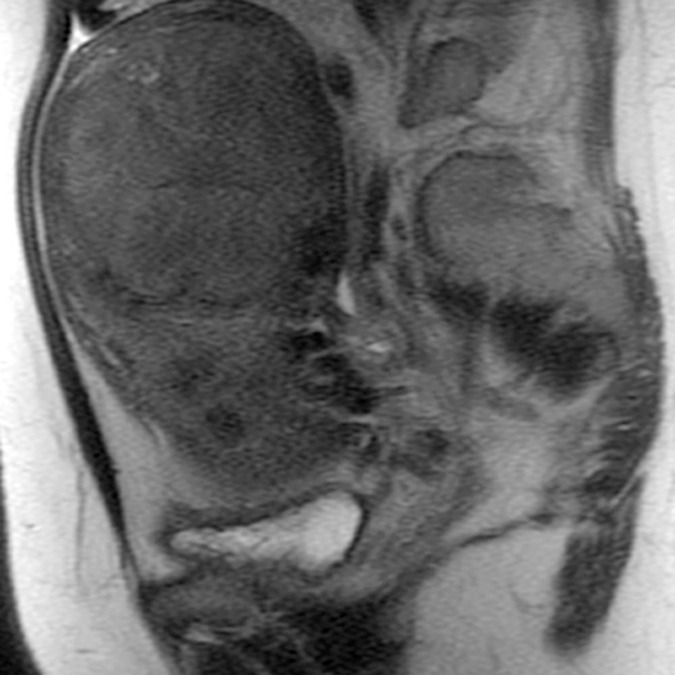
Figure 2d:

(a–c) Coronal oblique and (d–f) sagittal T2-weighted MR images obtained in a 55-year-old woman with a large, pathologically proven intramural leiomyoma demonstrate atypical intermediate signal intensity by using conventional SSFSE (a, d), vrfSSFSE (b, e), and FSE (c, f) imaging. A small, typically appearing intramural leiomyoma is medially adjacent, and an additional, atypically appearing intramural leiomyoma along the right uterine wall is partially included in the plane of imaging. Images were cropped to 20 cm; the SSFSE and vrfSSFSE images were acquired with a 42-cm (coronal oblique) or 30-cm (sagittal) field of view, and the FSE images were acquired with a 22-cm field of view. Note the improved black-blood appearance in vessels with vrfSSFSE imaging compared with conventional SSFSE imaging, owing to its increased sensitivity to flow-related signal loss. Artifacts on the FSE images that are projecting over the uterus are from respiratory motion.
