Figure 2.
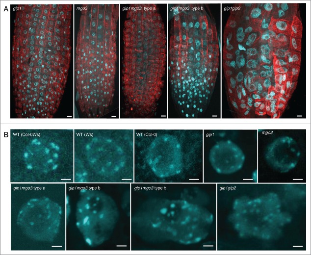
Analysis of root meristematic nuclei from gip1, mgo3, gip1mgo3 compared with gip1gip2. (A) Detection of chromatin by DAPI staining (blue) and microtubules by immuno-labeling with antibodies directed against α-tubulin (red) performed on whole mount meristems of the different seedlings (gip1, mgo3, gip1mgo3 and gip1gip2). Images were captured by confocal microscopy and correspond to Z-stack projections of focal planes. Bars = 10 µm. (B) Meristematic nuclei representative for different WT backgrounds (Ws Col-0), Col-0Ws) were compared with gip1, mgo3, gip1mgo3 type a and b and gip1gip2. Images were captured by confocal microscopy and correspond to Z-stack projections of focal planes. Bars = 2 µm. For Z-stacks, slides were acquired in 0.35 µm intervals.

