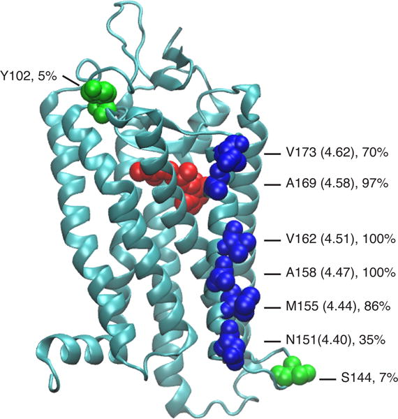
Figure 2.

Structure of bovine Rho (PDB 1GZM) with 11-cis-retinal (red) and the tested amino acid residues (blue: TM4 sites; green: sites reported earlier[10c]). For the TM4 sites, the Ballesteros-Weinstein notation is shown in parentheses.[52] The hydrophobicity of the local environment at these sites is estimated by the percent of lipid contacts versus water contacts. The values give the fraction of the surface exposure of these residues to lipids as observed in molecular dynamics simulations of Rho in a phospholipid bilayer membrane.[15]
