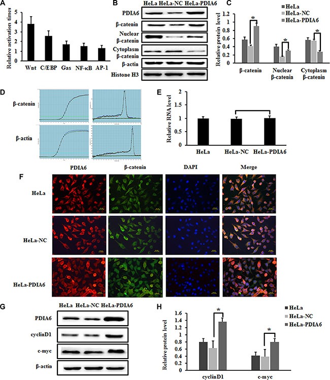
Figure 2. PDIA6 activates the canonical Wnt/β-catenin signaling in HeLa cells.

(A) PDIA6 significantly increased the Top- flash reporter activity of Wnt pathway for more than tripled in HeLa cells. (B) Western blot analysis of total β-catenin, nuclear β-catenin, cytoplasmic β-catenin expression in PDIA6-overexpression cells. Nuclear β-catenin was normalized to Histone H3, other genes were normalized to β-actin. (C) The relative expression of these proteins was normalized to β-actin or Histone H3. *p < 0.05. (D) Quantitative real-time PCR demonstrated that PDIA6 overexpression did not cause a significant change in β-catenin level. (E) The quantitative analysis of β-catenin RNA level. (F) The immunofluorescence of PDIA6 is indicated as red fluorescence, β-catenin is indicated as green fluorescence, and nuclear stained with DAPI as blue. (G) The representative blots of c-myc and cyclinD1 expression were shown. (H) The quantitative analysis of c-myc and cyclinD1 was normalized to β-actin. *p < 0.05.
