Abstract
Microorganisms dominate most Antarctic marine ecosystems, in terms of biomass and taxonomic diversity, and play crucial role in ecosystem functioning due to their high metabolic plasticity. Admiralty Bay is the largest bay on King George Island (South Shetland Islands, Antarctic Peninsula) and a combination of hydro-oceanographic characteristics (bathymetry, sea ice and glacier melting, seasonal entrance of water masses, turbidity, vertical fluxes) create conditions favoring organic carbon deposition on the seafloor and microbial activities. We sampled surface sediments from 15 sites across Admiralty Bay (100–502 m total depth) and the adjacent North Bransfield Basin (693–1147 m), and used the amplicon 454-sequencing of 16S rRNA gene tags to compare the bacterial composition, diversity, and microbial community structure across environmental parameters (sediment grain size, pigments and organic nutrients) between the two areas. Marine sediments had a high abundance of heterotrophic Gammaproteobacteria (92.4% and 83.8% inside and outside the bay, respectively), followed by Alphaproteobacteria (2.5 and 5.5%), Firmicutes (1.5 and 1.6%), Bacteroidetes (1.1 and 1.7%), Deltaproteobacteria (0.8 and 2.5%) and Actinobacteria (0.7 and 1.3%). Differences in alpha-diversity and bacterial community structure were found between the two areas, reflecting the physical and chemical differences in the sediments, and the organic matter input.
Keywords: marine sediments, microbial diversity, bacterial community structure, Antarctica, polar microbiology
Introduction
Marine microbial communities in sediments play a critical role in ecosystems functioning and are the main drivers of biogeochemical cycling of carbon, nitrogen and sulfur. They constitute a huge biomass portion of the Earth, are highly taxonomic diverse, and responsible for the majority of metabolic activity in the ocean including the polar regions (Parkes et al., 2000; Azam and Malfatti, 2007; Fuhrman, 2012). Various physical and chemical parameters can affect the marine sediment communities, in particular the organic matter. Studies conducted in the sediments of the Ross Sea (Carr et al., 2013) and the Drake Passage at the Antarctic Polar Front (Ruff et al., 2014) showed that the organic matter can increase the microbial abundance based on phospholipids and DNA sequencing, respectively. The organic matter can also influence communities composed by lithotrophic and heterotrophic microbes, associated with degradation of organics from sinking particles and fecal pellets in the Western Antarctica (Learman et al., 2016).
As consequences of climate change and rapid warming registered in the Western Antarctic Peninsula, the sea ice declines and glacial inputs increase. These conditions can favor phytoplankton and seaweed blooms (Meredith and King, 2005; Clarke et al., 2007; Montes-Hugo et al., 2009; Pearson et al., 2015), which then increase the quantity of organic matter transported from surface layers to the ocean floor (ex. Ducklow et al., 2006; Gillies et al., 2012). The quantity and type of carbon could also be altered due to the introduction of more terrestrial carbon into marine and benthic environments in polar systems (Learman et al., 2016).
Admiralty Bay is the largest bay on King George Island (South Shetlands Archipelago), located within the maritime Antarctic region. It was designated an Antarctic Specially Managed Area no. 1 (ASMA 1) and contains the Antarctic Specially Protected Area 128 (ASPA 128, former SSSI N° 8). Its hydrology is complex, as it receives different contributions from water masses originating in the Bransfield Strait, and also from ice melt within the bay (Szafrański and Lipski, 1982). Depending on the bathymetry, regional water circulation, winds and seasonal regime, waters from the Bransfield Strait that penetrate the bay originate from the adjacent warm and low saline Bellingshausen Sea (normally in the summer) or the cold and saline waters of Weddell Sea (in the winter) (Gordon and Nowlin, 1978; Tokarczyk, 1987; Huneke et al., 2016). The fjord-like shape of Admiralty Bay reflects freshwater input due to a strong glacial influence and high water column turbidity caused by suspension of soft sediments (Pichlmaier et al., 2004). All this organic matter falls to the seafloor, providing organic carbon to the reservoir, which is partially consumed by the cold-adapted microorganisms from the sediments, leading to low oxygen conditions (Orcutt et al., 2011).
To understand how the contribution of the organic matter from different sources can shape the microbial community in the Admiralty Bay and surrounding areas, in this study the bacterial diversity and community structure of the sediment samples from Admiralty Bay (AB) and adjacent areas of North Bransfield Basin (NBB) were compared. The results contribute to a better understanding of the sequenced microbial community structure in this polar area, and show differences in richness and community structure between the two sites across physical-chemical characteristics of the sediments.
Materials and Methods
Sampling Strategy
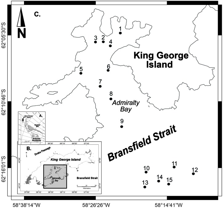
Surface sediment samples were collected along a bathymetric gradient in AB – King George Island (stations 1–9, depths ranging from 100 to 502 m) and NBB – Bransfield Strait (stations 10–15, depths 693–1,147 m), located in the Northwestern Antarctic Peninsula (Figure 1). Sampling was conducted by the Brazilian Navy vessel NApOc Ary Rongel during the austral summer, December 2008. In total, 15 samples of marine sediments were collected using a Mini Box Corer (MBC) at different depths. The top 5 cm of sediments were transferred to sterile Whirl-Pack sample bags (Nasco, WI, USA) and stored frozen onboard (-20°C). Samples were shipped to the University of São Paulo (USP) after 4 months of sampling.
FIGURE 1.
Sampling map with 15 selected stations, 1–9 inside Admiralty Bay and 10–15 in the North Bransfield Basin, Northwestern Antarctic Peninsula.
Environmental Parameters
Grain size was determined by laser diffraction (SALD 3101, Shimadzu, Japan), following the Wentworth scale (Suguio, 1973), yielding the mean and standard deviation of particle size (Folk and Ward, 1957), and classified into categories (clay, sand and silt). Concentrations of chlorophyll and phaeopigments were estimated according to Lorenzen (1967) and Plante-Cuny (1978), adapted for sediments (Gheller, 2014). Percentages of total organic carbon (TOC), total nitrogen, organic matter and carbonates were obtained using the CHNSO elemental analyzer (Elemental Combustion System 4010, Costech Analytical Technologies, USA).
DNA Extraction, 16S rRNA Gene Amplification and Sequencing
Genomic DNA was extracted from 0.25 g of surface sediment in quadruplicate using a PowerSoil DNA Kit (MoBio, Carlsbad, CA, USA), according to the manufacturer’s instructions. Microbial 16S rRNA gene fragments were amplified using a set of primers designed by adding a 10-nucleotide barcode to the forward primer, 519F, (5′-CAGCMGCCGCGGTAATWC-3′) and reverse primer 1068R (5′-CTGACGRCRGCCATGC-3′) (Wang and Qian, 2009). The amplification reaction was carried out using the Accuprime pfx SuperMix (Thermo Scientific, USA) according to the manufacturer. PCR was performed with a thermal cycler (Thermo Scientific, USA) under the following conditions: 95°C for 5 min, 26 cycles of 95°C for 15 s, 59°C for 30 s and 68°C for 1 min. The PCR products were purified by using a DNA clean & concentrator kit (Zymo Research, USA). The amplicons from each sample were mixed at equimolar concentrations and then sequenced using GSFLX titanium instruments and reagents (Roche 454, Life Sciences, USA) at the Center for Advanced Technologies in Genomics (University of São Paulo, Brazil). All sequence data have been deposited in the National Center for Biotechnology Information Sequence Read Archives (SRA) under BioProject ID PRJNA335729.
Sequencing Data Analyses
Raw sequence reads were filtered to eliminate the effect of the random sequencing using the Mothur 454 SOP (Schloss et al., 2011). The primer and barcodes of each read were removed and trimmed. Sequences shorter than 150 nucleotides with ambiguous bases or homopolymer regions were excluded. Sequences were clustered into operational taxonomic units (OTUs) by setting a 97% similarity. OTUs occurring once (singletons) were removed from dataset. Quality-filtered sequences were classified using the RDP Naïve Bayesian Classifier (Wang et al., 2007). For each sample, alpha-diversity indexes (Simpson diversity index and abundance-based coverage estimators - ACE index) were calculated using Mothur (version 1.35.1), and differences in alpha-diversity estimates between groups of samples were tested using Student t-test in R (version 3.3.2). The amplicon reads were normalized using the package DESeq2 (Love et al., 2014) following the general procedure for normalization using a variance stabilization transformation. DESeq2 normalized reads were used for all downstream analyses. The number of shared OTUs between samples were visualized using ggplot2 package in R (Wickham, 2009). Beta-diversity between samples was examined using Bray-Curtis dissimilarity matrix and ordinated by non-metric multidimensional scaling (nMDS) in R, with fitting of the environmental parameters applying the envfit function from the vegan package (Oksanen et al., 2013). To test the significance of differences between groups of samples (AB vs. NBB), analysis of similarity (adonis) was used.
Results
Environmental Parameters
Sediments in the area of study were mainly composed of silt and clay (representing 70.4 to 100.0% of sediment composition). In 12 of 15 samples, granulometry was dominated by silt, with the remaining three dominated by clay. The silt fraction in sediments varied from 39.1 to 64.9%, followed by clay (23.1–59.3%) and sand (0–29.5%) (Table 1). Stations located in the NBB had higher sand contents (6.6–20.6%) when compared to AB samples (0–10.1%), with the exception of station 5 (29.5%), located in front of Lange glacier.
Table 1.
Environmental parameters measured for each sample in the sediments of Admiralty Bay and North Bransfield Basin, Western Antarctic Peninsula. Granulometry: Sand (%), Silt (%), Clay (%), Silt + Clay (%).
| Sample | Sand(%) | Silt(%) | Clay(%) | Silt + Clay(%) | Chlorophyll(mg.m-2) | Phaeopigment (mg.m-2) | Chloro/PhaeoRatio | CPE | OM (%) | Carbonate (%) | TOC (%) | Total N (%) | C/NRatio |
|---|---|---|---|---|---|---|---|---|---|---|---|---|---|
| 1 | 0.00 | 48.93 | 51.07 | 100.00 | 1.25 | 41.68 | 0.03 | 42.93 | 3.09 | 17.46 | 0.56 | 0.07 | 7.15 |
| 2 | 1.13 | 53.13 | 45.74 | 98.87 | 4.13 | 45.09 | 0.11 | 49.22 | 2.41 | 16.61 | 0.25 | 0.07 | 3.62 |
| 3 | 0.10 | 43.52 | 56.38 | 99.90 | 0.34 | 84.53 | 0.00 | 84.87 | 5.10 | 19.31 | 0.83 | 0.12 | 6.46 |
| 4 | 9.28 | 57.38 | 33.35 | 90.72 | 2.54 | 55.80 | 0.05 | 58.34 | 2.91 | 14.93 | 0.30 | 0.08 | 4.10 |
| 5 | 29.57 | 45.35 | 25.09 | 70.43 | 3.82 | 51.66 | 0.08 | 55.48 | 2.72 | 14.38 | 0.40 | 0.05 | 7.96 |
| 6 | 1.64 | 57.06 | 41.30 | 98.36 | 3.95 | 58.52 | 0.08 | 62.47 | 3.94 | 14.47 | 0.25 | 0.06 | 4.36 |
| 7 | 2.27 | 51.52 | 46.21 | 97.73 | 5.83 | 52.50 | 0.11 | 58.32 | 2.90 | 14.33 | 0.21 | 0.06 | 3.40 |
| 8 | 2.74 | 50.80 | 46.46 | 97.26 | 0.17 | 60.17 | 0.00 | 60.34 | 3.65 | 17.52 | 0.43 | 0.08 | 5.53 |
| 9 | 1.54 | 39.11 | 59.35 | 98.46 | 8.25 | 42.92 | 0.20 | 51.17 | 2.70 | 12.23 | 0.35 | 0.06 | 6.27 |
| 10 | 10.17 | 58.23 | 31.60 | 89.83 | 4.58 | 26.57 | 0.17 | 31.15 | 2.85 | 13.74 | 0.51 | 0.06 | 8.96 |
| 11 | 20.62 | 56.20 | 23.17 | 79.38 | 1.90 | 48.01 | 0.04 | 49.91 | 2.61 | 13.19 | 0.40 | 0.06 | 6.64 |
| 12 | 6.61 | 64.94 | 28.45 | 93.39 | 0.37 | 49.22 | 0.01 | 49.59 | 3.49 | 16.73 | 0.44 | 0.07 | 6.31 |
| 13 | 16.16 | 55.50 | 28.35 | 83.84 | 0.31 | 64.41 | 0.01 | 64.72 | 6.03 | 16.51 | 0.70 | 0.08 | 8.69 |
| 14 | 17.79 | 56.85 | 25.36 | 82.21 | 0.00 | 46.51 | 0.00 | 46.51 | 3.53 | 15.30 | 0.63 | 0.08 | 7.98 |
| 15 | 20.30 | 54.48 | 25.22 | 79.70 | 0.00 | 44.45 | 0.00 | 44.45 | 3.37 | 16.21 | 0.62 | 0.07 | 8.69 |
Pigments: Chlorophyll (mg.m-2), Phaeopigment (mg.m-2), Chloro/Phaeo, Chlorophyll/Phaeopigment Ratio; CPE, Chloroplast Pigment Equivalent. OM, Organic Matter (%); Carbonate (%), TOC, Total Organic Carbon (%); Total N, Total Nitrogen (%); C/N, Total Carbon to Nitrogen Ratio.
The sediment chlorophyll concentrations varied from zero (stations 14 and 15) to 8.25 mg.m-2 (station 9), and phaeopigment concentrations from 41.68 (station 1) to 84.53 mg.m-2 (station 3), indicating a predominance of degraded organic matter and small quantities of recent organic matter in the sediments. Stations in AB showed higher concentrations of chlorophyll than in the NBB, indicating the presence of higher quantities of recent organic matter to be degraded in the bay. Station 13 presented the highest organic matter concentration (6.0%) and C/N ratio (8.6).
Community Composition
In this study, we used massively parallel signature sequencing technologies to obtain a total of 117,267 sequences (range 1000–26178 reads per sample) from 15 sediment samples from depths varying from 100 to 1.147 m. At the phylum level, all OTUs could be classified and belonged to 22 formally described bacterial phyla and 18 candidate phyla.
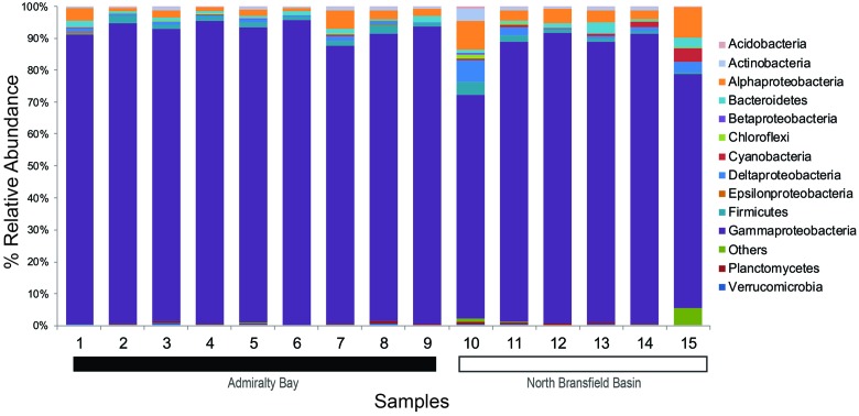
The abundance analysis showed that eight phyla accounted for more than 97% of the total amplicons: Proteobacteria (89%), Firmicutes (1.5%), Bacteroidetes (1.4%), Actinobacteria (0.9%), Chloroflexi (0.7%), Planctomycetes (0.4%), Verrucomicrobia (0.3%) and Acidobacteria (0.1%) (Figure 2).
FIGURE 2.
Taxonomic composition and relative abundance of 16S rRNA sequences based on bacterial phyla and proteobacterial classes. Samples 1–9 belong to Admiralty Bay, and 10–15 are from North Bransfield Basin.
Considering the total of 40 classes identified, only 10 were found in all samples and accounted for 99.4% of the total tags. Based on the relative abundance, Gammaproteobacteria was the top dominant class in both sites, varying from 87.1 to 95.7% in AB and 70.5 to 91.4% in NBB. Alphaproteobacteria was the second dominant class, accounting for 0.9–5.9% in AB and 2.4–9.6% in NBB. Firmicutes (0.4–3.0% in AB and 0.4–4.0% in NBB) and Bacteroidetes (0.3–2.2% in AB and 0.6–3.4% in NBB) were also present in all sediment samples at similar rages. Other groups were more abundant in NBB in comparison with AB, such as Deltaproteobacteria (2.5 × 0.7%), Actinobacteria (1.3 × 0.7%) and Cyanobacteria (1.4 × 0.2%).
At the genus level, Psychrobacter showed high abundance in all 15 samples, varying from 79.3% (station 15) to 95.4% (station 4). Other nine abundant genera were Psychromonas (0–6.1%), Gillisia (0–4.1%), Loktanella (0–3.9%), Paenisporosarcina (0–2.4%), Bacillariophyta (0–1.8%), Carnobacterium (0–1.4%), Planococcus (0–1.1%), Filomicrobium (0–0.8%) and Blastopirellula (0–0.7%).
Alpha Diversity
In general, alpha diversity values were higher in NBB when compared to AB. The number of observed OTUs per sample ranged from 36 (station 1) to 417 (station 10) (Table 2). The ACE index used for richness varied from 119.10 (station 1) to 1794.19 (station 11). Simpson’s diversity index varied from 0.40 (station 15) to 0.84 (station 9). Stations 11, 12, 15 and 10 (descending order), located in the NBB, showed the highest richness, and stations 15 and 10 presented the highest diversity. For ACE index, it was verified a significant difference between the two sites (t-test = 2.95, p = 0.02).
Table 2.
Values of alpha-diversity estimates, using number of OTUs, ACE, Abundance-based Coverage Estimator and Simpson Index.
| Sample | Latitude (°S) | Longitude (°W) | Depth (m) | Reads | Number of OTUs | ACE | Simpson Index | Coverage |
|---|---|---|---|---|---|---|---|---|
| 1 | 62° 5′ 11.800′′ | 58° 22′ 19.300′′ | 100 | 1000 | 36 | 119.10 | 0.65 | 0.97 |
| 2 | 62° 6′ 14.000′′ | 58° 24′ 38.000′′ | 158 | 4728 | 80 | 247.32 | 0.73 | 0.98 |
| 3 | 62° 5′ 56.000′′ | 58° 26′ 15.000′′ | 105 | 8718 | 204 | 726.23 | 0.58 | 0.98 |
| 4 | 62° 6′ 15.800′′ | 58° 23′ 49.700′′ | 277 | 5004 | 76 | 261.54 | 0.79 | 0.99 |
| 5 | 62° 8′ 27.800′′ | 58° 28′ 33.200′′ | 296 | 8508 | 169 | 684.96 | 0.82 | 0.98 |
| 6 | 62° 8′ 17.700′′ | 58° 24′ 8.100′′ | 305 | 7970 | 108 | 378.77 | 0.77 | 0.99 |
| 7 | 62° 9′ 30.945′′ | 58° 25′ 25.501′′ | 493 | 11776 | 245 | 1217.65 | 0.73 | 0.98 |
| 8 | 62° 10′ 26.000′′ | 58° 23′ 41.001′′ | 502 | 5600 | 166 | 855.39 | 0.72 | 0.96 |
| 9 | 62° 12′ 38.888′′ | 58° 21′ 54.300′′ | 483 | 1894 | 50 | 288.52 | 0.84 | 0.98 |
| 10 | 62° 16′ 21.300′′ | 58° 18′ 3.003′′ | 693 | 5599 | 417 | 1520.40 | 0.42 | 0.95 |
| 11 | 62° 16′ 28.900′′ | 58° 10′ 10.800′′ | 724 | 14419 | 378 | 1794.19 | 0.75 | 0.98 |
| 12 | 62° 15′ 59.900′′ | 58° 13′ 29.800′′ | 708 | 26178 | 332 | 1744.01 | 0.80 | 0.99 |
| 13 | 62° 17′ 11.800′′ | 58° 15′ 50.002′′ | 1054 | 4722 | 141 | 464.45 | 0.72 | 0.98 |
| 14 | 62° 17′ 33.360′′ | 58° 18′ 12.600′′ | 1147 | 6161 | 194 | 706.26 | 0.72 | 0.97 |
| 15 | 62° 17′ 15.400′′ | 58° 14′ 26.100′′ | 1140 | 4990 | 341 | 1613.40 | 0.40 | 0.94 |
| T-TESTa | ||||||||
| Average Admiralty Bay | 83.56 | 364.63 | 0.74 | |||||
| Average North Bransfield Basin | 154.83 | 614.59 | 0.63 | |||||
| t-statistics | 3.45 | 2.95 | 1.32 | |||||
| P-value | 2.31 | 0.02 | 0.24 | |||||
aResults of Student t-test, testing whether samples means of Admiralty Bay vs. North Bransfield Basin are different. P-value < 0.05 is significant.
Microbial Community Structure and the Influence of Environmental Parameters
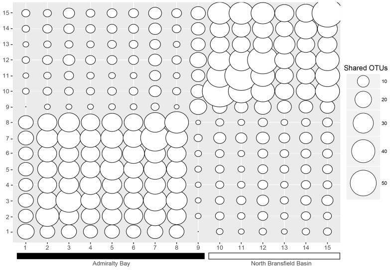
When analyzing the shared OTUs, it was found that more OTUs were shared within samples inside the bay versus samples outside the bay (Figure 3), except for station 9. Stations 2–8 shared more OTUs among each other, with sharing percentages varying from 48.3 to 92.8%. Station 9, placed at the entrance of the bay, shared 7–20 OTUs with stations 1–8, and 41–53 OTUs with stations 10–15, corresponding to 11.7–33.3% and 68.3–88.3%, respectively, and showing as a transitional environment. It was also evident that stations 10–15 shared more OTUs between each other (42.6–88.3%) than with stations inside the bay (3.8–33.3%).
FIGURE 3.
Shared OTUs between the samples. The numbers on the x and y axes correspond to the sample numbers (1–15). The smaller the circles, the less OTUs are shared, and the bigger the circles, the more OTUs are shared.
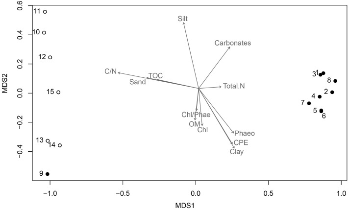
To determine the distribution of the bacterial community (beta-diversity), the relative abundance of the different phyla and proteobacterial classes were analyzed in relation to the sampling locations and the sediment physical-chemical parameters using nMDS. It was revealed a clear distinction between samples collected inside AB and in the NBB, where stations 10 and 15 were more distant, which was statistically supported by adonis analysis (r2 = 0.20, p = 0.002). Only station 9, which is a transitional sampling point located at the entrance of the bay, was more related to the second group. Gammaproteobacteria was the dominant taxon for all samples, but others influenced station 10 (Firmicutes, Actinobacteria, Alphaproteobacteria, and Deltaproteobacteria) and station 15 (Deltaproteobacteria and others).
The environmental factors associated with the sediment characteristics influenced the samples dissimilarity and the taxonomic composition. Sediments composed by clay, as well as relatively higher concentrations of phaeopigments were prevalent inside the bay and positively correlated with nMDS axis 1 (r = 0.62), whereas sediments constituted by sand and silt, and higher concentrations of total organic carbon and C/N ratio were predominant outside the bay (NBB), and negatively correlated with nMDS axis 1 (r = -0.98 for both parameters) (Table 1, Figure 4).
FIGURE 4.
Non-metric multidimensional scaling (nMDS) ordination. Black dots represent stations 1–9 (Admiralty Bay), and white dots are stations 10–15 (North Bransfield Basin). Each arrow is significantly correlated to the ordination (envfit, p < 0.05), and represents the direction and strength of the environmental gradient (Clay, Silt, Sand, Phaeo, Phaeopigments; Chl, Chlorophyll; CPE, Chloroplast Pigment Equivalent, Carbonate; OM, Organic Matter; TOC, Total Organic Carbon; Total N, Total Nitrogen; C/N, Carbon to Nitrogen ratio).
Discussion
The present study compared the bacterial diversity and community structure found in marine sediments of AB and NBB. Although similar taxonomic groups were identified, mostly related to heterotrophic metabolism, their relative abundances, as well as alpha-diversity values and bacterial community structure have differed between the two sampling regions, possibly reflecting the distinct physical-chemical characteristics of the sediments (e.g., grain size, organic matter, chlorophyll, carbonate percentages, carbon and nitrogen concentrations). Additionally, hydro-oceanographic conditions such as bathymetry, regional water circulation and winds regime, continental inputs from glaciers melting, contribution of primary producers, seasonal entrance of water masses, and turbidity, might have favored the organic matter supply in the area. Indeed, a combination of physical and chemical parameters can influence the marine sediment community structure (e.g., Austen et al., 2002; Schauer et al., 2010; Zinger et al., 2011; Bienhold et al., 2012; Nguyen and Landfald, 2015; Learman et al., 2016).
The most abundant phylum (Proteobacteria) was represented by the classes Gamma-, Alpha- and Deltaproteobacteria in different proportions between the samples. This supports the results from previous studies showing that this phylum has a wide phylogenetic and phenotypic diversity in several marine and benthic environments (Sogin et al., 2006; Danovaro et al., 2010; Williams et al., 2010; Zinger et al., 2011; Lyra et al., 2013; Sun et al., 2013). Similarly, Proteobacteria has been reported from sediments of Antarctic Polar Front (Ruff et al., 2014), Ross Sea (Baldi et al., 2010) and Western Antarctic Peninsula (Learman et al., 2016).
The report of Psychrobacter as the dominant genus of Gammaproteobacteria in Antarctic sediments has not been previously reported. These heterotrophic versatile microorganisms include piezophilic and halophilic species (Kawasaki et al., 2002; Nogi et al., 2002), found in shallow and deep-sea sediments, in the water column, as part of the fish and krill microbiomes, and in association with brown macroalgae (Bozal et al., 2003; Lee et al., 2006; Dang et al., 2008; Teske et al., 2011; Tropeano et al., 2012). Most species of this genus can adapt to cold conditions, such as polar permafrost and ice, and are capable of reproducing at temperatures ranging from -10°C to 40°C (Rodrigues et al., 2009). They often produce low temperature-adaptive lipases (Bozal et al., 2003; Yumoto et al., 2003) and play essential roles in fat decomposition reactions (Li et al., 2013). The polar environment therefore constitutes an ecological niche for Psychrobacter strains (Bozal et al., 2003).
Alphaproteobacterial sequences affiliated with Loktanella, Litorimicrobium and Hoefla, which play a common role in the nitrogen cycle, were present in all samples, although in higher relative abundances outside the bay. They are able to reduce nitrate to nitrite, degrade aromatic compounds, and oxidize sulfur, ammonia, carbon monoxide, iron and manganese (Van Trappen et al., 2004a; Ivanova et al., 2005; Jin et al., 2011; Jung et al., 2013).
Flavobacteria, generally a major clade of Bacteroidetes in marine environments, was the third most abundant class. The clade was mainly represented by the genus Gillisia, specifically in samples 15 (4.1%) and 13 (3.4%), both located in NBB. Several strains have previously been isolated from marine and polar environments, such as Antarctic lakes (Van Trappen et al., 2004b). These heterotrophs are especially important, as they break down complex organic matter using exoenzymes to degrade algal cells and algal-detrital particles (Kirchman, 2002; Gómez-Pereira et al., 2012; Teeling et al., 2012). In fact, the presence of such distinct taxa in particular at stations placed outside the bay, indicated by alpha-diversity and community composition, helps to explain the differences in bacterial community between the two study areas.
The microbial community composition found in AB and NBB showed the prevalence of microorganisms related to heterotrophic metabolism. High relative abundance of heterotrophs can be explained by the high input of organic matter from different sources. The phytodetritus in deep sediments are allochthonous and can be derived from microphytobenthos, abundant in shallow areas of AB, or deposition of phytoplankton and macroalgae fragments (Gheller, 2014). Macroalgae can cover about 30% of the seabed surface of AB, and produce ca. 74,000 tons of wet biomass (Zielinski, 1990; Nedzarek and Rakusa-Suszczewski, 2004), resulting in substrates that favor the presence of these heterotrophs.
Phytoplankton blooms can also contribute to the transfer of organic matter to the seafloor. They normally occur in the study area during the austral summer due to the advection of continental input of ice melts (Nedzarek, 2008) and coastal upwelling (Brandini and Rebello, 1994; Schloss et al., 2002). This rapidly transported phytoplankton-derived organic carbon will be recycled by heterotrophic microbes fueling a diverse microbial community in deep sediments, as previously shown by Ruff et al. (2014). Moreover, the system of currents in the study area, influenced by the warmer and less saline waters from Bellingshausen Sea and the cold and saline waters from Weddell, may also indirectly influence microbial composition. The currents within the bay are more intense when compared to those in the inlets (Gordon and Nowlin, 1978; Huneke et al., 2016), thus enabling the transport, deposition and homogenization of the organic matter that drives the heterotrophic bacterial community.
Conclusion
The next generation DNA sequencing data of 15 samples of seafloor sediments provides the first results characterizing the sediment microbial communities of the Northwestern Antarctic Peninsula, in a transect from AB to the NBB. Our study revealed high prevalence of heterotrophic gammaproteobacterial phylotypes, and differences in bacterial diversity and community structure between the two sites. A combination of conditions that favor the organic matter input, like regional water circulation and winds regime, bathymetry, continental influence from glaciers melting, inputs from primary producers of the euphotic zone or continental areas, water masses carrying nutrients, besides the grain size and sediment characteristics (organic matter, carbon and nitrogen concentrations), may contribute to shape the marine sediment communities in Antarctica.
Author Contributions
DF, RD, and CS analyzed data and prepared figures and tables. All authors contributed to writing the paper.
Conflict of Interest Statement
The authors declare that the research was conducted in the absence of any commercial or financial relationships that could be construed as a potential conflict of interest.
Acknowledgments
We thank the Brazilian Antarctic Program (PROANTAR). We also thank Dr. Paula F. Gheller and Dr. Thaïs Corbisier for sharing the environmental data. This work was supported by the Brazilian National Council for Scientific and Technological Development – CNPq (MABIREH/IPY/CAML Project n. 520293/2006-1). DF was supported by the CAPES-Master‘s fellowship.
References
- Austen M. C., Lambshead P. J. D., Hutchings P. A., Boucher G., Snelgrove P. V. R., Heip C., et al. (2002). Biodiversity links above and below the marine sediment-water interface that may influence community stability. Biodivers. Conserv. 11 113–136. 10.1023/A:1014098917535 [DOI] [Google Scholar]
- Azam F., Malfatti F. (2007). Microbial structuring of marine ecosystems. Nat. Rev. Microbiol. 5 782–791. 10.1038/nrmicro1747 [DOI] [PubMed] [Google Scholar]
- Baldi F., Marchetto D., Pini F., Fani R., Michaud L., Giudice A. L., et al. (2010). Biochemical and microbial features of shallow marine sediments along the Terra Nova Bay (Ross Sea, Antarctica). Cont. Shelf Res. 30 1614–1625. 10.1016/j.csr.2010.06.009 [DOI] [Google Scholar]
- Bienhold C., Boetius A., Ramette A. (2012). The energy-diversity relationship of complex bacterial communities in Arctic deep-sea sediments. ISME J. 6 724–732. 10.1038/ismej.2011.140 [DOI] [PMC free article] [PubMed] [Google Scholar]
- Bozal N., Montes M. J., Tudela E., Guinea J. (2003). Characterization of several Psychrobacter strains isolated from Antarctic environments and description of Psychrobacter luti sp. nov. and Psychrobacter fozii sp. nov. Int. J. Syst. Evol. Microbiol. 53 1093–1100. 10.1099/ijs.0.02457-0 [DOI] [PubMed] [Google Scholar]
- Brandini F. P., Rebello J. (1994). Wind field effect on hydrography and chlorophyll dynamics in the coastal pelagial of Admiralty Bay. King George Island, Antarctica. Antarct. Sci. 6 433–442. [Google Scholar]
- Carr S. A., Vogel S. W., Dunbar R. B., Brandes J., Spear J. R., Levy R., et al. (2013). Bacterial abundance and composition in marine sediments beneath the Ross Ice Shelf, Antarctica. Geobiology 11 377–395. 10.1111/gbi.12042 [DOI] [PubMed] [Google Scholar]
- Clarke A., Murphy E. J., Meredith M. P., King J. C., Peck L. S., Barnes D. K., et al. (2007). Climate change and the marine ecosystem of the western Antarctic Peninsula. Philos. Trans. R. Soc. Lond. B Biol. Sci. 362 149–166. 10.1098/rstb.2006.1958 [DOI] [PMC free article] [PubMed] [Google Scholar]
- Dang H., Zhu H., Wang J., Li T. (2008). Extracellular hydrolytic enzyme screening of culturable heterotrophic bacteria from deep-sea sediments of the Southern Okinawa Trough. World J. Microbiol. Biotechnol. 25 71–79. 10.1007/s11274-008-9865-5 [DOI] [Google Scholar]
- Danovaro R., Company J. B., Corinaldesi C., D’Onghia G., Galil B., Gambi C., et al. (2010). Deep-sea biodiversity in the Mediterranean Sea: the known, the unknown, and the unknowable. PLoS ONE 5:e11832 10.1371/journal.pone.0011832 [DOI] [PMC free article] [PubMed] [Google Scholar]
- Ducklow H. W., Fraser W., Karl D. M., Quetin L. B., Ross R. M., Smith R. C., et al. (2006). Water-column processes in the West Antarctic Peninsula and the Ross Sea: interannual variations and foodweb structure. Deep Sea Res. II 53 834–852. 10.1016/j.dsr2.2006.02.009 [DOI] [Google Scholar]
- Folk R. L., Ward W. C. (1957). Brazos River Bar: a study in the significance of grain size parameters. J. Sediment. Petrol. 27 3–27. 10.1306/74D70646-2B21-11D7-8648000102C1865D [DOI] [Google Scholar]
- Fuhrman J. A. (2012). Metagenomics and its connection to microbial community organization. F1000 Biol. Rep. 4:15 10.3410/B4-15 [DOI] [PMC free article] [PubMed] [Google Scholar]
- Gheller P. F. (2014). A Meiofauna da baía do Almirantado e Áreas Adjacentes no Estreito de Bransfield (Antártica), com Ênfase em Nematoda. Ph.D. Tese de Doutorado, Instituto Oceanográfico, Universidade de São Paulo, São Paulo; 10.11606/T.21.2014.tde-09042015-145736 [DOI] [Google Scholar]
- Gillies C. L., Stark J. S., Johnstone G. J., Smith S. D. A. (2012). Carbon flow and trophic structure of an Antarctic coastal benthic community as determined by d13C and d15N. East Coast Shelf Sci. 97 44–57. 10.1016/j.ecss.2011.11.003 [DOI] [Google Scholar]
- Gómez-Pereira P. R., Schüler M., Fuchs B. M., Bennke C., Teeling H., Waldmann J., et al. (2012). Genomic content of uncultured Bacteroidetes from contrasting oceanic provinces in the North Atlantic Ocean. Environ. Microbiol. 14 52–66. 10.1111/j.1462-2920.2011.02555.x [DOI] [PubMed] [Google Scholar]
- Gordon A. L., Nowlin W. D., Jr. (1978). The basin waters of the bransfield strait. J. Phys. Oceanogr. 8 258–264. [DOI] [Google Scholar]
- Huneke W. G. C., Huhn O., Schröeder M. (2016). Water masses in the bransfield strait and adjacent seas, austral summer 2013. Polar Biol. 39 789–798. 10.1007/s00300-016-1936-8 [DOI] [Google Scholar]
- Ivanova E. P., Zhukova N. V., Lysenko A. M., Gorshkova N. M., Sergeev A. F., Mikhailov V. V., et al. (2005). Loktanella agnita sp. nov. and Loktanella rosea sp. nov., from the north-west Pacific Ocean. Int. J. Syst. Evol. Microbiol. 55 2203–2207. 10.1099/ijs.0.63461-0 [DOI] [PubMed] [Google Scholar]
- Jin H. M., Lee H. J., Kim J. M., Park M. S., Lee K., Jeon C. O. (2011). Litorimicrobium taeanense gen. nov., sp. nov., isolated from a sandy beach. Int. J. Syst. Evol. Microbiol. 61 1392–1396. 10.1099/ijs.0.025007-0 [DOI] [PubMed] [Google Scholar]
- Jung M. Y., Shin K. S., Kim S., Kim S. J., Park S. J., Kim J. G., et al. (2013). Hoeflea halophila sp. nov., a novel bacterium isolated from marine sediment of the East Sea, Korea. Antonie Leeuwenhoek 103 971–978. 10.1007/s10482-013-9876-6 [DOI] [PubMed] [Google Scholar]
- Kawasaki K., Nogi Y., Hishinuma M., Nodasaka Y., Matsuyama H., Yumoto I. (2002). Psychromonas marina sp. nov., a novel halophilic, facultatively psychrophilic bacterium isolated from the coast of the Okhotsk Sea. Int. J. Syst. Evol. Microbiol. 52 1455–1459. 10.1099/00207713-52-5-1455 [DOI] [PubMed] [Google Scholar]
- Kirchman D. L. (2002). The ecology of Cytophaga-Flavobacteria in aquatic environments. FEMS Microbiol. Ecol. 39 91–100. 10.1111/j.1574-6941.2002.tb00910.x [DOI] [PubMed] [Google Scholar]
- Learman D. R., Henson M. W., Thrash J. C., Temperton B., Brannock P. M., Santos S. R., et al. (2016). Biogeochemical and microbial variation across 5500 km of antarctic surface sediment implicates organic matter as a driver of benthic community structure. Front. Microbiol. 7:284 10.3389/fmicb.2016.00284 [DOI] [PMC free article] [PubMed] [Google Scholar]
- Lee Y. K., Jung H. J., Lee H. K. (2006). Marine bacteria associated with the Korean brown alga, Undaria pinnatifida. J. Microbiol. 694–708. [PubMed] [Google Scholar]
- Li A., Chu Y., Wang X., Ren L., Yu J., Liu X., et al. (2013). A pyrosequencing-based metagenomic study of methane-producing microbial community in solid-state biogas reactor. Biotechnol. Biofuels 6:3 10.1186/1754-6834-6-3 [DOI] [PMC free article] [PubMed] [Google Scholar]
- Lorenzen C. J. (1967). Determination of chlorophyll and phaeopigments: spectrophotometric equations. Limnol. Oceanogr. 12 343–346. 10.4319/lo.1967.12.2.0343 [DOI] [Google Scholar]
- Love M. I., Huber W., Anders S. (2014). Moderated estimation of fold change and dispersion for RNA-seq data with DESeq2. Genome Biol. 15:550 10.1186/s13059-014-0550-8 [DOI] [PMC free article] [PubMed] [Google Scholar]
- Lyra C., Sinkko H., Rantanen M., Paulin L., Kotilainen A. (2013). Sediment bacterial communities reflect the history of a Sea Basin. PLoS ONE 8:e54326 10.1371/journal.pone.0054326 [DOI] [PMC free article] [PubMed] [Google Scholar]
- Meredith M. P., King J. C. (2005). Rapid climate change in the ocean west of the Antarctic Peninsula during the second half of the 20th century. Geophys. Res. Lett. 32. 10.1029/2005GL024042 [DOI] [Google Scholar]
- Montes-Hugo M., Doney S. C., Ducklow H. W., Fraser W., Martinson D., Stammerjohn S. E., et al. (2009). Recent changes in phytoplankton communities associated with rapid regional climate change along the western Antarctic Peninsula. Science 323 1470–1473. 10.1126/science.1164533 [DOI] [PubMed] [Google Scholar]
- Nedzarek A. (2008). Sources, diversity and circulation of biogenic compounds in Admiralty Bay, King George Island, Antarctica. Antarct. Sci. 20 135–145. 10.1017/S0954102007000909 [DOI] [Google Scholar]
- Nedzarek A., Rakusa-Suszczewski S. (2004). Decomposition of macroalgae and the release of nutrients in Admiralty Bay, King George Island, Antarctica. Polar Biosci. 17 16–35. [Google Scholar]
- Nguyen T. T., Landfald B. (2015). Polar front associated variation in prokaryotic community structure in Arctic shelf seafloor. Front. Microbiol. 6:17 10.3389/fmicb.2015.00017 [DOI] [PMC free article] [PubMed] [Google Scholar]
- Nogi Y., Kato C., Horikoshi K. (2002). Psychromonas kaikoae sp. nov., a novel piezophilic bacterium from the deepest cold seep sediments in the Japan Trench. Int. J. Syst. Evol. Microbiol. 52 1527–1532. 10.1099/ijs.0.01911-0 [DOI] [PubMed] [Google Scholar]
- Oksanen J., Blanchet F. G., Kindt R., Legendre P., Minchin P. R., O’Hara R. B., et al. (2013). Vegan: Community Ecology Package. R package version 2.0-8. Available at: http://CRAN.R-project.org/package=vegan [Google Scholar]
- Orcutt B. N., Sylvan J. B., Knab N. J., Edwards K. J. (2011). Microbial ecology of the Dark Ocean above, at, and below the Seafloor. Microbiol. Mol. Biol. Rev. 75 361–422. 10.1128/MMBR.00039-10 [DOI] [PMC free article] [PubMed] [Google Scholar]
- Parkes R., Barry J., Cragg A., Wellsbury P. (2000). Recent studies on bacterial populations and processes in subseafloor sediments: a review. Hydrogeol. J. 8 11–28. 10.1007/PL00010971 [DOI] [Google Scholar]
- Pearson G. A., Lago-Leston A., Cánovas F., Cox C. J., Verret F., Lasternas S., et al. (2015). Metatranscriptomes reveal functional variation in diatom communities from the Antarctic Peninsula. ISME J. 9 2275–2289. 10.1038/ismej.2015.40 [DOI] [PMC free article] [PubMed] [Google Scholar]
- Pichlmaier M., Aquino F. E., Da-Silva C. S., Braun M. (2004). Suspended sediments in Admiralty Bay, King George Island (Antarctica). Pesq. Antárt. Bras. 4 77–85. [Google Scholar]
- Plante-Cuny M. R. (1978). Pigments photosynthétiques et production primaire dês fonds meubles néritiques d’une region tropicale (Nosy-Bé, Madagascar). J. Rech. Océanogr. 96 1–114. [Google Scholar]
- Rodrigues D. F., da C Jesus E., Ayala-del-Río H. L., Pellizari V. H., Gilichinsky D., Sepulveda-Torres L., et al. (2009). Biogeography of two cold-adapted genera: Psychrobacter and Exiguobacterium. ISME J. 3 658–665. 10.1038/ismej.2009.25 [DOI] [PubMed] [Google Scholar]
- Ruff S. E., Probandt D., Zinkann A. C., Iversen M. H., Klaas C., Würzberg L., et al. (2014). Indications for algae-degrading benthic microbial communities in deep-sea sediments along the Antarctic Polar Front. Deep Sea Res. Part II Top. Stud. Oceanogr. 108 6–16. 10.1016/j.dsr2.2014.05.011 [DOI] [Google Scholar]
- Schauer R., Bienhold C., Ramette A., Harder J. (2010). Bacterial diversity and biogeography in deep-sea surface sediments of the South Atlantic Ocean. ISME J. 4 159–170. 10.1038/ismej.2009.106.? [DOI] [PubMed] [Google Scholar]
- Schloss I. R., Ferreyra G. A., Ruiz-Pino D. (2002). Phytoplankton biomass in Antarctic shelf zones: a conceptual model based on Potter Cove, King George Island. J. Marine Syst. 36 129–143. 10.1016/S0924-7963(02)00183-5 [DOI] [Google Scholar]
- Schloss P. D., Gevers D., Westcott S. L. (2011). Reducing the effects of PCR amplification and sequencing artifacts on 16S rRNA-based studies. PLoS ONE 6:e27310 10.1371/journal.pone.0027310 [DOI] [PMC free article] [PubMed] [Google Scholar]
- Sogin M. L., Morrison H. G., Huber J. A., Welch D. M., Huse S. M., Neal P. R., et al. (2006). Microbial diversity in the deep sea and the underexplored “rare biosphere.” Proc. Natl. Acad. Sci. U.S.A. 103 12115–12120. 10.1073/pnas.0605127103 [DOI] [PMC free article] [PubMed] [Google Scholar]
- Suguio K. (1973). Introdução à Sedimentologia. São Paulo: Edgard Bluecher, Editora da Universidade de São Paulo, 317. [Google Scholar]
- Sun M. Y., Dafforn K. A., Johnston E. L., Brown M. V. (2013). Core sediment bacteria drive community response to anthropogenic contamination over multiple environmental gradients. Environ. Microbiol. 15 2517–2531. 10.1111/1462-2920.12133 [DOI] [PubMed] [Google Scholar]
- Szafrański Z., Lipski M. (1982). Characteristics of water temperature and salinity at Admiralty Bay (King George Island, South Shetland Islands, Antarctica) during austral summer 1978/1979. Pol. Polar Res. 3 7–24. [Google Scholar]
- Teeling H., Fuchs B. M., Becher D., Klockow C., Gardebrecht A., Bennke C. M., et al. (2012). Substrate-controlled succession of marine bacterioplankton populations induced by a phytoplankton bloom. Science 336 608–611. 10.1126/science.1218344 [DOI] [PubMed] [Google Scholar]
- Teske A., Durbin A., Ziervogel K., Cox C., Arnosti C. (2011). Microbial community composition and function in permanently cold seawater and sediments from an arctic fjord of svalbard. Appl. Environ. Microbiol. 77 2008–2018. 10.1128/AEM.01507-10 [DOI] [PMC free article] [PubMed] [Google Scholar]
- Tokarczyk R. (1987). Classification of water masses in the Bransfield Strait and Southern part of the Drake Passage using a method of statistical multidimensional analysis. Pol. Polar Res. 8 333–366. [Google Scholar]
- Tropeano M., Coria S., Turjanski A., Cicero D., Bercovich A., Mac Cormack W., et al. (2012). Culturable heterotrophic bacteria from potter cove, Antarctica, and their hydrolytic enzymes production. Polar Res. 31 18507–18517 10.3402/polar.v31i0.18507 [DOI] [Google Scholar]
- Van Trappen S., Mergaert J., Swings J. (2004a). Loktanella salsilacus gen. nov., sp. nov., Loktanella fryxellensis sp. nov. and Loktanella vestfoldensis sp. nov., new members of the Rhodobacter group, isolated from microbial mats in Antarctic lakes. Int. J. Syst. Evol. Microbiol. 54 1263–1269. 10.1099/ijs.0.03006-0 [DOI] [PubMed] [Google Scholar]
- Van Trappen S., Vandecandelaere I., Mergaert J., Swings J. (2004b). Gillisia limnaea gen. nov., sp. nov., a new member of the family Flavobacteriaceae isolated from a microbial mat in Lake Fryxell, Antartica. Int. J. Syst. Evol. Microbiol. 54 445–448. 10.1099/ijs.0.02922-0 [DOI] [PubMed] [Google Scholar]
- Wang Q., Garrity G. M., Tiedje J. M., Cole J. R. (2007). Naïve Bayesian classifier for rapid assignment of rRNA sequences into the new bacterial taxonomy. Appl. Environ. Microbiol. 73 5261–5267. 10.1128/AEM.00062-07 [DOI] [PMC free article] [PubMed] [Google Scholar]
- Wang Y., Qian P.-Y. (2009). Conservative fragments in bacterial 16S rRNA genes and primer design for 16S ribosomal DNA amplicons in metagenomic studies. PLoS ONE 4:e7401 10.1371/journal.pone.0007401 [DOI] [PMC free article] [PubMed] [Google Scholar]
- Wickham H. (2009). ggplot2: Elegant Graphics for Data Analysis. New York, NY: Springer-Verlag. [Google Scholar]
- Williams K. P., Gillespie J. J., Sobral B. W. S., Nordberg E. K., Snyder E. E., Shallom J. M., et al. (2010). Phylogeny of Gammaproteobacteria. J. Bacteriol. 192 2305–2314. 10.1128/JB.01480-09 [DOI] [PMC free article] [PubMed] [Google Scholar]
- Yumoto I., Hirota K., Sogabe Y., Nodasaka Y., Yokota Y., Hoshino T. (2003). Psychrobacter okhotskensis sp nov., a lipase-producing facultative psychrophile isolated from the coast of the Okhotsk Sea. Int. J. Syst. Evol. Microbiol. 53 1985–1989. 10.1099/ijs.0.02686-0 [DOI] [PubMed] [Google Scholar]
- Zielinski K. (1990). Bottom macroalgae of admiralty bay (King George Island, South Shetland Islands, Antarctic). Dep. Polar Res. 11 95–131. [Google Scholar]
- Zinger L., Amaral-Zettler L. A., Fuhrman J. A., Horner-Devine M. C., Huse S. M., Welch D. B., et al. (2011). Global patterns of bacterial beta-diversity in seafloor and seawater ecosystems. PLoS ONE 6:e24570 10.1371/journal.pone.0024570 [DOI] [PMC free article] [PubMed] [Google Scholar]


