Abstract
Introduction:
Osteoporosis is a significant health-care problem characterized by excessive skeletal fragility, susceptibility to low-trauma fractures in men as well as women. Any abnormality of the bone that reduces the strength of the bone predisposes it to mechanical failure during normal activity or with minimum trauma. The mechanical failure manifests itself as a fracture, and this fracture must be recognized as a pathological fracture if the patient is to be treated properly. Osteoporosis is one of the leading causes of such pathological fractures and accounts for 1.5 million fractures annually. In the following case report, we present a 56-year-old postmenopausal female patient with bilateral pathological subtrochanteric fracture femurii due to intake of bisphosponates for 4 years for osteoporosis. Bilateral pathological subtrochanteric femurii fractures are extremely uncommon injuries which occur in adults who sustain injuries due to trivial trauma. A variety of management modalities has been tried to treat this complex fracture pattern. Standard fixation treatment is intramedullary nailing.
Case Report:
A postmenopausal female of rheumatoid arthritis aged 56 years, presented to our emergency department with a history of trivial fall at home. Following the fall, she was unable to bear weight on bilateral feet and complained of deformity. History revealed consumption of bisphosphonates (tablet alendronate 10 mg) for the last 4 years and glucocorticoids for rheumatiod arthritis. Radiographs were taken, which revealed bilateral pathological subtrochanteric fracture femurii. After obtaining necessary fitness, the patient was taken up for surgery. Closed reduction and Internal fixation with long proximal femoral nail were done. Bisphosphonate intake was stopped and teriparatide 20 µg/day subcutaneously given for 3 months. Fracture healed after 3 months and patient resumed her daily activities.
Conclusion:
In people taking long-term bisphoshponate therapy, symptomatic cortical stress reactions accompanied by evidence of a stress line across the cortical thickening suggest an increased risk of a complete stress fracture. The patients should be informed about the prodromal symptoms like pain and swelling, following this bisphosphonates should be stopped and teriparatide has to be started for osteoporosis.
Keywords: Osteoporotic fracture, trivial trauma, bisphosphonates, glucocorticosteroids, long proximal femoral nail, stress fracture
What to Learn from this Article?
Long-term treatment with alendronate causes atypical femoral fractures and it is recommended that orthopedic surgeons remain vigilant in assessing their patients treated with alendronate (bisphosphonates) for osteoporosis. In such cases, alendronate should be stopped and teriparatide started.
Introduction
Treatment of osteoporosis by Bisphosphonates for 3-4 years is shown to decrease spine, hip and other nonvertebral fracture risk, increase bone mineral density (BMD) and to decrease bone remodeling rate [1, 2, 3, 4, 5, 6]. Treatment with bisphosphonates significantly reduces the risk of fractures in men and women with osteoporosis. The evidence is based on high-quality Phase III randomized controlled trials with fracture as an end point.
The benefit of bisphosphonates also extends to other disorders of bone metabolism such as glucocorticoid-induced osteoporosis, Paget’s disease, and bone metastases. The number of atypical subtrochanteric fractures in association with bisphosphonate is an estimated one per 1,000 per year. It is recommended that physicians remain vigilant in assessing their patients treated with bisphosphonates for the treatment or prevention of osteoporosis and advice patients of the potential risks. Minor features were that fractures were commonly preceded by prodromal pain and, on radiographs, there appeared breaking of the cortex on one side and bilateral thickened diaphyseal cortices. This fracture pattern has often been referred to as an “atypical subtrochanteric fracture.” It is worth noting that, on radiography, the appearance of atypical subtrochanteric fractures is similar to that of stress fractures, including a periosteal reaction, linear areas of bone sclerosis and a transverse fracture line. Prodromal pain before diagnosis is also common.
Case Report
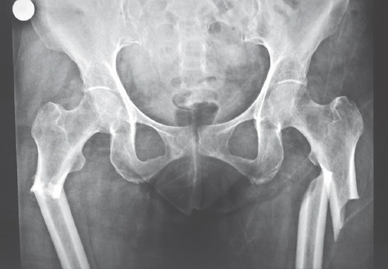
A postmenopausal female of rheumatoid arthritis aged 56 years, presented to our emergency department with a history of trivial fall at home. Following the fall, she was unable to bear weight on bilateral feet and complained of deformity. History revealed consumption of bisphosphonates (tablet alendronate 10 mg) for the last 4 years and glucocorticoids for rheumatiod arthritis. Radiographs were taken, which revealed bilateral pathological subtrochanteric fracture femurii (Fig. 1). After obtaining necessary fitness, the patient was taken up for surgery. Closed reduction and internal fixation with long proximal femoral nail were done (Fig. 2). Bisphosphonate intake was stopped and teriparatide 20 µg/day subcutaneously given for 3 months. Fracture healed after 3 months and patient resumed her daily activities.
Figure 1.

Pre-operative radiograph.
Figure 2.

Post-operative radiograph.
Discussion
Osteoporosis is defined as a disease of the bone characterized by reduced mass of the bone. When bone mass falls below the required level for mechanical support, fracture occurs. Osteoporosis is a condition which until recently thought of as incurable, at best a preventable illness. Today, it is not only preventable but also treatable [7]. It is also an extremely common illness worldwide. It is next only to hypertension, and diabetes. Its frequency is common particularly in postmenopausal women. Osteoporosis is responsible for 1.5 million fractures annually. Among them, more than half a million are vertebral fractures. 3, 00,000 are hip fractures. 2,00,000 wrist fractures. 3,00,000 fractures of other bones. Approximately, 37,000 people die each year from complications related to fractures caused by osteoporosis [8, 9]. 70% of affected people have genetic predisposition that includes how a person responds to external stress causing factors. Estrogen deficiency patients - women who have had early menopause, those who had their uterus and ovaries removed surgically at an age much earlier than that of normal menopause.
Bisphosphonates are very commonly used for the treatment of osteoporosis. The pathophysiology of atypical low-trauma subtrochanteric fractures following bisphosphonate use is not known. However, pre-clinical and clinical studies of the effects of bisphosphonates on bone suggest that there are several possible mechanisms that work alone or in tandem. The organic matrix of the bone determines its toughness, and this matrix is partly made up of bone collagen, which impacts on the bone’s mechanical properties. Bisphosphonates use may negatively affect collagen by preventing or reducing its maturation [10, 11, 12], although this finding has not been consistently replicated [13]. Bisphosphonates may also affect bone mineralization density distribution. The more heterogeneous the BMDD, the slower that cracks in the bone will develop and lower the risk of new cracks and fracture forming [14]. As bisphosphonate treatment reduces bone turnover, the increase in overall mineralization leads to more homogenous bone - As evidenced by a narrow BMDD [15, 16] and thus an increased risk of cracks and fractures. Reduced bone turnover also increases the accumulation of microdamage, as cracks are not repaired, and reduces bone toughness, which contributes to the increased susceptibility of bone to new cracks [17, 18, 19]. Finally, bisphosphonates have differentiating impacts on different type of fractures. Acute fractures of long bone are not affected by bisphosphonates in the initial healing stages [20, 21, 22], as they heal via endochondral ossification. However, stress fractures heal by normal bone remodeling, and thus, bisphoshphonates may prevent or delay healing, increasing the likelihood of a complete fracture with little or no trauma. Several reports have reported on bone quality in people with low trauma fractures taking bisphosphonate therapy. Our patient is a case of rheumatoid arthritis on the treatment of corticosteroid, the patient then developed generalized osteoporosis. It is questionable whether osteoporosis has developed due to rheumatoid arthritis or is corticosteroid-induced.
Furthermore, the patient was on bisphosphonates, i.e., alendronate for a period of 4-year and had a bilateral sub trochanteric atypical fracture, but literature reports that bisphosphonate induced fractures usually occur after a period of 5-year of intake.
Conclusion
In people taking long-term bisphoshponate therapy, symptomatic cortical stress reactions accompanied by evidence of a stress line across the cortical thickening suggest an increased risk of a complete stress fracture. The patients should be informed about the prodromal symptoms like pain and swelling, following this bisphosphonates should be stopped and teriparatide has to be started for osteoporosis.
Clinical Message.
A sense of proportion may be helpful in alleviating the concerns of the medical community. A plausible scenario is that long-term exposure to bisphosphonates (more than 5 years) increases the risk of subtrochanteric femoral fractures 2 fold. Thus, the available evidence does not suggest that the well-known benefits of bisphosphonates are outweighed by the risk of these rare, atypical, and low-trauma subtrochanteric fractures. Nevertheless, it is recommended that physicians remain vigilant in assessing their patients treated with bisphosphonates for osteoporosis or associated conditions. They should continue to follow the recommendations on the drug label when prescribing bisphosphonates and advise patients of the potential risks. Patients with pain in the hips, thighs or femur should be radiologically assessed and, where a stress fracture is evident, the physician should decide whether bisphosphonate therapy should be discontinued pending a full evaluation, based on an individual benefit-risk assessment. The radiographic changes should be evaluated for orthopedic intervention - since surgery prior to fracture completion might be advantageous- or to be closely monitored. There are hardly any studies mentioning alendronate as a cause of fractures.
Alendronate now includes a special warning/precaution, advising discontinuation of bisphosphonate therapy in patients with stress fractures pending evaluation, based on an individual benefit-risk assessment. Alendronate is the only bisphosphonate for osteoporosis treatment that carries this warning [23]. Long-term therapy with bisphosphonates can be complicated by atypical femoral fractures. The purpose of this study is to demonstrate an association between alendronate use and specific pattern of low energy subtrochanteric femur fractures. The previous studies show conflicting results regarding the possible excess risk of atypical fractures of subtrochanteric associated with alendronate use. A curative effect of teriparatide has been suggested [24].
Biography


Footnotes
Conflict of Interest: Nil
Source of Support: None
References
- 1.Bartucci EJ, Gonzalez MH, Cooperman DR, Freedberg HI, Barmada R, Laros GS. The effect of adjunctive methylmethacrylate on failures of fixation and function in patients with intertrochanteric fractures and osteoporosis. J Bone Joint Surg Am. 1985;67(7):1094–1107. [PubMed] [Google Scholar]
- 2.Dobozi WR, Dvonch VM, Saltzman ML, Beigler DF, Belich P. Treatment of impending pathological fractures of the femur with flexible intramedullary nails. Orthopedics. 1984;7(11):1682–1688. doi: 10.3928/0147-7447-19841101-05. [DOI] [PubMed] [Google Scholar]
- 3.Douglass HO, Jr, Shukla SK, Mindell E. Treatment of pathological fractures of long bones excluding those due to breast cancer. J Bone Joint Surg Am. 1976;58(8):1055–1061. [PubMed] [Google Scholar]
- 4.Fidler M. Incidence of fracture through metastases in long bones. Acta Orthop Scand. 1981;52(6):623–627. doi: 10.3109/17453678108992157. [DOI] [PubMed] [Google Scholar]
- 5.Harrington KD. Problems of pathological fractures. Complications Orthop. 1987;2(1):4. [Google Scholar]
- 6.Heisterberg L, Johansen TS. Treatment of pathological fractures. Acta Orthop Scand. 1979;50(6 Pt 2):787–790. doi: 10.3109/17453677908991310. [DOI] [PubMed] [Google Scholar]
- 7.National Institutes of Health. National Institute of Arthritis and Musculoskeletal and Skin Diseases. Washington, DC: National Institutes of Health; 1994. [Google Scholar]
- 8.Ray NF, Chan JK, Thamer M, Melton LJ., 3rd Medical expenditures for the treatment of osteoporotic fractures in the United States in 1995: Report from the National Osteoporosis Foundation. J Bone Miner Res. 1997;12(1):24–35. doi: 10.1359/jbmr.1997.12.1.24. [DOI] [PubMed] [Google Scholar]
- 9.Cummings SR, Black DM, Thompson DE, Applegate WB, Barrett-Connor E, Musliner TA, et al. Effect of alendronate on risk of fracture in women with low bone density but without vertebral fractures:Results from the Fracture Intervention Trial. JAMA. 1998;280(24):2077–2082. doi: 10.1001/jama.280.24.2077. [DOI] [PubMed] [Google Scholar]
- 10.Merck Sharp & Dohme Limited. Fosavance Summary of Product Characteristics. Hertforshire: Merck Sharp & Dohme; 2009. [Google Scholar]
- 11.Merck Sharp & Dohme Limited. Fosavance Summary of Product Characteristics. Hertforshire: Merck Sharp & Dohme; 2010. [Google Scholar]
- 12.Durchschlag E, Paschalis EP, Zoehrer R, Roschger P, Fratzl P, Recker R, et al. Bone material properties in trabecular bone from human iliac crest biopsies after 3- and 5-year treatment with risedronate. J Bone Miner Res. 2006;21(10):1581–1590. doi: 10.1359/jbmr.060701. [DOI] [PubMed] [Google Scholar]
- 13.Boskey AL, Spevak L, Weinstein RS. Spectroscopic markers of bone quality in alendronate-treated postmenopausal women. Osteoporos Int. 2009;20(5):793–800. doi: 10.1007/s00198-008-0725-9. [DOI] [PMC free article] [PubMed] [Google Scholar]
- 14.Turner CH, Burr DB. Principles of bone biomechanics. In: Lane NE, Sambrook PN, editors. Osteoporosis and the Osteoporosis of Rheumatic Disease. Philadelphia, PA: Mosby Elseivier; 2006. pp. 41–53. [Google Scholar]
- 15.Boivin GY, Chavassieux PM, Santora AC, Yates J, Meunier PJ. Alendronate increases bone strength by increasing the mean degree of mineralization of bone tissue in osteoporotic women. Bone. 2000;27(5):687–694. doi: 10.1016/s8756-3282(00)00376-8. [DOI] [PubMed] [Google Scholar]
- 16.Roschger P, Rinnerthaler S, Yates J, Rodan GA, Fratzl P, Klaushofer K. Alendronate increases degree and uniformity of mineralization in cancellous bone and decreases the porosity in cortical bone of osteoporotic women. Bone. 2001;29(2):185–191. doi: 10.1016/s8756-3282(01)00485-9. [DOI] [PubMed] [Google Scholar]
- 17.Allen MR, Burr DB. Three years of alendronate treatment results in similar levels of vertebral microdamage as after one year of treatment. J Bone Miner Res. 2007;22(11):1759–1765. doi: 10.1359/jbmr.070720. [DOI] [PubMed] [Google Scholar]
- 18.Allen MR, Iwata K, Phipps R, Burr DB. Alterations in canine vertebral bone turnover, microdamage accumulation, and biomechanical properties following 1-year treatment with clinical treatment doses of risedronate or alendronate. Bone. 2006;39(4):872–879. doi: 10.1016/j.bone.2006.04.028. [DOI] [PubMed] [Google Scholar]
- 19.Allen MR, Reinwald S, Burr DB. Alendronate reduces bone toughness of ribs without significantly increasing microdamage accumulation in dogs following 3 years of daily treatment. Calcif Tissue Int. 2008;82(5):354–360. doi: 10.1007/s00223-008-9131-8. [DOI] [PMC free article] [PubMed] [Google Scholar]
- 20.Iwata K, Allen MR, Phipps R, Burr DB. Microcrack initiation occurs more easily in vertebrae from beagles treated with alendronate than with risedronate. Bone. 2006;38(Suppl):42. [Google Scholar]
- 21.MacDonald MM, Schindeler A, Little DG. Bisphosphonate treatment and fracture repair. Bonekey Osteovision. 2007;4:236–251. [Google Scholar]
- 22.Martinez MD, Schmid GJ, McKenzie JA, Ornitz DM, Silva MJ. Healing of non-displaced fractures produced by fatigue loading of the mouse ulna. Bone. 2010;46(6):1604–1612. doi: 10.1016/j.bone.2010.02.030. [DOI] [PMC free article] [PubMed] [Google Scholar]
- 23.Rizzoli R, Akesson K, Bouxsein M, Kanis JA, Napoli N, Papapoulos S, et al. Sub-trochanteric fractures after log-term treatment with bisphosphonates:A European society on clinical and economic aspects of osteoporosis and osteoarthritis, and international osteoporosis foundation Working Group Report. Osteoporos Int. 2011;22(2):373–390. doi: 10.1007/s00198-010-1453-5. [DOI] [PMC free article] [PubMed] [Google Scholar]
- 24.Reid DM, Devogelaer JP, Saag K, Roux C, Lau CS, Reginster JY, et al. Zoledronic acid and risedronate in the prevention and treatment of glucocorticoid-induced osteoporosis (HORIZON):A multicentre, double-blind, double-dummy, randomised controlled trial. Lancet. 2009;373:1253–63. doi: 10.1016/S0140-6736(09)60250-6. [DOI] [PubMed] [Google Scholar]


