Abstract
BACKGROUND
The use of wide pore lightweight polypropylene mesh to improve anatomical outcomes in the surgical repair of prolapse has been hampered by mesh complications. One of the prototype prolapse meshes has been found to negatively impact the vagina by inducing a decrease in smooth muscle volume and contractility and the degradation of key structural proteins (collagen and elastin), resulting in vaginal degeneration. Recently, bioscaffolds derived from extracellular matrix have been used to mediate tissue regeneration and have been widely adopted in tissue engineering applications.
OBJECTIVE
Here we aimed to: (1) define whether augmentation of a polypropylene prolapse mesh with an extracellular matrix regenerative graft in a primate sacrocolpopexy model could mitigate the degenerative changes; and (2) determine the impact of the extracellular matrix graft on vagina when implanted alone.
STUDY DESIGN
A polypropylene-extracellular matrix composite graft (n = 9) and a 6-layered extracellular matrix graft alone (n = 8) were implanted in 17 middle-aged parous rhesus macaques via sacrocolpopexy and compared to historical data obtained from sham (n = 12) and the polypropylene mesh (n = 12) implanted by the same method. Vaginal function was measured in passive (ball-burst test) and active (smooth muscle contractility) mechanical tests. Vaginal histomorphologic/ biochemical assessments included hematoxylin-eosin and trichrome staining, immunofluorescent labeling of α-smooth muscle actin and apoptotic cells, measurement of total collagen, collagen subtypes (ratio III/ I), mature elastin, and sulfated glycosaminoglycans. Statistical analyses included 1-way analysis of variance, Kruskal-Wallis, and appropriate posthoc tests.
RESULTS
The host inflammatory response in the composite mesh-implanted vagina was reduced compared to that following implantation with the polypropylene mesh alone. The increase in apoptotic cells observed with the polypropylene mesh was blunted in the composite (overall P < .001). Passive mechanical testing showed inferior parameters for both polypropylene mesh alone and the composite compared to sham whereas the contractility and thickness of smooth muscle layer in the composite were improved with a value similar to sham, which was distinct from the decreases observed with polypropylene mesh alone. Biochemically, the composite had similar mature elastin content, sulfated glycosaminoglycan content, and collagen subtype III/I ratio but lower total collagen content when compared to sham (P = .011). Multilayered extracellular matrix graft alone showed overall comparable values to sham in aspects of the biomechanical, histomorphologic, or biochemical end-points of the vagina. The increased collagen subtype ratio III/I with the extracellular matrix graft alone (P = .033 compared to sham) is consistent with an ongoing active remodeling response.
CONCLUSION
Mesh augmentation with a regenerative extracellular matrix graft attenuated the negative impact of polypropylene mesh on the vagina. Application of the extracellular matrix graft alone had no measurable negative effects suggesting that the benefits of this extra-cellular matrix graft occur when used without a permanent material. Future studies will focus on understanding mechanisms.
Keywords: apoptosis, extracellular matrix, function, graft, mechanical properties, pelvic organ prolapse, regeneration, rhesus macaque, smooth muscle, structure, synthetic mesh, vagina
Introduction
Lightweight polypropylene mesh used in the surgical repair of pelvic organ prolapse has been associated with significant mesh-related complications, most commonly mesh exposure and pain.1-4 Gynemesh PS (Ethicon, Sommersville, NJ), one of the most widely implanted polypropylene meshes, has been shown to induce a robust foreign-body response and degenerative changes in the vagina when implanted via sacrocolpopexy including a decrease in smooth muscle contractility and volume, a loss of mechanical integrity, a decrease in matrix structural proteins (collagen and elastin) accompanied by an increase in glycosaminoglycan (GAG) content, and an increase in the amount and activity of matrix-degrading proteases.5-8
In theory, these negative effects could be overcome with the use of regenerative biomaterials either in combination with Gynemesh PS as a composite graft or alone with the elimination of permanent materials. However, given that the sacrocolpopexy is not an anatomical surgery and regenerative matrices are designed to restore or repair injured tissues in a site-specific manner, it is not clear whether the use of a regenerative matrix in sacrocolpopexy surgery would be successful.
Products derived from extracellular matrices (ECM) have been widely adopted in tissue-engineering applications and are considered a novel tool to facilitate tissue regeneration. It has been shown that these grafts, when placed in the appropriate micro- and loading environments, can promote site-specific functional tissue remodeling by limiting destructive inflammatory responses, recruiting requisite cell populations including progenitor cells, and supporting cell attachment, proliferation, and matrix production.9,10
We chose to use MatriStem Surgical Matrix RS (MatriStem RS, ACell Inc, Columbia, MD), a noncross-linked degradable acellular porcine urinary bladder matrix, because it facilitates reconstructive remodeling of smooth muscle and connective tissue as shown in its application in cardiac defect repair, esophagus reinforcement, and chronic wound repairs.11-14 It is important to distinguish regenerative matrices such as MatriStem (noncross-linked) from biologic materials that have been chemically cross-linked (eg, Pelvicol, Bard, Murray Hill, NJ) to provide mechanical support similar to polypropylene mesh. Permanent cross-links can elicit more robust proinflammatory foreign-body responses resulting in encapsulation and fibrosis.15,16 Consequently, cross-linked matrices have been associated with comparable or increased complication rates relative to that for the polypropylene mesh in prolapse surgeries.17,18
In this study, we used a 2-ply MatriStem in our composite graft to counteract the degenerative changes induced by Gynemesh PS (referred to as Gynemesh going forward) with minimal impact on its mechanical properties. To evaluate the potential of using MatriStem alone in prolapse repair, we chose a 6-layered product (MatriStem pelvic floor matrix) with mechanical properties in the range of those of polypropylene mesh19 and slowed degradation rate for replacement by host tissue. We hypothesized that an ECM graft, via its innate antiinflammatory and regenerative properties, would limit the negative impact of Gynemesh, and/or act as a physical barrier between the native tissue and the mesh. We further hypothesized that an ECM graft alone would maintain/restore the structural and functional properties of the vagina and support a novel tissue bridge between the vagina and the sacrum. The Gynemesh-MatriStem composite and MatriStem alone grafts were implanted in a rhesus macaque model via sacrocolpopexy.6,7 The grafted vaginal tissues were evaluated 3 months after surgery.
Materials and Methods
Animals
Middle-aged rhesus macaques were maintained and treated according to experimental protocols approved by the Institutional Animal Care Use Committee of the University of Pittsburgh (no. 13081928). Age, weight, parity, and Pelvic Organ Prolapse Quantification (POP-Q) stage were collected prior to and after surgery. The POP-Q stage was evaluated according to a modified POP-Q examination.20
Surgical procedures
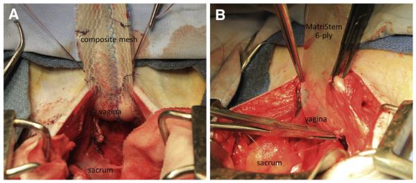
Sterile samples of Gynemesh, and 2-ply and 6-ply MatriStem Surgical Matrix were obtained and trimmed into straps appropriate for sacrocolpopexy (3-cm wide × 10cm long). Seventeen middle-aged parous rhesus macaques were implanted with a composite mesh comprised of Gynemesh plus 2-ply MatriStem (n = 9) or 6-ply MatriStem alone (n = 8) via sacrocolpopexy after hysterectomy (Figure 1). For the composite, the precut MatriStem, trimmed to the size of Gynemesh, was sutured to the mesh with absorbable 3-0 Vicryl (Ethicon) before mesh insertion. At implantation, the composite mesh was placed with the MatriStem side facing the vaginal lumen, forming a layer between the Gynemesh and the vagina. The implantation and postoperative care were performed as previously described.6 Twelve weeks after surgery, the graft-vagina complex (GVC) was harvested for functional, morphological, and biochemical analyses. Sham (animals underwent the identical surgery without mesh insertion, n = 12) and Gynemesh implanted (animals underwent the identical surgery with insertion of Gynemesh, n = 12) tissues, properly stored from a previous study,7 were used for measurement of elastin content and collagen type III/I ratio. For histomorphometrics, biomechanical testing, collagen, and GAG content, previously published data on sham and Gynemeshimplanted vagina were used for comparison.6,7
FIGURE 1. Surgical implantation of Gynemesh-MatriStem composite mesh and MatriStem via sacrocolpopexy.
Surgical implantation of A, Gynemesh-MatriStem (2-ply) composite mesh and B, MatriStem (6-ply) via sacrocolpopexy in rhesus macaque.
Ball-burst test for biomechanical properties
The passive mechanical properties of GVC were determined by the ball-burst test.6 All GVC samples used for ball-burst testing were obtained from the posterior vagina. Fifteen samples from composite mesh (n = 7) and MatriStem alone (n = 8) groups were tested whereas 2 GVC samples from the composite mesh group were too small to test (vagina in these animals was small). Structural properties including stiffness (N/ mm), ultimate load (N), ultimate elongation (mm), and energy absorbed (N/ mm) were determined.6 The contribution of soft tissues associated with the GVC was determined for ultimate load (loadtissue) and stiffness (ktissue) by sub-tracting the average corresponding values of Gynemesh obtained ex vivo.6 For samples that were grafted with MatriStem alone, the tissue contribution was equal to those values obtained for the entire complex. Since noncross-linked ECM grafts degrade rapidly in vivo (10-ply 14C-labeled ECM was completely degraded by 3 months),21 we assumed that MatriStem grafts were completely degraded during the implantation period (3 months) and replaced by host connective tissue, and thus, the ECM graft did not contribute to the mechanical integrity at the time of tissue procurement.
Functional test for smooth muscle contractility
The contractile response to a single dose of potassium chloride (KCl, 120 mmol/L) was tested within 30 minutes of tissue harvest as described previously.22 To normalize for differences in tissue quantity, the data were expressed as force per tissue volume of each strip (mN/ mm3).
Immunofluorescent labeling of α-smooth muscle actin and in situ terminal deoxynucleotidyl transferase dUTP nick end labeling (TUNEL) labeling of cell apoptosis
GVCs were cryosectioned (7 mm) perpendicular to the longitudinal axis of the vagina. Hematoxylin-eosin and Masson trichrome staining were performed following manufacturer’s protocols (Sigma-Aldrich, St Louis, MO). For analysis, specimens were required to contain all layers of vagina, and those obliquely sectioned with subepithelium >1039 μm (mean of sham values that were sectioned perpendicularly plus 2SD) were excluded. One sample from composite mesh was excluded for not containing all layers of vagina.
Immunofluorescent labeling of α-smooth muscle actin and in situ TUNEL labeling of apoptotic cells were used to measure thickness of the subepithelial and smooth muscle layers, and the percentage of apoptotic cells in each vaginal layer as previously described.7
Biochemical assays
Total collagen content and sulfated GAG content were measured as previously described,23,24 and expressed as percentage of tissue dry weight.
Mature elastin content was measured as the total content of desmosine and isodesmosine by ultra high-performance liquid chromography (UPLC).25 Briefly, hydrolyzed tissue was applied to Oasis mixed-mode cation-exchange (MCX) solid-phase extraction (SPE) columns (Waters, Milford, MA), and eluted with ammonium hydroxide/ water/methanol (10/40/50). Stock solutions of desmosine, isodesmosine, and S-[beta-(4-pyridyl)ethyl]L-cysteine (S-LC, internal standard) (Santa Cruz Biotechnology Inc, Dallas, TX) were diluted to series concentration to create standard curves. S-LC (2 μmol) was added to each sample and standards as an internal control. A total of 3 μL of the prepared samples and standards were injected onto a prepared Waters Acquity UPLC ethylene bridged hybrid (BEH) C18 column and monitored by measuring absorbance at 270-nm wavelength. The elution time of isodesmosine was 1.4 minutes, desmosine was 1.9 minutes, and S-LC was 2.4 minutes. The amount of mature elastin in each sample was represented by the sum of desmosine and isodesmosine normalized by that for bovine elastin (Sigma-Aldrich).
Ratios of collagen subtype III/I in the tissue of GVC were determined as previously described8,26 and calculated as a1(III) ×2/α1(I) ×3 normalized by the ratio of purified collagen I/III.
Statistical analysis
Sample size was determined using previously published mechanical data (ultimate load and stiffness) for sham and Gynemesh, in which at least 8 animals were needed in each group to achieve significance with a error of 0.05 and power of 0.80. Data were tested for distribution before statistical analysis. For normally distributed data, a 1-way analysis of variance was used followed by the appropriate post-hoc tests including Dunnett for comparison to sham and pairwise test using Bonferroni multiple comparisons procedure between all groups. For nonparametric data, Kruskal-Wallis was used to compare overall significance and Mann-Whitney to compare between groups.
Results
Animals in the Gynemesh-MatriStem composite group were younger than those in the sham group (P = .007) but had similar parity, weight, and POP-Q scores (Table 1). Multivariable regression modeling showed that age did not impact any of the experimental outcomes (P > .15) except the mechanical endpoint elongation at failure (P =.029), for which the results were subsequently adjusted for age. None of the animals in the study had prolapse beyond the hymen.
TABLE 1.
Demographics of nonhuman primates in study
| Groups | Age, y | Parity | Weight, kg | POP-Q stage |
|---|---|---|---|---|
| Shama | 14.5 (10.0, 15.0) | 4 (2, 6) | 7.5 ±1.3 | 0 (0, 1) |
|
| ||||
| Gynemeshb | 13.0 (11.5, 14.0) | 4 (2, 5) | 7.9 ±1.6 | 0 (0, 0) |
|
| ||||
| Composite | 8.0 (7.0, 11.0)c | 2 (2, 5) | 7.7 ±1.8 | 0 (0, 0) |
|
| ||||
| MatriStem | 13.5 (12.0, 14.0) | 4 (3, 4) | 8.5 ±1.4 | 0 (0, 0) |
|
| ||||
| P b | .049 | .949 | .597 | .225 |
Composite: Gynemesh-MatriStem (2-ply) (n = 9); MatriStem: MatriStem (6-ply) (n = 8).
Data for sham (n = 12) and Gynemesh (n = 12) were obtained from previous study.7
Results are expressed as mean ± SD for weight or median (first quartile, third quartile) for age, parity and POP-Q stage.
POP-Q, Pelvic Organ Prolapse Quantification.
Historical data;
Overall comparison of P value among groups;
P < .05 compared with sham.
Gross appearance
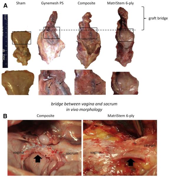
Gross inspection of samples implanted with the Gynemesh-MatriStem composite mesh revealed newly formed tissue on the surface of mesh. Gynemesh within the composite appeared flatter with open pores (Figure 2, A). This is in contrast to the collapsed pores, buckling, shrinkage, and poor tissue ingrowth observed in Gynemesh alone implanted specimens. The graft bridge extending from the vagina to the sacrum also appeared thicker and flatter with newly incorporated vascularized tissue in the composite as compared to Gynemesh alone. In the MatriStem alone group, a newly formed tissue bridge was present in all specimens extending from the vagina to the sacrum at the site where the original graft had been placed (Figure 2, B).
FIGURE 2. Gross morphology of graft-implanted vagina and graft bridges at 12 weeks postsurgery.
A, Gross appearance of sham and graft-vagina complex after implantation via sacrocolpopexy at 12 weeks. Graft bridge describes portion of graft-tissue extending from vagina to sacrum. All images were shown with same scale with magnified portion below showing appearance of grafts following implantation. *Area where tissue ingrowth was poor. Arrows point to area showing mesh buckling and shrinkage with collapsed pores in Gynemesh and flat surface with open pores in composite. B, In vivo morphology of graft bridges (arrows) following implantation of Gynemesh-MatriStem composite mesh and MatriStem alone.
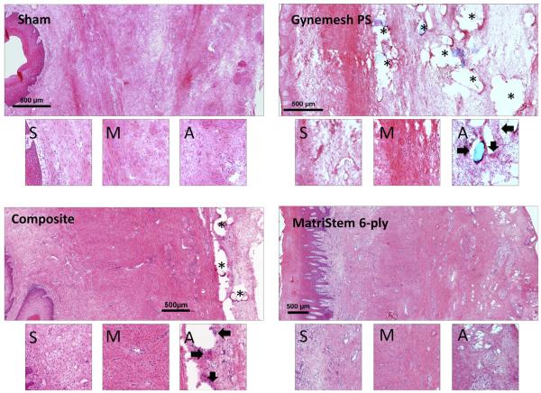
Histomorphology/morphometrics Overall, vaginal morphology improved in the Gynemesh-MatriStem composite mesh as compared to Gynemesh alone. Microscopic appearance of the vagina implanted with MatriStem alone was not different from sham (Figure 3). In contrast to the pronounced infiltration of inflammatory cells and tissue destruction in the adventitia observed after implantation with Gynemesh (Figure 3), the amount and intensity of the inflammatory response in the Gynemesh-MatriStem composite group appeared to be decreased. The clustering of mesh fibers in Gynemesh as shown in Figure 3 is consistent with a mesh following implantation. The MatriStem alone group had little to no inflammatory cells in the adventitia with an appearance similar to sham.
FIGURE 3. Hematoxylin-eosin images of grafted vagina at 12 weeks postsurgery.
Hematoxylin-eosin staining of vagina in sham and graft-implanted groups. For each group, upper image shows full-thickness vagina with epithelium placed on the left and adventitia on the right. Lower images show magnified portion of subepithelium (S), muscularis (M), and adventitia (A) of upper images. Arrows point to mesh area with intensified inflammatory response. Asterisk indicates mesh fiber occupied areas.
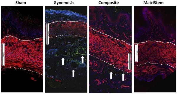
Compared to the disorganization and thinning of the smooth muscle layer observed with Gynemesh, the composite showed an overall more orderly smooth muscle appearance and distribution (Figure 4). Semiquantitative measurements showed that the thickness of the smooth muscle layer in the composite was preserved with values similar to sham and increased 89% compared to Gynemesh (P < .001) (Table 2). MatriStem alone did not have a negative impact on the smooth muscle (Figure 4 and Table 2). Interestingly, the thickness of vaginal subepithelium in all groups was similar (P = .38) (Table 2), indicating that the primary impact was occurring within the muscularis. The variance observed in the thickness of subepithelium in individual samples may be due to the presence of vaginal folds/rugae.
FIGURE 4. IF labeling of vaginal muscularis and cell apoptosis following graft implantations.
Immunofluorescent labeling of vaginal smooth muscle and apoptotic cells in sham and graft-implanted vagina at 12 weeks postsurgery. Red represents positive labeling of α-smooth muscle actin; green overlapping blue represents positive labeling of apoptotic cells; blue represents labeling of nuclei; arrows point to mesh fibers. Solid line demarcates border between subepithelium and muscularis. Broken line demarcates border between muscularis and adventitia. Scales equal 1000 μm.
TABLE 2.
Functional and morphometric analysis of vagina after implantation of Gynemesh-MatriStem composite mesh (n = 8) or MatriStem alone (n = 8) as compared to historical data for sham-operated and Gynemesh alone6,7
| Function |
Morphometrics |
Cell apoptosis, % of total cell number |
||||
|---|---|---|---|---|---|---|
| Contractility, mN/mm3 |
Subepithelium thickness, μm |
Muscularis thickness, μm |
Subepithelium | Muscularis | Adventitia | |
| Shama | 0.26 ±0.11 | 522 ±189 | 1557 ±499 | 0.2 (0.1, 0.7) | 0.1 (0.0, 0.1) | 1.2 (0.4, 2.3) |
|
| ||||||
| Gynemesha | 0.08 ± 0.03c | 517 ±180 | 866±210c | 4.2c (2.1, 10.6) | 0.5c (0.3, 0.9) | 12.2c (8.2, 25.2) |
|
| ||||||
| Composite | 0.14 ±0.11 | 710 ±366 | 1635 ± 403d | 0.1d (0.0, 0.3) | 0.0d (0.0, 0.1) | 2.3d (1.0, 5.3) |
|
| ||||||
| MatriStem | 0.17 ±0.18 | 596 ±178 | 1311 ±187 | 0.1d (0.1, 0.2) | 0.0d (0.0, 0.1) | 0.5d (0.2, 3.2) |
|
| ||||||
| P b | .009 | .380 | .001 | .000 | .000 | .000 |
Composite: Gynemesh-MatriStem RS (2-ply); MatriStem: MatriStem pelvic floor matrix (6-ply).
Results are expressed as mean ± SD for contractility, subepithelium, and muscularis thickness, or median (first quartile, third quartile) for cell apoptosis quantification.
Historical data;
Overall comparison of P value among groups;
P < .05 compared with sham;
P < .05 compared with Gynemesh.
Cell apoptosis
As compared with Gynemesh, for which implantation resulted in a significantly higher percentage of cells undergoing apoptosis than sham, the Gynemesh-MatriStem composite demonstrated an overall 83% decrease in the percentage of apoptotic cells (P = .009). Specifically, apoptotic cells decreased by 99% in subepithelium (P = .005), 96% in muscularis (P = .016), and 81% in adventitia (P = .016) (Figure 4 and Table 2). In the presence of MatriStem alone, the percentage of cells undergoing apoptosis in all layers was similar to sham and lower than Gynemesh (all values with P <.001) (Table 2).
Biochemical analysis of matrix proteins and ratio of collagen subtype III to I
Total collagen content in the Gynemesh-MatriStem composite group did not change significantly when compared to Gynemesh and remained approximately 18% lower than sham (P = .017) (Table 3). In the MatriStem alone group, collagen content was not statistically different from sham (P = .149) or Gynemesh (P = 1.000) (Table 3). The decrease in the content of mature elastin observed following implantation with Gynemesh was not observed in the composite mesh or MatriStem alone (P = .64 and P = .09 as compared to sham, respectively) (Table 3). Similarly, the increase in GAG content (a marker of tissue injury) in the Gynemesh group relative to sham (P = .02) was not observed in the composite mesh group or MatriStem alone group (P = .65 and P = .875 as compared to sham, respectively) (Table 3).
TABLE 3.
Biochemical analysis of vaginal wall after implantation of Gynemesh-MatriStem composite mesh (n = 9)orMatriStem alone (n = 8)ascompared to sham-operated and implantation of Gynemesh alone7
| Biochemistry analysis |
||||
|---|---|---|---|---|
| Collagen,a % of dry weight |
Elastin, normalized | GAG,a % of dry weight |
Collagen III/I ratio, normalized |
|
| Sham | 48.3 ± 8.6 | 1.51 ±0.28 | 1.35 ±0.33 | 0.14 ±0.05 |
|
| ||||
| Gynemesh | 38.5 ± 5.2c | 1.26 ±0.24 | 1.71 ±0.38c | 0.22 ± 0.04c |
|
| ||||
| Composite | 39.4 ± 8.7 | 1.35 ±0.96 | 1.57 ±0.32 | 0.18±0.03c,d |
|
| ||||
| MatriStem | 43.1 ± 5.4 | 1.25 ±0.30 | 1.50 ±0.18 | 0.20 ± 0.05d |
|
| ||||
| P b | .011 | .749 | .069 | .000 |
Composite: Gynemesh-MatriStem RS (2-ply); MatriStem: MatriStem pelvic floor matrix (6-ply).
Results are expressed as mean ± SD.
GAG, glycosaminoglycan.
Historical data for sham and Gynemesh;
Overall comparison of P value among groups;
P < .05 compared with sham;
P < .05 compared with Gynemesh.
Finally, the increased ratio of collagen subtype III/I observed following implantation with Gynemesh relative to sham (57% higher than sham, P < .001) was also present in composite mesh (28% higher than sham, P = .025) and MatriStem alone (43% higher than sham, P = .01); however, the magnitude of the increase was less, indicating that MatriStem decreased the negative impact on the vagina.
Biomechanical properties
While the ultimate load at failure and the stiffness of Gynemesh-MatriStem composite mesh were not different from Gynemesh (P = .234 and P = .731, respectively), both the ultimate elongation and energy absorbed at failure in the composite mesh were increased by 33% and 51% (P = .022 and P = .014, respectively) (Table 4). However, when adjusting for age, the difference in ultimate elongation was no longer present. Similar to Gynemesh, the estimated contribution of tissue alone to the overall load and stiffness (represented as loadtissue and stiffness [ktissue]) of GVC remained 45% and 75% lower in the composite than those for sham (P =.001 and P = .051, respectively). In the MatriStem alone group, all of the passive mechanical properties of GVC were similar to sham indicating an absence of a negative impact on the vagina (Table 4).
TABLE 4.
Biomechanical analysis of vaginal wall after implantation of Gynemesh-MatriStem composite mesh (n = 7) or MatriStem alone (n = 8) as compared to historical data for sham and Gynemesh alone6
| Stiffness, N/mm | Ultimate load, N | Elongation, mm | Energy absorbed, N/mm |
Ktissue, N/mm | Loadtissue, N | |
|---|---|---|---|---|---|---|
| Shama | 35.8 (30.6, 49.5) | 154.9 (95.8, 172.2) | 11.7 (10.0, 12.8) | 413.5 (248.1, 495.7) | 35.8 (30.6, 49.5) | 154.9 (95.8, 172.2) |
|
| ||||||
| Gynemesha | 28.1 (27.1, 48.1) | 170.0 (140.3, 192.2) | 10.2 (9.3, 11.7) | 508.9 (425.0, 547.8) | 2.0c (1.0, 22.0) | 58.0c (28.3, 80.1) |
|
| ||||||
| Composite | 34.9 (31.0, 44.2) | 196.4 (175.5, 226.4) | 13.6d (12.5, 15.6) | 769.2c,d (651.2, 859.0) | 8.8c (4.9, 18.1) | 84.4c (63.5, 114.4) |
|
| ||||||
| MatriStem | 28.6 (23.9, 39.3) | 145.6 (108.0, 161.3) | 13.7 (11.8, 15.1) | 438.3 (347.1, 469.0) | 28.6d (23.9, 39.3) | 145.8d (108.0, 161.3) |
|
| ||||||
| P b | .570 | .155 | .025 | .001 | .001 | .006 |
Composite: Gynemesh-MatriStem RS (2-ply); MatriStem: MatriStem pelvic floor matrix (6-ply). Results are expressed as median (first quartile, third quartile).
Historical data;
Overall comparison of P value among groups;
P < .05 compared with sham;
P < .05 compared with Gynemesh.
Vaginal smooth muscle contractility
The profound decrease in vaginal smooth muscle contractility observed following the implantation of Gynemesh did not occur in the Gynemesh-MatriStem composite group with values not significantly different from sham (P = .155) (Table 2). The results, however, were highly variable indicating a mitigation of the negative impact but not fully protective of vaginal smooth muscle function. With MatriStem alone, vaginal contractility was preserved with no difference from sham (P = .297) (Table 2).
Comment
MatriStem, when used in sacrocolpopexy as a composite mesh with Gynemesh, mitigated the host inflammatory response, decreased cell apoptosis, and had an overall protective effect on vaginal smooth muscle structure and function. In addition, MatriStem favorably impacted vaginal elastin content, collagen subtype III/I ratios, and GAG content. When used alone, multilayered MatriStem effectively formed a novel tissue bridge between the vagina and the sacrum. Thus, the multilayered graft, with out-of-the-package mechanical properties in the range of those of currently widely used polypropylene mesh, did not negatively impact the vagina.
In this study, 2-ply MatriStem decreased the infiltration of inflammatory cells surrounding Gynemesh in gross morphology and significantly suppressed overall apoptotic activity in the vagina, signifying a reduced inflammatory response to the implant. Such results may be related to the immune modulation by the host in response to ECM grafts.15,27,28 Although it is not completely clear how MatriStem mitigates the intensity and duration of host immune responses to foreign materials, it has been suggested that ECM graft materials like MatriStem hasten the transition of macrophage phenotypes from a proinflammatory M1 phenotype to a tissue-reconstructive remodeling M2 phenotype.15,28 A detailed study on the immune responses and macrophage dichotomization in the vagina following the implantation of composite mesh and MatriStem is ongoing in our laboratory.
Additionally, the degradation products from MatriStem are critical for tissue-constructive remodeling and recruitment of progenitor cells to injury sites to facilitate tissue regeneration.9 For example, the release of cytokines and growth factors, with basic fibroblast growth factor (bFGF) and transforming growth factor (TGF)-μ1 being the most abundant,29 may play important roles in promoting cell proliferation, matrix production, and angiogenesis. These bioactive factors may account for at least some of the positive effects observed with MatriStem in the current study.
Smooth muscle forms a critical layer in the vaginal wall (vaginal muscularis) that is integral to structural integrity and function. With aging and the development of pelvic organ prolapse, this layer becomes thinned and/or functionally impaired.30,31 Our fiding that MatriStem, as a composite with Gynemesh or alone, preserved the morphology and function of muscularis is highly significant in that it suggests that MatriStem has the potential to protect/preserve the structural integrity and contractile function of the vagina. The beneficial effect of MatriStem may be derived from a less pronounced inflammatory response or reduced mechanical mismatches with loading, or MatriStem may simply be acting as a physical barrier. Indeed, a layer of collagen foil interposed between polypropylene mesh and viscera in hernia repairs considerably decreased the intensity of foreign-body reaction, tissue necrosis, and extent of adhesion.32 Further studies are needed to compare the impact of ECM used in sheets vs that applied as a coating to polypropylene similar to what has been done previously in a rat abdominal hernia model.33
Our fiding that the energy absorbed at failure was increased in the Gynemesh-MatriStem composite mesh relative to Gynemesh alone indicates that a tissue component in the composite mesh-implanted vagina allowed for more energy to be absorbed prior to failure. This biomechanical phenomenon may be attributable to the preservation of mature elastin content in the composite mesh as opposed to the Gynemesh-implanted vagina. The failure of the strength of the tissue to improve, as shown by the lack of a significant difference in the tissue contributions to ultimate load and stiffness in the composite mesh and Gynemesh, is consistent with the decreased collagen content in these 2 groups.
The primary limitation of the study is that only a limited number of animals was used in the composite mesh and MatriStem only groups due to the high expense of nonhuman primates. The relatively small sample size may reduce the predictability of the multivariable regression modeling. In our study, the only parameter that may have been impacted by age was elongation at failure as predicted by the model (P = .029). Yet, since there was no significant difference in elongation at failure between composite mesh and Gynemesh after adjustment for age, the decrease of accuracy in predictability of multivariable regression modeling would thus not affect our conclusion. Secondly, it could be argued that absolute continuity between the 2 materials comprising the composite, ie, MatriStem and polypropylene mesh, is important to ensure that no separation of the 2 phases occurs when they are mechanically loaded. While it is unclear whether such a separation occurred in vivo, our in vitro testing of the composite mesh assembled as described did not show material separation under tensile loading conditions.
In conclusion, mesh augmentation with MatriStem attenuated the degenerative impact of Gynemesh on the vagina, particularly in regards to the vaginal muscularis. In addition, the application of ECM graft alone in sacrocolpopexy did not negatively impact the functional, morphological, and biochemical properties of the vagina. Future studies will focus on mechanisms.
Acknowledgment
We are grateful for technical support from Dr Zegbeh C. Jallah for vaginal smooth muscle contractility testing.
This work was supported by National Institutes of Health (NIH) award R01 HD061811 and an institute cooperative research agreement with ACell Inc (Columbia, MD). The content is solely the responsibility of the authors and does not necessarily represent the official views of NIH or ACell Inc. The funding source had no involvement in the study design, collection of data, analysis of data, interpretation of data, writing of the report, or the decision to submit for publication.
Footnotes
Disclosure: R.L., A.N., S.P., S.A., and P.A.M. received partial salary support from their institute cooperative research agreement with ACell Inc (Columbia, MD). K.K., W.B., and R.W.P. have no conflicts of interest to declare.
Presented at the 2014 and 2015 American Urogynecologic Society/International Urogynecological Association Joint Scientific Meeting in Washington, DC, July 22-26, 2014, and Seattle, WA, Oct. 13-17, 2015.
References
- 1.Maher C, Feiner B, Baessler K, Schmid C. Surgical management of pelvic organ prolapse in women. Cochrane Database Syst Rev. 2013;4:CD004014. doi: 10.1002/14651858.CD004014.pub5. [DOI] [PubMed] [Google Scholar]
- 2.Committee on Gynecologic Practice Vaginal placement of synthetic mesh for pelvic organ prolapse. Committee opinion no. 513. Obstet Gynecol. 2011;118:1459–64. doi: 10.1097/AOG.0b013e31823ed1d9. [DOI] [PubMed] [Google Scholar]
- 3.Maher CF, Qatawneh AM, Dwyer PL, Carey MP, Cornish A, Schluter PJ. Abdominal sacral colpopexy or vaginal sacrospinous colpopexy for vaginal vault prolapse: a prospective randomized study. Am J Obstet Gynecol. 2004;190:20–6. doi: 10.1016/j.ajog.2003.08.031. [DOI] [PubMed] [Google Scholar]
- 4.Nygaard IE, McCreery R, Brubaker L, et al. Abdominal sacrocolpopexy: a comprehensive review. Obstet Gynecol. 2004;104:805–23. doi: 10.1097/01.AOG.0000139514.90897.07. [DOI] [PubMed] [Google Scholar]
- 5.Brown BN, Mani D, Nolfi AL, Liang R, Abramowitch SD, Moalli PA. Characterization of the host inflammatory response following implantation of prolapse mesh in rhesus macaque. Am J Obstet Gynecol. 2015;213:668. doi: 10.1016/j.ajog.2015.08.002. e1-10. [DOI] [PMC free article] [PubMed] [Google Scholar]
- 6.Feola A, Abramowitch S, Jallah Z, et al. Deterioration in biomechanical properties of the vagina following implantation of a high-stiffness prolapse mesh. BJOG. 2013;120:224–32. doi: 10.1111/1471-0528.12077. [DOI] [PMC free article] [PubMed] [Google Scholar]
- 7.Liang R, Abramowitch S, Knight K, et al. Vaginal degeneration following implantation of synthetic mesh with increased stiffness. BJOG. 2013;120:233–43. doi: 10.1111/1471-0528.12085. [DOI] [PMC free article] [PubMed] [Google Scholar]
- 8.Liang R, Zong W, Palcsey S, Abramowitch S, Moalli PA. Impact of prolapse meshes on the metabolism of vaginal extracellular matrix in rhesus macaque. Am J Obstet Gynecol. 2015;212:174. doi: 10.1016/j.ajog.2014.08.008. e1-7. [DOI] [PMC free article] [PubMed] [Google Scholar]
- 9.Badylak SF, Freytes DO, Gilbert TW. Reprint of: extracellular matrix as a biological scaffold material: structure and function. Acta Biomater. 2015;23(Suppl):S17–26. doi: 10.1016/j.actbio.2015.07.016. [DOI] [PubMed] [Google Scholar]
- 10.Badylak SF, Gilbert TW. Immune response to biologic scaffold materials. Semin Immunol. 2008;20:109–16. doi: 10.1016/j.smim.2007.11.003. [DOI] [PMC free article] [PubMed] [Google Scholar]
- 11.Afaneh C, Abelson J, Schattner M, et al. Esophageal reinforcement with an extracellular scaffold during total gastrectomy for gastric cancer. Ann Surg Oncol. 2015;22:1252–7. doi: 10.1245/s10434-014-4125-4. [DOI] [PubMed] [Google Scholar]
- 12.Fleming ME, Bharmal H, Valerio I. Regenerative medicine applications in combat casualty care. Regen Med. 2014;9:179–90. doi: 10.2217/rme.13.96. [DOI] [PubMed] [Google Scholar]
- 13.Remlinger NT, Gilbert TW, Yoshida M, et al. Urinary bladder matrix promotes site appropriate tissue formation following right ventricle outflow tract repair. Organogenesis. 2013;9:149–60. doi: 10.4161/org.25394. [DOI] [PMC free article] [PubMed] [Google Scholar]
- 14.Sasse KC, Brandt J, Lim DC, Ackerman E. Accelerated healing of complex open pilonidal wounds using MatriStem extracellular matrix xenograft: nine cases. J Surg Case Rep. 2013:4. doi: 10.1093/jscr/rjt025. [DOI] [PMC free article] [PubMed] [Google Scholar]
- 15.Brown BN, Ratner BD, Goodman SB, Amar S, Badylak SF. Macrophage polarization: an opportunity for improved outcomes in biomaterials and regenerative medicine. Biomaterials. 2012;33:3792–802. doi: 10.1016/j.biomaterials.2012.02.034. [DOI] [PMC free article] [PubMed] [Google Scholar]
- 16.Cavallo JA, Greco SC, Liu J, Frisella MM, Deeken CR, Matthews BD. Remodeling characteristics and biomechanical properties of a crosslinked versus a non-crosslinked porcine dermis scaffolds in a porcine model of ventral hernia repair. Hernia. 2015;19:207–18. doi: 10.1007/s10029-013-1070-2. [DOI] [PMC free article] [PubMed] [Google Scholar]
- 17.Quiroz LH, Gutman RE, Shippey S, et al. Abdominal sacrocolpopexy: anatomic outcomes and complications with Pelvicol, autologous and synthetic graft materials. Am J Obstet Gynecol. 2008;198:557. doi: 10.1016/j.ajog.2008.01.050. e1-5. [DOI] [PubMed] [Google Scholar]
- 18.Gomelsky A, Dmochowski RR. Biologic materials for pelvic floor reconstruction. Curr Bladder Dysfunct Rep. 2012;7:201–9. [Google Scholar]
- 19.Shepherd JP, Feola AJ, Abramowitch SD, Moalli PA. Uniaxial biomechanical properties of seven different vaginally implanted meshes for pelvic organ prolapse. Int Urogynecol J. 2012;23:613–20. doi: 10.1007/s00192-011-1616-8. [DOI] [PMC free article] [PubMed] [Google Scholar]
- 20.Feola A, Abramowitch S, Jones K, Stein S, Moalli P. Parity negatively impacts vaginal mechanical properties and collagen structure in rhesus macaques. Am J Obstet Gynecol. 2010;203:595. doi: 10.1016/j.ajog.2010.06.035. e1-8. [DOI] [PMC free article] [PubMed] [Google Scholar]
- 21.Gilbert TW, Stewart-Akers AM, Simmons-Byrd A, Badylak SF. Degradation and remodeling of small intestinal submucosa in canine Achilles tendon repair. J Bone Joint Surg Am. 2007;89:621–30. doi: 10.2106/JBJS.E.00742. [DOI] [PubMed] [Google Scholar]
- 22.Jallah Z, Liang R, Feola A, et al. The impact of prolapse mesh on vaginal smooth muscle structure and function. BJOG. 2016;123:1076–85. doi: 10.1111/1471-0528.13514. [DOI] [PMC free article] [PubMed] [Google Scholar]
- 23.Blumenkrantz N, Asboe-Hansen G. An automated procedure for quantitative determination of hydroxyproline. Clin Biochem. 1974;7:251–7. doi: 10.1016/s0009-9120(74)92204-8. [DOI] [PubMed] [Google Scholar]
- 24.Muller G, Hanschke M. Quantitative and qualitative analyses of proteoglycans in cartilage extracts by precipitation with 1,9-dimethylmethylene blue. Connect Tissue Res. 1996;33:243–8. doi: 10.3109/03008209609028881. [DOI] [PubMed] [Google Scholar]
- 25.Shiraishi K, Matsuzaki K, Matsumoto A, Hashimoto Y, Iba K. Development of a robust LC-MS/MS method for determination of desmosine and isodesmosine in human urine. J Oleo Sci. 2010;59:431–9. doi: 10.5650/jos.59.431. [DOI] [PubMed] [Google Scholar]
- 26.Sykes B, Puddle B, Francis M, Smith R. The estimation of two collagens from human dermis by interrupted gel electrophoresis. Biochem Biophys Res Commun. 1976;72:1472–80. doi: 10.1016/s0006-291x(76)80180-5. [DOI] [PubMed] [Google Scholar]
- 27.Brown BN, Valentin JE, Stewart-Akers AM, McCabe GP, Badylak SF. Macrophage phenotype and remodeling outcomes in response to biologic scaffolds with and without a cellular component. Biomaterials. 2009;30:1482–91. doi: 10.1016/j.biomaterials.2008.11.040. [DOI] [PMC free article] [PubMed] [Google Scholar]
- 28.Londono R, Badylak SF. Biologic scaffolds for regenerative medicine: mechanisms of in vivo remodeling. Ann Biomed Eng. 2015;43:577–92. doi: 10.1007/s10439-014-1103-8. [DOI] [PubMed] [Google Scholar]
- 29.Liang R, Fisher M, Yang G, Hall C, Woo SL. Alpha1,3-galactosyltransferase knockout does not alter the properties of porcine extracellular matrix bioscaffolds. Acta Biomater. 2011;7:1719–27. doi: 10.1016/j.actbio.2011.01.001. [DOI] [PubMed] [Google Scholar]
- 30.Liang R, Fawcett M, Shaffer R, Palcsey S, Moalli P. Impact of aging on vaginal smooth muscle: a morphometric study. Female Pelvic Med Reconstr Surg. 2014;20(Suppl):S128. [Google Scholar]
- 31.Northington GM, Basha M, Arya LA, Wein AJ, Chacko S. Contractile response of human anterior vaginal muscularis in women with and without pelvic organ prolapse. Reprod Sci. 2011;18:296–303. doi: 10.1177/1933719110392054. [DOI] [PMC free article] [PubMed] [Google Scholar]
- 32.Schonleben F, Reck T, Tannapfel A, Hohenberger W, Schneider I. Collagen foil (TissuFoil E) reduces the formation of adhesions when using polypropylene mesh for the repair of experimental abdominal wall defects. Int J Colorectal Dis. 2006;21:840–6. doi: 10.1007/s00384-006-0091-z. [DOI] [PubMed] [Google Scholar]
- 33.Faulk DM, Londono R, Wolf MT, et al. ECM hydrogel coating mitigates the chronic inflammatory response to polypropylene mesh. Biomaterials. 2014;35:8585–95. doi: 10.1016/j.biomaterials.2014.06.057. [DOI] [PMC free article] [PubMed] [Google Scholar]


