Abstract
The growing attention to the luminescent nanocarriers is strongly stimulated by their potential application as drug delivery systems and by the necessity to monitor their distribution in cells and tissues. In this communication we report on the synthesis of amphiphilic polypeptides bearing C-terminal phosphorescent label together with preparation of nanoparticles using the polypeptides obtained. The approach suggested is based on a unique and highly technological process where the new phosphorescent Pt-cysteine complex serves as initiator of the ring-opening polymerization of α-amino acid N-carboxyanhydrides to obtain the polypeptides bearing intact the platinum chromophore covalently bound to the polymer chain. It was established that the luminescent label retains unchanged its emission characteristics not only in the polypeptides but also in more complicated nanoaggregates such as the polymer derived amphiphilic block-copolymers and self-assembled nanoparticles. The phosphorescent nanoparticles display no cytotoxicity and hemolytic activity in the tested range of concentrations and easily internalize into living cells that makes possible in vivo cell visualization, including prospective application in time resolved imaging and drug delivery monitoring.
Bioimaging based on luminescent microscopy represents one of the most powerful analytical techniques in the life sciences because of its high sensitivity accompanied with simplicity and low cost. This visualization procedure can be carried out using water soluble organic and organometallic dyes, their conjugates with polymers and biomolecules1,2,3, as well as with luminescent nanoobjects4,5,6,7. The growing attention to the latter approach can be explained by well known practice of application of the nanoparticles to construct advanced drug delivery systems, which also make possible easy visualization of drug distribution in cells and tissues. During the last years, such smart combination of diagnostic and therapeutic properties caused enormous interest in biomedical research area.
Nowadays, the nanocarriers intended for a creation of drug delivery systems can be both of inorganic8 and organic nature9,10. Among inorganic nanoparticles applied for bioimaging and drug delivery such systems as dye-doped silica11, quantum dots12, metal nanoclusters13, lanthanide-doped nanoparticles14, etc. have got a particular attention. The luminescent properties of organic nanocarriers are usually associated with the native material emission characteristics or are the result of their labeling with emissive moieties. In particular, the materials based on photo-luminescent polyacrylonitrile can be mentioned as an example of label-free organic nanoparticles for bioimaging15. However, both the covalent labeling of organic nanoparticles or encapsulation of a dye inside the particles16,17 are the most common approaches. Encapsulation of dyes in a drug-carrying nanoparticle is usually aimed at synchronous release of drug and dye to signal about drug availability in biological system. Such a process can occur due to the biodegradation of nanoparticles18, or is a result of nanocarrier response to external stimuli (temperature, pH, or some others)19. Another way of monitoring of drug-carrier localization is preparation of nanoparticles containing covalently bound dye molecules, which represent a stable form of luminescent nanoobjects. Currently, there are well elaborated approaches to prepare functionalized luminophores19 and ways of their covalent binding to nanocontainers20,21, which however needs a special chemical procedures to link the probe to a certain chemical function of the nanocontainer like for example conjugation of polymer material functional groups with the reactive moiety of the dye to give the luminescent nanoobjects22. This approach normally gives an even distribution of labels on the surface of nanocarrier. In contrast, modification of polymer end-functional group allows for preparation of uniformed nanoparticles with strictly defined localization of luminescent molecule23.
One of the prospective groups of nanoparticles, which are widely considered as drug delivery systems24,25,26, as well as materials for the realization of two-photon fluorescence bioimaging27,28, is so called soft materials. These objects mostly represent self-assembled nanostructures obtained from amphiphilic precursors, in which the hydrophobic part is responsible for self-aggregation in aqueous media, whereas hydrophilic segment is responsible for both solubility and chemical functionality.
In the present communication we report on a novel approach to the synthesis of amphiphilic polypeptides bearing C-terminal phosphorescent label followed by preparation of luminescent nanoparticles. Taking into account the biodegradability, biocompatibility and wide variability of suitable functional groups, the amphiphilic polypeptides are very attractive candidates for preparation of nanoparticles of different morphology targeted to the biomedical applications29,30,31. The synthesis of amphiphilic co-polypeptides was carried out using ring-opening polymerization strategy (ROP) of N-carboxyanhydrides (NCAs) of α-amino acids. Traditionally, such polymerization is initiated with amines, but only primary amines provide narrow molecular weight distribution of resulting polymer product32,33. In our research we suggested an original approach based on the use of NH2-bearing luminescent organometallic complex as initiator for NCA polymerization. In particular, a luminescent Pt-cysteine complex with emission in green area of visible spectrum was specially designed and applied as polymerization initiator. The advantages of the application of phosphorescent organometallic probes over fluorescent organic dyes are higher photostability of the complexes and higher sensitivity of time resolved imaging experiments due to the opportunity to cut off background emission in time-gated regime34. Moreover, the diversity of the complexes properties determined by easy variation of their ligand environment and the nature of the metal center allows for the rational choice of the complexes with appropriate absorption-emission characteristics35,36,37,38.
The developed approach made possible attachment of the luminescent label at C-terminal position of the synthesized polypeptide that strictly determined location of the label on the nanoparticle outer (hydrophilic) surface after amphiphilic copolymer self-assembling. This design of the nanocarrier is extremely important to increase brightness of the probe and consequently sensitivity of the imaging experiments.
Experimental Section
Materials
L-cysteine, γ-benzyl-L-glutamate ((Bzl)Glu), ɛ-Z-L-lysine ((Z)Lys), L-leucine (Leu), triphosgene, α-pinene, trifluoromethanesulfonic acid (TFMSA), trifluoroacetic acid (TFA), 2-(p-tolyl)-pyridine (NCtpy) and other reagents were purchased from Sigma–Aldrich (Germany) and used as received. 1,4-Dioxane, n-hexane were purchased from Vecton Ltd. (Russia) and distilled prior to use. Dimethyl sulfoxide (DMSO), also purchased from Vecton Ltd. (Russia), was dried under molecular sieves 4 Ǻ and distilled under vacuum. Ethyl acetate purchased from Panreac (Spain) was distilled before application. All solvents were purified and distilled using standard procedures.
HEK 293 (human embryonic kidney cells), HELF (human embryonic lung fibroblasts) and U937 (human lymphoblast lung cells) cell lines were purchased from BioloT (Russia). The cells were cultured in Dulbecco’s Modified Eagle Medium (BioloT, Russia) and grown in Dulbecco’s Modified Eagle’s Medium containing 10% (v/v) heat-inactivated fetal bovine serum (FBS, HyClone Laboratories, UT, USA), 1% L-glutamine, 1% sodium pyruvate, 50 U/mL penicillin and 50 μg/mL streptomycin (BioloT).
Instruments
The solution 1H NMR spectra were recorded on a Bruker Avance 400 spectrometer. Mass spectra were recorded on a Bruker microTOF 10223 instrument at ESI+ mode (solvent – MeOH). UV/Vis absorption and emission spectra were recorded with a Shimadzu UV-1800 spectrophotometer (Japan), a HORIBA Scientific FluoroLog-3 spectrofluorometer and Avantes spectrometer. Gel-permeation chromatography (GPC) was performed using Shimadzu LC-20 Prominence system equipped with refractometric RID 10-A detector (Japan) and 7.8 × 300 mm Styragel Column HMW 6E, 15–20 μm bead size (Waters, USA). The GPC calculations were fulfilled with GPC LC Solutions software (Shimadzu, Japan). Amino acid HPLC analysis was carried out using an LCMS-8030 Shimadzu system with Mass-detection (LC-MS) (Shimadzu, Japan), equipped with 2 × 150 mm Luna C18 column, packed with 5 μm particles. Dynamic light scattering (DLS) and zeta potential measurements of colloids were performed using a Zetasizer ZS device (Malvern, Great Britain) at angle 113°.
Synthesis of (cis)-[Pt(NCtpy)(1,3-dibenzylbenzimidazol-2-yliden)Cl] (1)
The platinum(II) complex [Pt(NCtpy)(DMSO)Cl] and 1,3-dibenzyl-1H-benzo[d]imidazol-3-ium chloride were prepared according to the published procedures39,40. To synthesize 1 the compound [Pt(NCtpy)(DMSO)Cl] (200 mg, 0.418 mmol), 1,3-dibenzylbenzimidzolium chloride (154 mg, 0.460 mmol) and K2CO3 (288 mg, 2.09 mmol) were added to 40 ml of acetone and the mixture was stirred overnight at 40 °C. The resulting mixture was evaporated to dryness, redissolved in dichloromethane, filtered to remove potassium salts and evaporated to dryness again. The crude residue was purified by flash column chromatography (Silica, eluent – dichloromethane) to afford two fractions which were evaporated to give 1 (123 mg, 43%) and its trans-isomer 1* (147 mg, 51%) as yellow solids.
1*: 1Н NMR (acetone-d6, ambient temperature): δ (ppm) 8.12 (d with broad 195Pt satellites, 3JHH = 6.4 Hz, 1H, H1), 8.00 (s with broad 195Pt satellites, 1H, H8), 7.94 (m, 1H, H3), 7.87 (m, 1H, H4), 7.66 (m, 4H, H12), 7.55–7.51 (m, 3H, H5 and H10), 7.30–7.28 (m, 2H, H11), 7.25−7.19 (m, 6H, H13 and H14), 6.92 (d, 3JHH = 7.9 Hz, 1H, H6), 6.83 (m, 1H, H2), 6.23 (d, 2JHH = 15.2 Hz, 2H, H9), 6.10 (d, 2JHH = 15.2 Hz, 2H, H9’), 2.34 (s, 3H, H7). Proton numbering scheme is given in Fig. S1.
1: 1Н NMR (acetone-d6, ambient temperature): δ (ppm) 9.63 (d with broad 195Pt satellites, 3JHH = 5.3 Hz, 1H, H1), 8.04 (m, 1H, H3), 7.98 (m, 1H, H4), 7.73 (m, 4H, H12), 7.61 (d, 3JHH = 7.7 Hz, 1H, H5), 7.43 (m, 3H, H2 and H10), 7.30−7.22 (m, 8H, H11, H13 and H14), 6.92 (d, 3JHH = 7.7 Hz, 1H, H6), 6.63 (m, 3JH-Pt = 34.6 Hz, 1H, H8), 6.22 (d, 2JHH = 15.5 Hz, 2H, H9), 6.10 (d, 2JHH = 15.5 Hz, 2H, H9’), 2.13 (s, 3H, H7). Proton numbering scheme is given in Fig. S2. ESI + (m/z): [M − Cl]+ 661.20, [M + K]+ 735.32, [M + K + acetone]+ 793.37.
Synthesis of (cis)-[Pt(NCtpy)(1,3-dibenzylbenzimidazol-2-yliden)(L-cysteine)] (2)
The complex 1 (17 mg, 0.0244 mmol) and L-cysteine (9 mg, 0.0742 mmol) were dissolved in 4 ml of the dichloromethane/methanol (1:1 v/v) mixture. Triethylamine (30 μl, 21.9 mg, 0.216 mmol) was added via syringe and the mixture was stirred overnight at room temperature. The reaction completion was monitored by the 1H NMR referenced to L-cysteine resonances. The resulting solution was evaporated to dryness. The residue washed with 2 × 3 ml of dichloromethane, 3 × 5 ml of water and dried in nitrogen stream. The solid obtained was dissolved in 5 ml of methanol, filtered to remove insoluble impurities and the solution was evaporated to give 2 as yellow solid (10.3 mg, 54%)1.Н NMR (methanol-d4, ambient temperature): δ (ppm) 9.27 (d with broad 195Pt satellites, 3JHH = 5.3 Hz, 1H, H1), 7.93 (m, 1H, H3), 7.87 (m, 1H, H4), 7.54 (m, 5H, H12 and H5), 7.44 (m, 2H, H10), 7.24 (m, 9H, H2, H11, H13 and H14), 6.86 (d, 3JHH = 7.2 Hz, 1H, H6), 6.37 (t, 3JHPt = 28.9 Hz, 1H, H8), 6.30 (d, 2JHH = 15.4 Hz, 1H, H9), 6.29 (d, 2JHH = 15.3 Hz, 1H, H9*), 5.90 (d, 2JHH = 15.3 Hz, 1H, H9’), 5.86 (d, 2JHH = 15.4 Hz, 1H, H9*’), 3.47 (m, 1H, H15), 2.72 (m, 1H, H16), 2.58 (m, 1H, H16’), 2.04 (s, 3H, H7). Proton numbering scheme is given in Fig. S3. ESI + (m/z): C33H28N3Pt [M − Cys]+ 661.19.
Synthesis of N-carboxyanhydrides (NCA) of α-amino acids
NCA monomers of γ-benzyl-L-glutamate, ɛ-Z-L-lysine, L-leucine, were prepared as described elsewhere41. The structure and purity of NCAs obtained was proved by 1H NMR at 25 °C in СDCl3. 1Н NMR (methanol-d4, ambient temperature): (Bzl)Glu NCA: δ 7.44–7.30 (m, 5H), 6.43 (s, 1H), 5.14 (s, 2H), 4.37 (t, J = 6.0 Hz, 1H), 2.60 (t, J = 6.8 Hz, 2H), 2.29 (m, 1H), 2.12 (m, 1H); (Z)Lys NCA: δ 7.44–7.28 (m, 5H), 6.61 (s, 1H), 5.26–5.01 (m, 2H), 4.88 (s, 1H), 4.27 (s, 1H), 3.27–3.16 (m, 2H), 1.97 (m, 1H), 1.83 (m, 1H), 1.73–1.28 (m, 4H); Leu NCA: δ 7.14 (s, 1H), 4.35 (dd, J = 8.8, 3.6 Hz, 1H), 1.96–1.75 (m, 2H), 1.72–1.66 (м, 1H), 0.98 (dd, J = 8.3, 6.1 Hz, 6H).
Synthesis and characterization of amphiphilic polypeptides labeled with 2
P(Z)Lys and P(Bzl)Glu were synthesized by ROP of appropriate NCAs using complex 2 as initiator. The following amounts of 2 (0.015 g) and NCAs (0.26 g for (Bzl)Glu and 0.60 g for (Z)Lys) were dissolved in dried DMSO (4 wt%) and the solution was incubated for reaction to proceed at 35 °C for 48 h. The product was precipitated with an excess of diethyl ether. To remove oligomers and unreacted low molecular weight compounds, the precipitate was washed with CHCl3. Molecular-weight characteristics of acquired 2-P(Z)Lys and 2-P(Bzl)Glu were determined via GPC method. The analysis was performed at 60 °C using 0.1 M LiBr solution in DMF as eluent. The mobile phase flow rate was 0.3 mL/min. Molecular weights and molecular weight distributions for homopolymers were calculated using poly(methyl methacrylate) standards in Mw range from 17,000 to 250,000 with polydispersity lower than 1.14.
Labeled homopolymers 2-P(Z)Lys and 2-P(Bzl)Glu were applied as macroinitiators for polymerization of Leu. Solutions of NCA (1 wt%) in DMSO containing 0.17 g Leu NCA and 9.5 μmol of 2-P(Z)Lys or 0.13 g Leu NCA and 15 μmol of 2-P(Bzl)Glu were incubated at 35 °C for 48 h. The copolymers were precipitated and purified as described above for homopolymers. The hydrophobic block contribution was evaluated by amino acid analysis using reversed-phase HPLC. The polypeptide samples were initially hydrolyzed as follows: 1 mg of a sample was dissolved in 2 mL of 6 M HCl containing 0.0001% phenol. The mixture was heated in vacuum-sealed tube for 4 days at 110 °C. Then the reaction mixture was diluted with distilled water and evaporated in vacuo to remove HCl. For HPLC analysis, the isocratic elution mode was applied and 0.1% acetonitrile/НСООН in a ratio 5/95 wt% was used as eluent. The mobile phase flow rate was equal to 0.3 mL/min, sample volume 10 μL.
Nanoparticles preparation and characterization
The Bzl- and Z-protective groups of copolymers were removed by addition of 0.042 mL TFMSA and 0.028 mL of TFA to 0.07 g of copolymer in 2.8 mL of DMSO and further incubation of the reaction mixture at 22 °C for 5 hours. After removing protective groups, the block copolymer was purified via dialysis (MWCO 1000) against DMSO for 12 h. Polymer nanoparticles were prepared by solvent inversion during dialysis (MWCO 1000) against 0.1 M PBS (pH 7.4) during 24 h, followed by freeze drying and final dispersing for 2 hours under sonication in chosen buffer at concentration of 0.5 mg/mL. The colloids obtained were then characterized by DLS method for measurement of hydrodynamic diameter of the prepared nanoobjects. TEM images was carried out as described in literature31.
Cell experiments
Cytotoxicity assay of prepared particles was performed using the same protocol as described elsewhere42. The incubation time was 24 h.
Blood samples from healthy volunteer were collected from cubital vein into vacutainers containing 124 mmol/L sodium citrate. Separation of erythrocytes was performed by centrifugation at 2000× g for 5 min. Pellets of erythrocytes were washed 5 times with isotonic sodium chloride solution. After washing, the collected erythrocytes were resuspended in isotonic sodium chloride solution and test substances were added. The reference sample was prepared by suspending of erythrocytes in 250 μL deionized water. Aliquots 250 μL of polypeptide particles of different concentrations were added to 250 μL of resuspended erythrocyte colloids. The samples were incubated for 1 h at 37 °C in CO2-incubator with gentle shaking. At the end of incubation period, centrifugation was done for 5 min at 2000× g. The aliquots of 200 μL supernatants were transferred into 96-well transparent plate and analyzed spectrophotometrically at 415 nm. The percentage of hemolysis induced by deionized water was taken as 100%.
For visualization experiment, the U937 cell line was used. 500 μl containing 2 × 10⁵ cells were placed in a glass chamber (LabTec-II with CC2 treatment) and cultured at 37 °C in the CO2-incubator for 24 h. At the end of incubation period, the medium was exchanged with a fresh medium containing 0.5 mg/mL of polypeptide nanoparticles and samples were incubated for 1 h at 37 °C in the CO2-incubator. The cells were then washed with pre-warmed PBS buffer three times and 100μl of U937 cell suspension was pipetted on a microscope slide. A coverslip was placed over the object for viewing by scanning confocal microscope. The images were acquired at the excitation wavelengths varying from 375–400 nm and emission wavelengths were scanned from 500–530 nm, respectively.
Results and Discussion
Synthesis and characterization of Pt-cysteine luminescent complex
Complex 1 was synthesized by the reaction of [Pt(NCtpy)(DMSO)Cl] with 1,3-dibenzylbenzimidzolium chloride in a presence of K2CO3 (Fig. 1)39. The reaction results in formation of cis- and trans-isomers, which were chromatographically separated. The cis-isomer was chosen for the further studies because it displays higher emission quantum yield. Compound 2 was obtained by the reaction of 1 with L-cysteine in the presence of triethylamine (Fig. 1).
Figure 1. Synthesis of the complexes 1 and 2 (i – 1,3-dibenzylbenzimidzolium chloride, K2CO3, acetone, 40 °C; ii – L-cysteine, methanol/dichloromethane mixture, triethylamine, 22 °C).
The compounds 1 and 2 were characterized using the 1H NMR spectroscopy and ESI+ mass-spectrometry. The 1H NMR spectra of the complexes fit completely the structural patterns shown in Fig. 1. The low field part of the both 1 and 2 1H NMR spectra (from 9.27 to 5.86 ppm) displays a clearly resolved set of the signals corresponding to the cyclometallated and carbene ligands (see Figs S2 and S3, Supporting Information). In the spectrum of 2 the resonances in range from 3.49 to 2.35 ppm are assigned to S-coordinated deprotonated L-cysteine moiety. The ESI+ mass spectra of 1 and 2 contain the signal corresponding to the monocations [M−Cl]+ and [M−Cys]+. respectively (see Figs S4 and S5 of Supporting Information). The latter signal appears because the coordinated L-cysteine fragment can be easily protonated followed by dissociation from the metal center under the conditions of ESI+ experiment.
The complex 2 is expectedly luminescent in solution, absorption and emission spectra are shown in Fig. 2, spectroscopic parameters and photophysical data obtained are summarized in Table 1. In solution 2 displays an intense high-energy absorption in the 260–350 nm interval, which can be assigned to intraligand electronic transitions, whereas relatively weak absorption bands in the 360–450 nm range originate from MLCT transitions typical of cyclometallated PtII complexes43,44,45,46. Excitation of 2 at 380 nm both in acetone or dichloromethane gives typical green emission (Fig. 2) with quantum yield of ca. 6%. Large Stokes shift together with the lifetime value of 0.29 μs in degassed solution are indicative of triplet nature of the emission, i.e. phosphorescence. In the aerated solutions, the emission intensity decreases appreciably that clearly points to the effective luminescence quenching by molecular oxygen and provides further support to the triplet nature of emission. The data obtained for methanol solution (Table 1) indicates that this solvent effectively quenches the luminescence of compound 2. The emission bands of 2 display clearly resolved vibrational structure with the spacing of ca. 1370 cm−1 that fits well with the vibrational frequencies of phenylpyridine moiety and clearly points to the strong involvement of the cyclometallated ligand orbitals into emissive excited state47. According to the previous studies this emission is determined by the mixed 3IL(π→π*)/3MLCT(dπ→π*) transitions with a prevalence of the former43,44,45,46.
Figure 2. Absorption (CH2Cl2, dash line) and normalized emission (acetone, λexc = 380 nm, ambient temperature, solid line) spectra of 2.
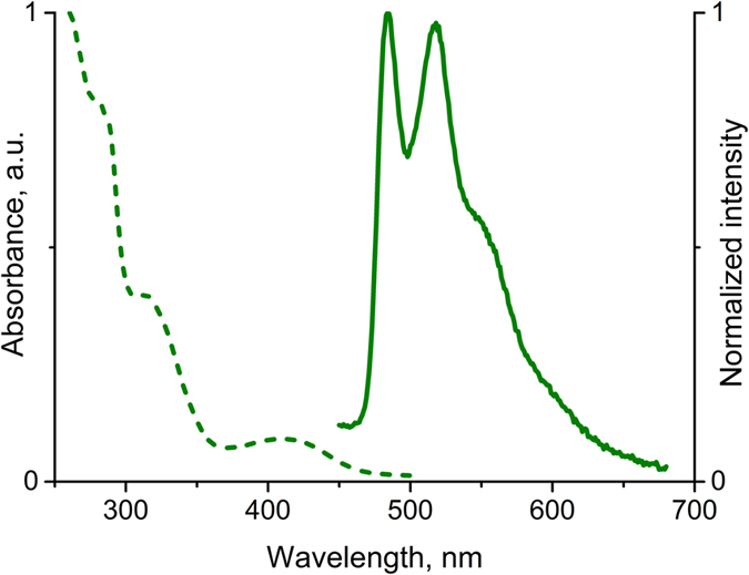
Table 1. Photophysical properties of 2 in aerated solution at ambient temperature.
| Solvent | λabs, nm (ε, 104 M−1cm−1) | λemi, nm | τ, μs | Q.Y., % |
|---|---|---|---|---|
| Methanol | 260(5.2), 285(2.4), 314(1.4), 410(0.35) | 483, 518, ~550 | 0.039 | <1 |
| Dichloromethane | 260(1.4), 285(1.1), 311(0.6), 415(0.13) | 484, 518, ~550 | 0.294 | 6.3 |
| Acetone | — | 483, 518, ~551 | 0.290 | 6.0 |
iλexit = 380 nm
Synthesis and characterization of labelled amphiphilic polypeptides
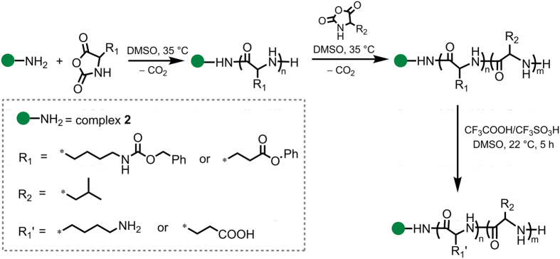
The synthesized luminescent compound 2 containing primary amino group was used as an initiator of ROP for both γ-(Bzl)Glu and ε-(Z)Lys NCAs. According to the known mechanism of NCA polymerization initiated by primary amines the reaction resulted in attachment of the initiator molecule to C-terminal position of the polypeptide chain32. The homopolypeptide obtained, also contains terminal NH2-group and can be applied as a macroinitiator for the synthesis of block-copolymer. The suggested strategy of the synthesis of amphiphilic polypeptides bearing C-terminal luminescent dye is illustrated in Fig. 3.
Figure 3. Synthesis of 2-bearing amphiphilic copolypeptides.
In principle the polymer chain bound to the phosphorescent chromophore may affect the luminescent properties of the label to give a partial or even a complete emission quenching depending on the length of the polymer. To evaluate this effect in the conjugates obtained, the conditions of synthesis were varied to prepare the polypeptides of different length. In compliance with the discussed step-wise synthesis, two amphiphilic polypeptide conjugates, 2-PLysn-b-PLeum and 2-PGlun-b-PLeum, were synthesized. In these macromolecular products, PGlu and PLys are hydrophilic polymers containing functional carboxylic and amino groups, respectively, whereas PLeu represents hydrophobic block. The molecular weight characteristics of the synthesized homopolymers and block copolymers are presented in Tables 2 and 3. Thus, the composition of the labeled copolypeptides can be described as follows: 2-PLys119-b-PLeu141 and 2-PGlu49-b-PLeu27.
Table 2. Molecular weight characteristics of labeled homopolypeptides.
| Sample | Mw | Mn | Mw/Mn | n |
|---|---|---|---|---|
| 2-P(Z)Lys | 44800 | 32000 | 1.4 | 119 |
| 2-P(Bzl)Glu | 12500 | 11500 | 1.1 | 49 |
Table 3. Molecular weight characteristics of labeled amphiphilic polypeptides.
| Sample | Hydrophilic block |
Hydrophobic block |
Copolymer | ||
|---|---|---|---|---|---|
| Mn | n | Mn | m | Mn | |
| 2-PLysn-b-PLeum | 16200 | 119 | 16000 | 141 | 32200 |
| 2-PGlun-b-PLeum | 7200 | 49 | 3150 | 27 | 10350 |
Investigation of the luminescent properties of 2-P(Z)Lys and 2-P(Bzl)Glu polypeptides in organic medium (CHCl3/DMSO) revealed the presence of emission bands typical for the complex 2 (Fig. 4) along with a weaker high energy emission component (400–450 nm), which can be assigned to organic matrix of polymers.
Figure 4. Emission spectra of 2-P(Z)Lys119 and 2-P(Bzl)Glu49 polypeptides (DMSO/chloroform 1/1; λexc = 385 nm, 22 °C), and 2-PGlu49-b-PLeu27 particles (borate buffer, pH = 9.4; λexc = 350 nm, 22 °C).
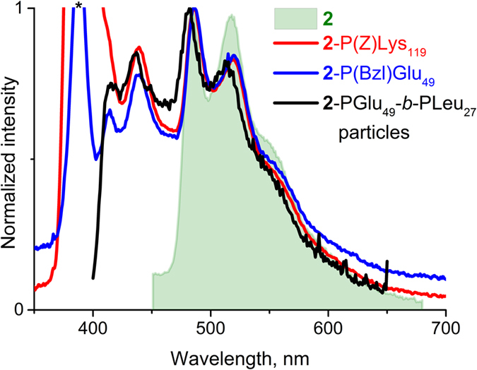
Excitation line at 385 nm is marked with an asterisk.
To estimate the content of the complex 2 in the samples of the synthesized polypeptides the thermogravimetric analyses (see Fig. S6 of Supporting Information) have been carried out. It was found that the content of metal complex with respect to theoretical one equals to 90.1% and 76.9% in 2-P(Bzl)Glu and 2-P(Z)Lys, respectively, that points to a rather high degree of the polymers labelling using the procedure suggested.
Characterization of self-assembled particles
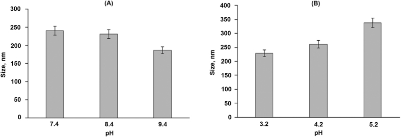
The synthesized amphiphilic polymers display a tendency to self-assembly in aqueous media that allowed for preparation of polymer particles using the phase inversion method. Since the hydrophilic blocks of the synthesized polypeptides had different functional groups, namely, amino and carboxylic ones, the optimal conditions for the 2-PLys119-b-PLeu141 and 2-PGlu49-b-PLeu27 self-assembly were essentially different. In particular, the alkaline medium evidently favors better ionization of carboxylic groups and, consequently, better solubility of 2-PGlu49-b-PLeu27. In turn, acidic conditions were preferable for the self-assembly of 2-PLys119-b-PLeu141. The dependence of the particle size on pH used in preparation of ther nanoparticles was established by DLS. The results obtained are presented in Fig. 5.
Figure 5.
The dependence of 2-PLys119-b-PLeu141 (A) and 2-PLys119-b-PLeu141 (B) particle size on pH applied for self-assembly.
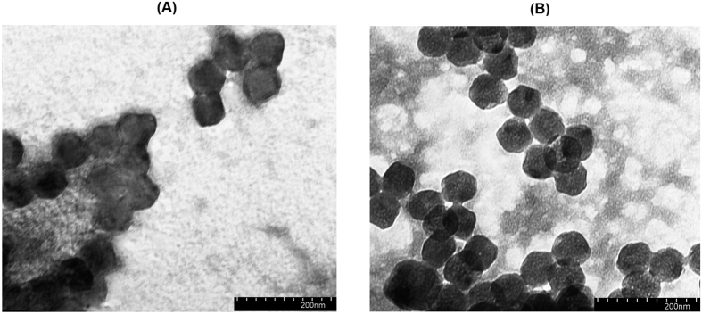
The average hydrodynamic diameter of the 2-PGlu49-b-PLeu27 particles was 241 ± 12 at pH 7.4 and 186 ± 24 at pH 9.4 that demonstrated more than 20% decrease in the particle size in alkaline media (Fig. 5A). Under the latter conditions the hydrophilic block (PGlu49) of the polymer is getting negatively charged because of acid residues deprotonation. This makes thermodynamically unfavorable self-assembly of the nanoparticles containing large number of like charged polymer chains that results in stabilization of smaller in “molecular weight” and size nanoparticles. A similar trend was observed for the 2-PLys119-b-pLeu141 particles in acidic media (Fig. 5B) where protonation of Lys amine group prevents formation of large nanoparticles due to repulsion between positively charge polymeric chains. In this case, the hydrodynamic diameter falls in the range 229 ± 14 at pH 3.2, and 338 ± 32 at pH 5.2. Additionally, the formation of the nanoparticles was studied by TEM (Fig. 6), which gives rather narrow particles size distribution. Even smaller size of nanoparticles (ca. 60–80 nm) measured by TEM method can be related to the deflation of water upon drying of particles on a microscope grid before TEM experiments.
Figure 6.
TEM images of 2-PLys119-b-PLeu141 (prepared at buffer, pH 3.2) (A) and 2-PLys119-b-PLeu141 (prepared at buffer, pH 9.4) (B) nanoparticles.
Zeta-potential is one of the key characteristics that affects the nanoparticles stability relative to aggregation in colloidal systems. The colloids are considered to be stable if ξ-potential is lower than –30 and higher than +30 mV48. Moreover, the high absolute value of zeta potential also favores the increase of drug loading efficiency and simultaneously improve the nanoparticles accumulation in target cells49. The particles obtained were characterized with ξ-potential values (in water) equal to −44 and +55 mV for 2-pGlu49-b-pLeu27 and 2-pLys119-b-pLeu141, respectively, that is a clear indication of their colloidal stability in aqueous media and high internalization rate. Stability of the particles in physiological solution was controlled by DLS, which showed the absence of aggregation within three weeks in the concentration range 0.005–2.000 mg/ml.
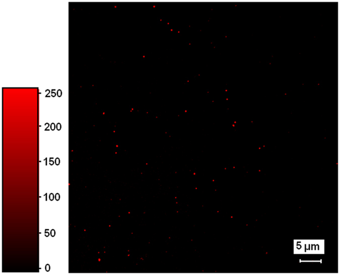
The labeled nanoparticles formed from 2-pGlu49-b-pLeu27 and 2-pLys119-b-pLeu141 display photoluminescence, see for example, the emission spectrum of 2-pGlu49-b-pLeu27 particles in buffer solution (pH 9.4), Fig. 4. Obviously, the emission band profile detected for the particles labeled with complex 2 are nearly identical to those measured for soluble label-bearing homopolymer. Additionally, the presence of complex 2 on the surface of polypeptide particles was proved by visualization of nanoobjects by two-photon confocal microscopy (Fig. 7). For better detection, the nanoparticles of ca. 600 nm were prepared by self-assembly of 2-PLys119-b-PLeu141 polypeptide in Na-phosphate buffer, pH 7.4, and used for analysis.
Figure 7. The confocal microscopy of 2-PLys119-b-PLeu141 colloids (buffer, pH 7.4; λex = 375–400 nm, λem = 500–530 nm).
Cell experiments: cytotoxicity, hemolysis and cell visualization
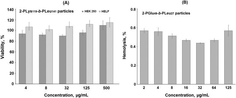
Recently it was observed that nanoparticles based on PGlu-b-PPhe copolymer did not affect cell metabolic activity31 that clearly points to the poly(amino acid) nanoparticles biocompatibility. It is reasonable to suppose that the PGlu-b-PLeu particles are not cytotoxic as well. However, the particles obtained in this study contain C-terminal Pt-complex that in principle may affect cell viability. To test the biocompatibility of the luminescent nanoparticles, MTT assay using HEK 293 and HELF cell lines was performed. The data shown in Fig. 8A indicate that there are no decrease in cell metabolic activity in the presence of 2-PLys119-b-PLeu141 during 24 h at the concentrations applied. A similar result was obtained for the 2-PGlu49-b-PLeu27 particles (Fig. S7 of Supporting Information).
Figure 8.
MTT (A) and hemolysis (B) assays of the luminescent polypeptide nanoparticles.
Another important test to prove biocompatibility of the nanoparticles is hemolysis of erythrocytes. The blood biocompatible substances and nanoobjects do not induce the destruction of external membrane of erythrocytes, which causes the release of intracell content. In this case, the toxicity of tested samples is evaluated by estimating the quantity of hemoglobin released. The results of hemolytic test revealed that the hemolysis rate for both 2-PLys119-b-PLeu141 and 2-PGlu49-b-PLeu27 particles was lower than 1%. As an example, the dependence of hemolysis rate on concentration of 2-PGlu49-b-PLeu27 particles is given in Fig. 8B. It is generally accepted that the materials are classified as non-hemolytic if the hemolysis rate is lower than 5%50. Thus, the nanoobjects studied can be considered as safe in the tested range of concentrations.
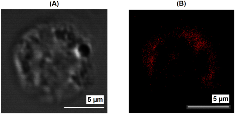
Finally, the luminescent nanoparticles obtained were tested for cell visualization. Fig. 9 and Fig. S8 show the cellular uptake of the 2-PLys119-b-PLeu141 nanoparticles by U937 cells after 1 hour of co-incubation, which indicates their applicability for luminescent visualization of living cells. The experiment displayed rapid cellular uptake that can be a result of relatively small size of the particles, as well as the positive charge of their surface. The confocal image shows a clear luminescent signal accumulation, which mostly distributed in the cytoplasm at the cell periphery.
Figure 9.
Bright-field (A) and confocal fluorescent (B); buffer, pH 7.4; λex = 375–400 nm, λem = 500–530 nm) images of U937 cell incubated with 2-PLys119-b-PLeu141 nanoparticles.
Conclusions
A novel luminescent PtII complex containing lateral amino group in the ligand environment was synthesized and its photophysical properties were studied. The complex obtained was used as effective ROP initiator to synthesize amphiphilic polypeptides, namely, PLys119-b-PLeu141 and PGlu49-b-PLeu27, with the luminescent label covalently bound to the C-terminal position of the polymer. It was shown that self-assembly of these polypeptides at optimal conditions led to the formation of nanoparticles of 250–350 nm size, which keep intact the luminescence characteristics of the starting platinum complex. The developed luminescent nanoparticles were found to be nontoxic and non-hemolytic in the tested concentration range and can be used for cell visualization with appreciable luminescence intensity in the green-yellow region of visible spectrum. Since such polypeptide-based soft materials are also considered as drug delivery systems, they have a great prospective in the application as duel-function nanoparticles with a combination of diagnostic and therapeutic properties.
Additional Information
How to cite this article: Vlakh, E. G. et al. Self-assemble nanoparticles based on polypeptides containing C-terminal luminescent Pt-cysteine complex. Sci. Rep. 7, 41991; doi: 10.1038/srep41991 (2017).
Publisher's note: Springer Nature remains neutral with regard to jurisdictional claims in published maps and institutional affiliations.
Supplementary Material
Acknowledgments
The authors greatly appreciate financial support of Russian Science Foundation. The part of work devoted to polymer synthesis, labelled-nanoparticles preparation and their investigation (including cell experiments) was supported by the RSF grant #14-50-00069. The synthesis, characterization of the Pt complexes 1, 2 and investigation of the photophysical properties of labeled polymers and nanoparticles were funded by RSF grant #16-43-03003. The authors are very grateful to Prof. E. Ruehl (Free University of Berlin) for his kind help with TEM measurements. The investigations were performed using the equipment of core facilities of St. Petersburg State University Research Park: Centre for Magnetic Resonance, Centre for Optical and Laser Materials Research, Centre for Chemical Analysis and Materials, Center on Development of Molecular and Cell Technologies as well as Thermogravimetric and Calorimetric Research Centre.
Footnotes
The authors declare no competing financial interests.
Author Contributions E. Vlakh – development of research concept and strategy, planning of experiments, data analysis; A. Mikhailova - experiments on polymer synthesis and nanoparticle preparation; A. Hubina – polymer and particle characterization, cell experiments; V. Sharoyko - cell experiments; T. Tennikova – general management of the project aimed on the development of novel biomedical nanomaterials; data analysis. E. Vlakh, T. Tennikova, S. Tunik – writing of the article. S. Tunik –management of the chemical part of the project (synthesis of the platinum complex, structural characterization, photophysics) and data analysis. D. Zhukovsky – synthesis of the platinum complex, samples preparation for NMR and photophysical experiments E. Grachova and J. Shakirova, NMR, mass spectroscopic, and photophysical measurements, interpretation of the data.
References
- Escobedo J. O., Rusin O., Lim S. & Strogin R. NIR dyes for bioimaging applications. Curr. Opin. Chem. Biol. 14, 64–70 (2010). [DOI] [PMC free article] [PubMed] [Google Scholar]
- Chelushkin P. S., Krupenya D. V., Tseng Y.-J., Kuo T.-Y., Chou P.-T., Koshevoy I. O., Burov S. V. & Tunik S. P. Water-Soluble Noncovalent Adducts of the Heterometallic Copper Subgroup Complexes and Human Serum Albumin with Remarkable Luminescent Properties. Chem. Comm. 50, 849–851 (2014). [DOI] [PubMed] [Google Scholar]
- Xu H., Wang L. & Fan C. Bioanalysis and Bioimaging with Fluorescent Conjugated Polymers and Conjugated Polymer Nanoparticles in Functional Nanoparticles for Bioanalysis, Nanomedicine and Bioelectronic Devices (eds. Hepel M., Zhong C.-J. ) 81–117 (ACS Publications: Washington, 2012).
- Liu Y., Miyoshi H. & Nakamura M. Nanomedicine for drug delivery and imaging: A promising avenue for cancer therapy and diagnosis using targeted functional nanoparticles. Int. J. Cancer. 120, 2527–2537 (2007). [DOI] [PubMed] [Google Scholar]
- Wolfbeis O. S. An overview of nanoparticles commonly used in fluorescent bioimaging. Chem. Soc. Rev. 44, 4743–4768 (2015). [DOI] [PubMed] [Google Scholar]
- Ma D.-L., He H.-Z., Leung K.-H., Chan D. & Leung C.-H. Bioactive Luminescent Transition-Metal Complexes for Biomedical Applications. Angew. Chem. Int. Ed. 52, 7666–7682 (2013). [DOI] [PubMed] [Google Scholar]
- Doulain P.-E., Decréau R., Racoeur C., Goncalves V., Dubrez L., Bettaieb A., Le Gendre P., Denat F., Paul K., Goze C. & Bodio E. Towards the elaboration of new gold-based optical theranostics. Dalton Trans, 44, 4874–4883 (2015). [DOI] [PubMed] [Google Scholar]
- Kim T. & Hyeon T. Applications of inorganic nanoparticles as therapeutic agents. Nanotechnol. 25, 012001 (2014). [DOI] [PubMed] [Google Scholar]
- Masood F. Polymeric nanoparticles for targeted drug delivery system for cancer therapy. Mater. Sci. Eng. C. 60, 569–578 (2016) [DOI] [PubMed] [Google Scholar]
- Allen T. M. & Cullis P. R. Liposomal drug delivery systems: from concept to clinical applications. Adv. Drug Deliv. Rev. 65, 36–48 (2013). [DOI] [PubMed] [Google Scholar]
- He X. X., Wang Y. S., Wang K. M., Chen M. & Chen S. Y. Fluorescence Resonance Energy Transfer Mediated Large Stokes Shifting Near-Infrared Fluorescent Silica Nanoparticles for in Vivo Small-Animal Imaging. Anal. Chem. 84, 9056–9064 (2012). [DOI] [PubMed] [Google Scholar]
- Roy M., Niu C. J., Chen Y. H., McVeigh P. Z., Shuhendler A. J., Leung M. K., Mariampillai A., DaCosta R. S. & Wilson B. C. Estimation of Minimum Doses for Optimized Quantum Dot Contrast-Enhanced Vascular Imaging In Vivo. Small. 8, 1780–1792 (2012). [DOI] [PubMed] [Google Scholar]
- Sharma J., Yeh H. C., Yoo H., Werner J. H. & Martinez J. S. Silver nanocluster aptamers: in situ generation of intrinsically fluorescent recognition ligands for protein detection. Chem. Comm. 47, 2294–2296 (2011). [DOI] [PubMed] [Google Scholar]
- Hemmer E., Venkatachalam N., Hyodo H., Hattori A., Ebina Y., Kishimoto H. & Soga K. Upconverting and NIR emitting rare earth based nanostructures for NIR-bioimaging. Nanoscale 5, 11339–11361 (2013). [DOI] [PubMed] [Google Scholar]
- Lee K. J., Oh W. K., Song J., Kim S., Lee J. & Jang J. Photoluminescent polymer nanoparticles for label-free cellular imaging. Chem. Comm. 46, 5229–5231 (2010). [DOI] [PubMed] [Google Scholar]
- Holowka E. P., Pochan D. J. & Deming T. J. Charged polypeptide vesicles with controllable diameter. J. Am. Chem. Soc. 127, 12423–12428 (2005). [DOI] [PubMed] [Google Scholar]
- Liu L., Fu L., Jing T., Ruan Z. & Yan L. Near-Infrared Polymeric Nanoparticles with High Content of Cyanine for Bimodal Imaging and Photothermal Therapy. ACS Appl. Mater. Interfaces. 8, 8980–8990 (2016). [DOI] [PubMed] [Google Scholar]
- Kolitz-Domb M., Grinberg I., Corem-Salkmon E. & Margel S. Engineering of near infrared fluorescent proteinoid-poly(L-lactic acid) particles for in vivo colon cancer detection. J. Nanotechnol. 12, 30, 13 pages (2014). [DOI] [PMC free article] [PubMed] [Google Scholar]
- Hermanson G. T. Bioconjugate techniques, Elsevier: Amsterdam, 2013, pp. 396–497. [Google Scholar]
- Gustafson T. P., Lim Y. H., Flores J. A., Heo G. S., Zhang F., Zhang S., Samarajeewa S., Raymond J. E. & Wooly K. L. Holistic Assessment of Covalently Labeled Core–Shell Polymeric Nanoparticles with Fluorescent Contrast Agents for Theranostic Applications. Langmuir. 30, 631–641 (2014). [DOI] [PMC free article] [PubMed] [Google Scholar]
- Robin M. P. & O’Reilly R. K. Strategies for preparing fluorescently labelled polymer nanoparticles. Polym. Int. 64, 174–182 (2015). [Google Scholar]
- Kim H., Park H. T., Tae Y. M., Kong W. H., Sung D. K., Hwang B. W., Kim K. S., Kim Y. K. & Hahn S. K. Bioimaging and pulmonary applications of self-assembled Flt1 peptide–hyaluronic acid conjugate nanoparticles. Biomaterials. 34, 8478–8490 (2013). [DOI] [PubMed] [Google Scholar]
- Tanisaka H., Kizaka-Kondoh S., Makino A., Tanaka S., Hiraoka M. & Kimura S. Near-Infrared Fluorescent Labeled Peptosome for Application to Cancer Imaging. Bioconjugate Chem. 19, 109–117 (2008). [DOI] [PubMed] [Google Scholar]
- Birch N. P. & Schiffman J. D. Characterization of self-assembled polyelectrolyte complex nanoparticles formed from chitosan and pectin. Langmuir. 30, 3441–3447 (2014). [DOI] [PubMed] [Google Scholar]
- Rahikkala A., Aseyev V., Tenhu H., Kauppinen E. I. & Raula J. Thermoresponsive Nanoparticles of Self-Assembled Block Copolymers as Potential Carriers for Drug Delivery and Diagnostics. Biomacromolecules. 16, 2750–2756 (2015). [DOI] [PubMed] [Google Scholar]
- Wiradharma N., Zhang Y., Venkataraman S., Hedrick J. L. & Yang Y. Y. Self-assembled polymer nanostructures for delivery of anticancer therapeutics. Nano Today. 4, 302–317 (2009). [Google Scholar]
- Kandoth N., Kirejev V., Monti S., Gref R., Ericson M. B. & Sortino S. Two-Photon Fluorescence Imaging and Bimodal Phototherapy of Epidermal Cancer Cells with Biocompatible Self-Assembled Polymer Nanoparticles. Biomacromolecules. 15, 1768–1776 (2014). [DOI] [PubMed] [Google Scholar]
- Wu T., Johnsen B., Qin Z., Morimoto M., Baillie D., Irie M. & Branda N. R. Two-colour fluorescent imaging in organisms using self-assembled nano-systems of upconverting nanoparticles and molecular switches. Nanoscale. 7, 11263–11266 (2015). [DOI] [PubMed] [Google Scholar]
- Toksoz S. & Guler M. O. Self-assembled peptidic nanostructures. Nano Today. 4, 458–469 (2009). [Google Scholar]
- Habibi N., Kamaly N., Memic A. & Shafiee H. Self-assembled peptide-based nanostructures: Smart nanomaterials toward targeted drug delivery. Nano Today. 11, 41–60 (2016). [DOI] [PMC free article] [PubMed] [Google Scholar]
- Vlakh E., Ananyan A., Zashikhina N., Hubina A., Pogodaev A., Volokitina M., Sharoyko V. & Tennikova T. Preparation, characterization, and biological evaluation of poly(glutamic acid)-b-polyphenylalanine polymersomes. Polymers. 8, 212, 14 pages (2016). [DOI] [PMC free article] [PubMed] [Google Scholar]
- Cheng J. & Deming T. J. Synthesis of polypeptides by ring-opening polymerization of α-amino acid N-carboxyanhydrides. Top. Curr. Chem. 310, 1–26 (2012). [DOI] [PubMed] [Google Scholar]
- Kim M. S., Dayananda K., Choi E. K., Park H. J., Kim J. S. & Lee D. S. Synthesis and characterization of poly(l-glutamic acid)-block-poly(l-phenylalanine). Polymer. 50, 2252–2257 (2009). [Google Scholar]
- Zhao Q., Huang C. & Li F. Phosphorescent heavy-metal complexes for bioimaging. Chem. Soc. Rev. 40, 2508–2524 (2011). [DOI] [PubMed] [Google Scholar]
- Fernández-Moreira V., Thorp-Greenwood F. L. & Coogan M. P. Application of d6 transition metal complexes in fluorescence cell imaging. Chem. Comm. 46, 186–202 (2010). [DOI] [PubMed] [Google Scholar]
- Chelushkin P. S., Nukolova N. V., Melnikov A. S., Serdobintsev P. Yu., Melnikov P. A., Krupenya D. V., Koshevoy I. O., Burov S. V. & Tunik S. P. HSA-based phosphorescent probe for two-photon in vitro visualization. J. Inorg. Biochem. 149, 108–111 (2015). [DOI] [PubMed] [Google Scholar]
- Koshel E. I., Chelushkin P. S., Melnikov A. S., Serdobintsev P. Yu., Stolbovaia A..Yu., Saifitdinova A. F., Shcheslavskiy V. I., Chernyavskiyh O., Gaginskaya E. R., Koshevoy I. O. & Tunik S. P. Lipophilic phosphorescent gold(I) clusters as selective probes for visualization of lipid droplets by two-photon microscopy. J. Photochem. Photobiol. A. 332, 122–130 (2017). [Google Scholar]
- Beljaev A. A., Krupenya D. V., Grachova E. V., Gurzhiy V. V., Melnikov A. S., Serdobintsev P..Yu., Sinitsyna E. S., Vlakh E. G., Tennikova T. B. & Tunik S. P. Supramolecular AuI-CuI complexes as new luminescent labels for covalent bioconjugation. Bioconjugate Chem. 27, 143–150 (2016). [DOI] [PubMed] [Google Scholar]
- Uesugi H., Tsukuda T., Takao K. & Tsubomura T. Highly emissive platinum(II) complexes bearing carbene and cyclometalated ligands. Dalton Trans. 42, 7396–7403 (2013). [DOI] [PubMed] [Google Scholar]
- Zhu Y., Cai C. & Lu G. N-Heterocyclic Carbene-Catalyzed α-Alkylation of Ketones with Primary Alcohols. Helvetica Chim. Acta. 97, 1666–1671 (2014). [Google Scholar]
- Wilder R. & Mobashery S. The use of triphosgene in preparation of N-carboxy α-amino acid anhydrides. J. Org. Chem. 57, 2755–2756 (1992). [Google Scholar]
- Hubina A. V., Pogodaev A. A., Sharoyko V. V., Vlakh E. G. & Tennikova T. B. Self-assembled spin-labeled nanoparticles based on poly(amino acids). React. Funct. Polym. 100, 173–180 (2016). [Google Scholar]
- Li C., Wang S., Huang Y., Zheng B., Tian Z., Wen Y. & Li F. Dalton Trans. 42, 4059–4067 (2013). [DOI] [PubMed] [Google Scholar]
- Bossi A., Rausch A. F., Leitl M. J., Czerwieniec R., Whited M. T., Djurovich P. I., Yersin H. & Thompson M. E. Synthesis, characterization and electrochemiluminescent properties of cyclometalated platinum(II) complexes with substituted 2-phenylpyridine ligands. Inorg. Chem. 52, 12403–12415 (2013). [DOI] [PubMed] [Google Scholar]
- Brooks J., Babayan Y., Lamansky S., Djurovich P. I., Tsyba I., Bau R. & Thompson M. E. Synthesis and characterization of phosphorescent cyclometalated platinum complexes. Inorg. Chem. 41, 3055–3066 (2002). [DOI] [PubMed] [Google Scholar]
- Solomatina A. I., Krupenya D. V., Gurzhiy V. V., Zlatkin I., Pushkarev A. P., Bochkarev M. N., Besley N. A., Bichoutskaia E. & Tunik S. P. Cyclometallated platinum(II) complexes containing NHC ligands: synthesis, characterization, photophysics and their application as emitters in OLEDs. Dalton Trans. 44, 7152–7162 (2015). [DOI] [PubMed] [Google Scholar]
- Matsuda Y., Kohra S., Katou K., Uemura T. & Yamashita K. Synthesis of an Annulenoannulenone, 3H-Benzo[e]cycl[3.3.2]azin-3-one. Heterocycles. 60, 405–411 (2003). [Google Scholar]
- Kratošová G., Dědková K., Vávra I. & Čiampor F. Investigation of Nanoparticles in Biological Objects by Electron Microscopy in Intracellular delivery II: Fundamentals and Applications (eds. Prokop A., Iwasaki Y., Harada A. ) 479 (Springer: Heidelberg, 2014).
- Pan J. & Feng S. S. Targeting and imaging cancer cells by Folate-decorated, quantum dots (QDs)- loaded nanoparticles of biodegradable polymers. Biomaterials. 30, 1176–1183 (2009). [DOI] [PubMed] [Google Scholar]
- Huang Z., Yang Y. & Fang J. Development and evaluation of lipid nanoparticles for camptothecin delivery: a comparison of solid lipid nanoparticles, nanostructured lipid carriers, and lipid emulsion. Acta Pharmacol. Sin. 29, 1094–1102 (2008). [DOI] [PubMed] [Google Scholar]
Associated Data
This section collects any data citations, data availability statements, or supplementary materials included in this article.


