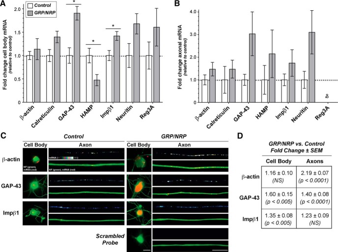
Figure 4.
Exposure to GRPs/NRPs alters the levels of growth-associated mRNA cell bodies and axons of adult DRG neurons. A, B, The modified coculture system from Figure 3 was used for analyses of RNA levels from the explanted ganglia (“cell body RNA”) and their extended axons (“axonal RNA”). Data from RT-ddPCR analyses with cell body and axonal RNA preparations for known axonal mRNAs are shown in A and B, respectively. Values are shown for the NRP/GRP coculture relative to control samples, as indicated, ±SD for biological replicates (N = 4; p ≤ 0.05 for indicated columns; &no positive droplets were detected for Reg3a amplification despite highest template input levels). C, Representative FISH/IF images of cell bodies and axon shafts are shown for indicated mRNAs for dissociated DRGs cultured under control or GRP/NRP-exposed conditions. For these studies, DRGs were cultured on coverslips over a bed of GRP/NRP cells. Image pairs for control and GRP/NRP conditions are exposure matched, and axonal mRNA signals are shown as a spectral intensity in the upper rows of axon image sequences and cell body, and lower axon panels show merged images for mRNA and NF. Scale bars: cell body panels, 25 µm; axon panels, 10 µm. D, Quantification of axonal FISH signal intensities from matched exposure images are shown for GRP/NRP coculture vs control cultures as the fold change ± SEM (n > 50 for axons and n > 25 for cell bodies in three culture preparations; p values are from ANOVA with Tukey post hoc analyses).

