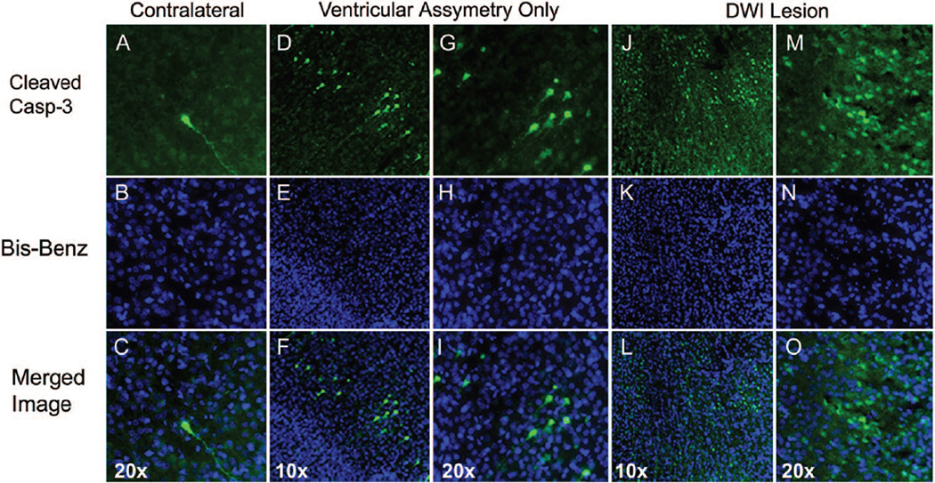
Figure 4.
Localization of cleaved caspase-3 24 hours post-H-I. A–C, Only occasional cells with cleaved caspase-3 are seen in hypoxic hemisphere in each brain studied. Cleaved caspase-3 is present in both cell body and processes of the shown cell (A, C, green). D–I, Scattered cells with cleaved caspase-3 are present in the cortex of pups in group 2. Many cells are in early apoptotic stages as seen from cell morphology (D and G) and the appearance of the nuclei (E and H). J–O, Marked number of cells with cleaved caspase-3 is seen ipsilateral in animals in group 1. C, F, I, L, and O, Overlay of cleaved caspase-3(green)/bis-benzamide (blue).

